

Testicular Appendage Ultrasound: Preparation, Procedure and Results
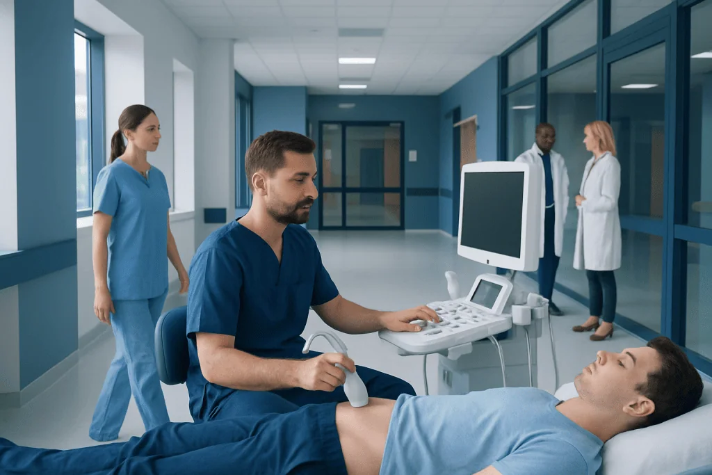
Testicular ultrasound does not use radiation and is usually painless. It can assess blood flow to the testicle, which is important when torsion is suspected.
Key Takeaways
- Testicular ultrasound does not use radiation and is usually painless.
- It can assess blood flow to the testicle, which is important when torsion is suspected.
- A twisted testicular appendage may appear as a small avascular structure near the upper testicle, but it is not always directly visible.
- No special fasting or medication changes are usually needed before the scan.
- Sudden or severe testicular pain needs urgent medical assessment because testicular torsion must be excluded.
A testicular appendage ultrasound is a noninvasive imaging test that uses sound waves to examine the testicles and nearby small structures, including the testicular appendages. It is commonly used when sudden scrotal pain needs prompt assessment, helping clinicians distinguish a benign appendage torsion from conditions that require urgent treatment.
Overview: what is a testicular appendage ultrasound?
A testicular appendage ultrasound is a scrotal ultrasound examination performed to investigate pain, swelling, tenderness, or a lump around a testicle. The testicular appendages are small, harmless remnants of embryonic tissue located near the upper part of the testicle or epididymis. They usually cause no symptoms, but one can twist, known as torsion of a testicular appendage, leading to localized pain and inflammation.
The scan uses high-frequency sound waves to create real-time images of the testicles, epididymis, scrotal tissues, and blood vessels. Doppler ultrasound is typically included to show blood flow. This is particularly valuable in acute scrotal pain because normal blood flow within the testicle supports an appendage-related problem, while reduced or absent flow can raise concern for testicular torsion, an emergency.
Ultrasound findings are interpreted alongside symptoms and a physical examination. The purpose is not only to look for an affected appendage, but also to evaluate other possible causes of discomfort, such as inflammation, infection, fluid around the testicle, injury, or a testicular mass.
How it works and who may need the scan
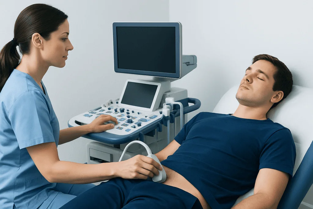
Ultrasound works by sending sound waves through a handheld device called a transducer. The waves reflect differently from fluid, soft tissue, and blood vessels, allowing the ultrasound system to form images on a screen. Color Doppler and spectral Doppler techniques assess the pattern and amount of blood flow within and around the testicles.
A clinician may request this examination for a child, teenager, or adult with one-sided scrotal pain, swelling, redness, tenderness, a new palpable change, or pain after an injury. Torsion of a testicular appendage is especially common in boys before puberty, but scrotal ultrasound is useful at any age when the cause of symptoms is unclear.
The test is also appropriate when an examination alone cannot safely rule out more urgent conditions. Although appendage torsion is usually self-limiting, symptoms can overlap with testicular torsion, epididymitis, and other conditions. Imaging should not delay emergency surgical assessment if clinicians strongly suspect testicular torsion.
How should I prepare for a testicular ultrasound?

Most people do not need special preparation for a testicular ultrasound. Fasting is not required, and regular meals, fluids, and prescribed medicines can usually be continued unless the healthcare team gives different instructions for another reason.
It may be helpful to wear comfortable, loose-fitting clothing that is easy to remove from the waist down. The patient should tell the sonographer or doctor about recent injury, surgery, symptoms of infection, medicines, allergies, or any previous scrotal imaging. A parent or trusted adult may usually accompany a child, according to the imaging department’s policies.
For people with significant pain, the clinician may recommend appropriate pain relief before or after the examination. It is best not to apply creams, powders, or topical products to the scrotum immediately before the scan, as these can make it harder for the gel and transducer to contact the skin properly.
Step by step: what happens during the procedure?
The examination is usually performed in an ultrasound department, emergency setting, or urology clinic. The patient lies on their back, and the scrotum is gently supported with a towel or positioned comfortably. Privacy is maintained, and only the area being examined is exposed.
A clear water-based gel is placed on the scrotal skin. The sonographer moves the transducer over both sides of the scrotum, taking images of each testicle, the epididymis, and nearby structures. Comparing both sides is important because it helps identify subtle differences in size, swelling, tissue appearance, and blood flow.
Images are collected in grayscale and with Doppler settings. The operator may apply light pressure or ask where the discomfort is most noticeable, but the scan should not be painful. A radiologist or appropriately trained clinician reviews the images and sends a report to the referring doctor, who explains what the findings mean in the context of the patient’s symptoms.
Can testicular appendages be seen on an ultrasound?
Testicular appendages can sometimes be seen on an ultrasound, particularly when one is enlarged or inflamed. A normal appendage is very small and may not be visible. When torsion has occurred, the affected appendage may appear as a small rounded or oval structure next to the upper pole of the testicle, often without internal blood flow.
However, a scan does not need to show the appendage directly to be useful. In suspected appendage torsion, ultrasound may show normal or increased blood flow inside the testicle, local inflammation, a small amount of surrounding fluid, or thickening of adjacent tissues. These findings can support the diagnosis while helping exclude impaired blood supply to the testicle.
The report may describe the result as consistent with torsion of an appendix testis or appendix epididymis, depending on its location. If imaging is inconclusive and symptoms suggest a time-sensitive problem, the clinical team will prioritize safety and may arrange urgent urology review.
What does a testicular appendage feel like?
A normal testicular appendage is typically too small to feel. It is not usually noticeable during everyday activities or self-examination. If an appendage twists and becomes inflamed, it may cause a tender spot near the upper part of one testicle, along with swelling or sensitivity of the scrotum.
Some children and teenagers with appendage torsion develop a small bluish discoloration under the scrotal skin, sometimes called a “blue dot” sign. This sign is not present in everyone and should not be used to diagnose the cause of pain at home. Pain and swelling can make it difficult to tell which structure is involved by touch alone.
A new hard lump within the testicle, persistent enlargement, or any unexplained scrotal change should be assessed by a qualified clinician. Self-examination can increase awareness of normal anatomy, but it cannot reliably distinguish benign changes from conditions needing medical care.
How long does a testicular ultrasound take?
A testicular ultrasound commonly takes around 15 to 30 minutes, although the total visit may be longer because of registration, preparation, waiting time, and review of urgent findings. The scan can take longer if the patient has severe pain, swelling, prior surgery, or findings that need additional Doppler images.
There is no recovery period after the examination. The gel is wiped away, and the patient can generally return to normal activities immediately. The procedure does not involve injections, sedation, radiation, or surgical instruments.
In an emergency assessment, preliminary results may be available quickly so the treating team can decide whether urgent care is needed. In non-urgent settings, the timing of the final report varies by facility and should be discussed with the referring clinician.
Results, benefits, limitations and next steps
The main benefit of a testicular appendage ultrasound is that it provides fast, radiation-free information about the structures inside the scrotum and their blood supply. It can support a diagnosis of appendage torsion and identify signs of alternative conditions, including epididymal inflammation, hydrocele, varicocele, trauma, or a testicular lesion.
Ultrasound is very safe. The gel may feel cool, and gentle probe pressure can be uncomfortable over an already tender area, but serious risks are not expected. Its main limitation is that a small appendage may not be seen clearly, and image findings must always be considered together with the clinical examination and the timing of symptoms.
When appendage torsion is diagnosed and testicular blood flow is normal, treatment commonly focuses on rest, scrotal support, and clinician-recommended pain relief. Symptoms generally improve over time. In contrast, suspected testicular torsion requires immediate urology assessment and may need urgent testicular torsion surgery to protect the testicle.
Acibadem International’s multidisciplinary urology, radiology, pediatric, and emergency specialists at JCI-accredited hospitals can assess acute scrotal symptoms and coordinate appropriate care for international patients.
When to seek medical care
Sudden, severe, or worsening pain in a testicle or the scrotum should be treated as urgent. Immediate medical evaluation is particularly important if pain begins abruptly, follows physical activity or sleep, or is accompanied by nausea, vomiting, marked swelling, redness, fever, or a testicle that appears higher or positioned differently than usual.
Medical care is also advisable for a new lump, persistent scrotal heaviness, discomfort that does not improve, pain after an injury, or urinary symptoms such as burning or discharge. These symptoms may have several causes, and an examination plus ultrasound can help guide appropriate treatment.
Parents and caregivers should seek urgent assessment for children with unexplained scrotal pain, even when the child seems otherwise well. It is not safe to assume that pain is caused by a twisted appendage without a medical evaluation, because prompt treatment can be critical when testicular torsion is possible.
Frequently asked questions
Is a testicular appendage ultrasound painful?
The ultrasound itself is usually painless. The transducer is moved gently over the scrotum, but pressure may feel uncomfortable if the area is already tender or swollen. The sonographer can adjust positioning and pressure to improve comfort.
Does a testicular ultrasound use radiation?
No. Ultrasound uses sound waves rather than X-rays or other ionizing radiation. It is widely used to assess scrotal conditions in children and adults.
Can ultrasound rule out testicular torsion?
Doppler ultrasound is an important tool for assessing blood flow to the testicle and can strongly support or reduce concern for torsion. However, no test replaces urgent clinical judgment when symptoms are strongly suggestive of testicular torsion. Emergency urology evaluation should not be delayed in a concerning situation.
What happens if torsion of a testicular appendage is found?
When the testicle has normal blood flow and appendage torsion is diagnosed, treatment is often conservative. A clinician may recommend rest, scrotal support, and suitable pain relief. Follow-up is advised if symptoms do not improve as expected or become worse.
Can a testicular appendage ultrasound find cancer?
Ultrasound can identify an abnormal area or mass within a testicle and helps clinicians decide whether further assessment is needed. It cannot determine every diagnosis on its own, so suspicious findings may lead to blood tests, specialist review, or additional imaging. Most scrotal pain is not caused by cancer, but a new testicular lump should always be evaluated.
Can I drive after a testicular ultrasound?
Most people can drive and resume usual activities immediately after the scan because no sedation is used. If severe pain, an acute illness, or emergency treatment is involved, the medical team may give different advice. Patients should follow the instructions provided for their individual situation.
References
- American College of Radiology
- American Urological Association
- Radiological Society of North America
- National Institute of Diabetes and Digestive and Kidney Diseases
- European Association of Urology
This article is for general information only and is not a substitute for professional medical advice. Please consult a qualified doctor about your individual situation.
Ultrasound in Turkey — costs, top hospitals & a free quote
JCI-accredited · board-certified surgeons · reply within 24h
Add us as a Preferred Source to see more of our trusted health content across Google Search, AI Overviews and Discover.

Check-up & Preventive Medicine
Comprehensive health screenings designed for early detection and prevention.
1 specialists in this unitMore from the Health Library



Related Specialists
Dr. İskender Dik
Internal Medicine
Dr. Cansu Hemşinlioğlu
Inpatient Clinic Physicians
Dr. Şelale Doğan
Anesthesiology
Prof. Dr. Erkin Sönmez
Neurosurgery