Ultrasound after Mammogram: Preparation, Procedure and Results
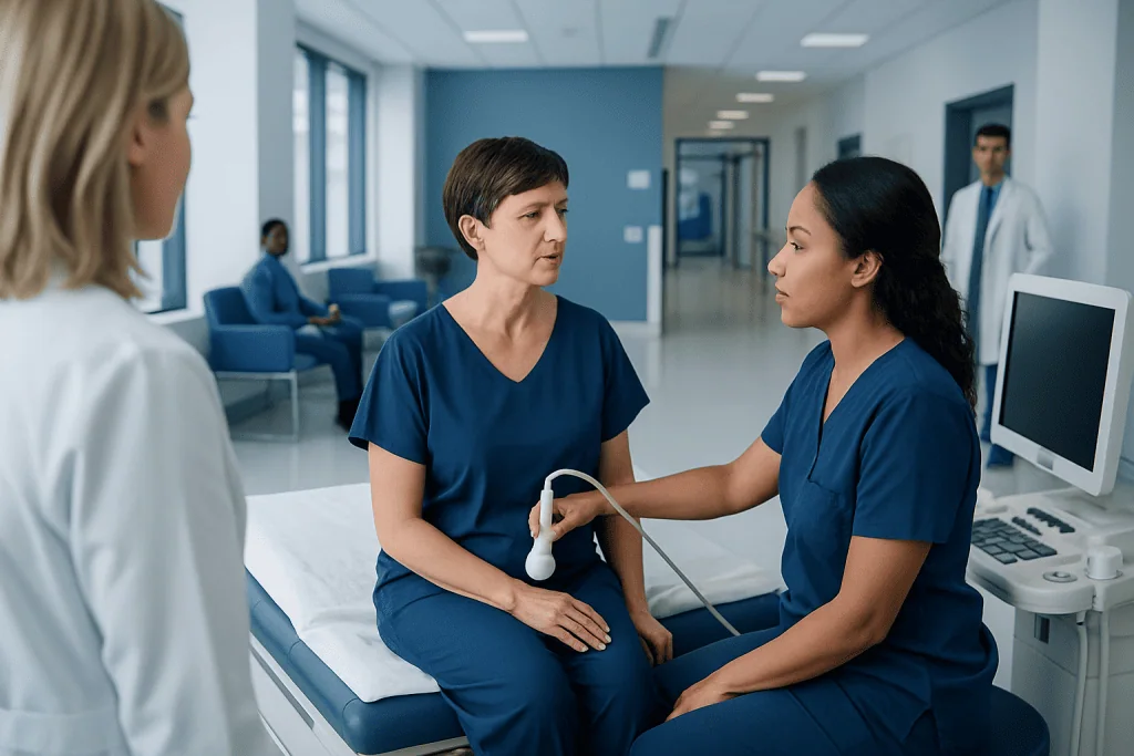
A follow-up ultrasound is common and does not by itself mean breast cancer is present. Breast ultrasound uses sound waves, involves no radiation and is usually painless.
Key Takeaways
- A follow-up ultrasound is common and does not by itself mean breast cancer is present.
- Breast ultrasound uses sound waves, involves no radiation and is usually painless.
- It is especially useful for evaluating a lump, a mammogram callback or dense breast tissue.
- Many findings are benign, including simple cysts and noncancerous breast changes.
- Results may be available on the day of testing or after review by a breast radiologist.
- A biopsy may be advised only when imaging cannot confidently identify a finding as benign.
An ultrasound after mammogram is commonly recommended to examine a specific breast area more closely. It uses sound waves rather than radiation and can help distinguish fluid-filled cysts from solid tissue or clarify an inconclusive mammogram finding.
Overview: Why an Ultrasound May Follow a Mammogram
An ultrasound after mammogram is a focused imaging examination used to provide more detail about an area that needs clarification. It is commonly requested after a screening mammogram shows an asymmetry, density, possible mass or other change that cannot be fully assessed on mammogram images alone. A callback for more imaging is not a diagnosis of cancer.
During breast ultrasound, a trained sonographer or radiologist moves a handheld device called a transducer over the breast. The device sends high-frequency sound waves into the tissue and creates real-time images. Unlike mammography, ultrasound does not use X-rays or compression of the breast.
The examination can show whether a finding is filled with fluid, such as a simple cyst, or is solid. It can also help assess the shape, edges and internal features of a breast area and may be used to examine nearby lymph nodes when appropriate.
Should I Worry if I Need an Ultrasound After a Mammogram?
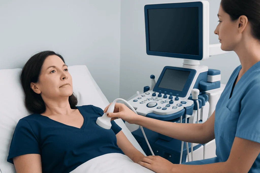
Most people do not need to assume the worst when an ultrasound is requested after a mammogram. Follow-up imaging is a routine part of careful breast screening, particularly when the original images show overlapping tissue, dense breast tissue or a small area that needs a closer look.
Many follow-up findings are benign. Examples include cysts, fibroadenomas, normal glandular tissue and stable areas of breast density. The purpose of the ultrasound is to obtain enough information for the radiologist to classify the finding confidently and recommend the next appropriate step.
Occasionally, ultrasound identifies features that need additional evaluation, such as short-interval follow-up imaging or a tissue biopsy. Even when a biopsy is recommended, it is a way to establish a clear diagnosis rather than a confirmation that cancer is present.
Anyone who has noticed a new lump, nipple discharge, skin change or persistent focal breast pain should tell the imaging team, even if the mammogram was performed as routine screening.
What Does It Mean if I Get a Callback for an Ultrasound After a Mammogram?

A callback means that the radiologist would like additional images before issuing a final assessment. Screening mammograms are designed to identify areas that may benefit from a closer examination; they are not always able to show every detail of breast tissue in a single set of images.
The next appointment may include diagnostic mammogram views, breast ultrasound or both. Diagnostic views use different positions or magnification to better evaluate the area seen on the screening study. Ultrasound may then help determine whether the finding is a cyst, a solid mass or simply normal tissue superimposed on the original images.
People with dense breasts may be called back more often because dense glandular tissue can make mammograms more difficult to interpret. Density is common and is not itself a cancer diagnosis, although it can modestly affect both breast cancer risk and mammogram sensitivity.
The radiologist generally assigns a standardized assessment category after completing the diagnostic work-up. This report explains whether routine screening, imaging follow-up or further investigation is recommended.
How Breast Ultrasound Works and Who May Need It
Ultrasound creates images from echoes produced when sound waves meet different types of tissue. Fluid allows sound waves to pass through differently from solid tissue, helping clinicians identify the characteristics of a breast finding. Doppler ultrasound may sometimes be used to assess blood flow in or around an area.
Breast ultrasound may be recommended after an abnormal or unclear mammogram, to investigate a symptom such as a new palpable lump, or to assess a focused area of pain or change. It can also be useful for people who are pregnant, younger patients with dense breast tissue, and patients who need image guidance during certain procedures.
Ultrasound is complementary to mammography rather than a universal replacement for it. Mammography is particularly good at detecting microcalcifications, which may not be visible on ultrasound. The most suitable imaging approach depends on age, symptoms, breast density, personal risk and previous imaging findings.
For a suspicious area that requires tissue sampling, ultrasound can guide the needle accurately. Breast biopsy is typically recommended only when imaging findings warrant confirmation under a microscope.
Preparing for and Having a Breast Ultrasound
Most people need little preparation for a breast ultrasound. It is helpful to wear a two-piece outfit, as clothing above the waist will need to be removed. Avoid applying lotion, powder, deodorant or perfume to the breasts or underarm area on the day of the examination if the imaging center advises this, as these products can occasionally affect mammogram images.
Previous mammogram images and reports are valuable because comparison with earlier studies can clarify whether a finding is new or unchanged. Patients should bring relevant prior images if they were performed at another facility, or arrange for them to be transferred before the appointment when possible.
During the procedure, the patient lies on an examination table, usually with one arm raised. Clear gel is placed on the skin to help transmit sound waves. The clinician moves the transducer over the breast and may take images of the underarm lymph nodes. Gentle pressure may be used, but the examination is generally well tolerated.
A typical targeted ultrasound is completed within a short outpatient visit, though timing varies according to the area being examined and whether additional imaging is needed. The gel is wiped away afterward, and normal activities can be resumed immediately.
How Long After a Mammogram and Ultrasound Will I Get Results?
Results are often available promptly when ultrasound is performed as part of a same-day diagnostic assessment. In many settings, the radiologist discusses the preliminary interpretation before the patient leaves or the referring clinician receives a report shortly afterward. Practices vary, so patients can ask the imaging center when and how results will be communicated.
If the ultrasound follows a screening mammogram, the final report may be sent after the radiologist has reviewed all mammogram and ultrasound images together. Comparison with prior examinations can sometimes take additional time, particularly if images must be obtained from another facility.
The report may describe the finding and provide a follow-up recommendation. This can include returning to regular screening, having another imaging study in a specified interval, seeing a breast specialist or arranging a biopsy. Patients should contact their doctor if they have not received results within the time frame provided by the imaging service.
It is reasonable to ask for an explanation of the assessment, the planned follow-up and whether any symptoms should prompt earlier review. Clear communication can reduce uncertainty while the diagnostic process is completed.
How Often Is Breast Cancer Found on Ultrasound?
Breast cancer can be identified on ultrasound, but the likelihood of cancer depends greatly on why the ultrasound was ordered, the person’s age, symptoms, individual risk factors and the appearance of the finding. Ultrasound performed after a screening callback often confirms a benign explanation or shows normal tissue rather than cancer.
Ultrasound is valuable because it can reveal cancers that may be less visible on mammography, including in dense breast tissue. However, it can also detect benign changes that need further imaging or, less commonly, biopsy. For this reason, results should be interpreted alongside mammography, clinical examination and prior imaging.
A radiologist considers several features, such as whether a mass is cystic or solid, its margins, orientation and changes over time. No single ultrasound feature should be interpreted by a patient without the full imaging report and clinical context.
If cancer is diagnosed, care is planned according to the specific type, stage and biological features of the tumor. Breast cancer evaluation commonly involves a coordinated team of breast imaging, pathology, surgery and oncology professionals.
Benefits, Limitations, Recovery and When to Seek Medical Care
The main benefits of breast ultrasound are that it uses no ionizing radiation, produces real-time images and can distinguish many cysts from solid masses. It is also widely used to guide procedures. There is no recovery period after a standard ultrasound, and side effects are very uncommon. Mild temporary tenderness may occur if an already sensitive area is examined with pressure.
Ultrasound has limitations. It may not show all types of breast abnormalities, including some microcalcifications that mammography detects well. Its accuracy can also depend on the reason for the test and the characteristics of the breast tissue. A normal ultrasound does not always replace other recommended imaging or clinical follow-up.
When to seek medical care: A person should arrange medical assessment for a new breast lump, a lump that is growing, dimpling or thickening of the skin, nipple inversion that is new, bloody or spontaneous nipple discharge, persistent one-sided breast changes, or swelling in the underarm area. These changes are often caused by noncancerous conditions, but timely assessment is important.
Acibadem International’s multidisciplinary specialists and JCI-accredited hospitals can coordinate diagnostic breast imaging and follow-up care for international patients. If further assessment is needed, options may include consultation with a breast specialist and breast cancer treatment planning based on confirmed findings.
Frequently asked questions
Should I worry if I need an ultrasound after a mammogram?
A follow-up ultrasound is common and does not mean that cancer has been found. It is usually requested to clarify an area that is unclear on mammography, particularly when breast tissue is dense or images show overlapping structures. Many findings turn out to be benign.
How long after a mammogram and ultrasound will I get results?
Some diagnostic imaging centers provide a preliminary explanation on the same day, especially when the mammogram and ultrasound are performed together. A final written report may follow after the radiologist reviews all images and any prior studies. Patients can ask the imaging center about its usual reporting process.
How often is breast cancer found on ultrasound?
There is no single percentage that applies to everyone because the chance of cancer depends on the reason for the test, symptoms, age, risk factors and imaging appearance. Most callbacks after screening do not result in a cancer diagnosis. Ultrasound findings are assessed together with mammogram results and medical history.
What does it mean if I get a callback for an ultrasound after a mammogram?
A callback means the radiologist needs more information before finalizing the screening result. The appointment may include extra mammogram views, ultrasound or both. It commonly identifies normal overlapping tissue, a cyst or another benign breast change.
Do I need to prepare for a breast ultrasound?
Usually, no special preparation is needed. Wearing a two-piece outfit can make the examination more convenient, and bringing prior breast imaging records can be helpful. Follow the imaging center’s instructions about skin products on the day of testing.
Can a breast ultrasound replace a mammogram?
Not usually. Ultrasound and mammography provide different information, and each has strengths and limitations. Mammography is particularly important for detecting certain changes, such as microcalcifications, while ultrasound can better characterize many masses.
References
- American College of Radiology
- American Cancer Society
- National Cancer Institute
- U.S. Food and Drug Administration
- World Health Organization
This article is for general information only and is not a substitute for professional medical advice. Please consult a qualified doctor about your individual situation.
Ultrasound in Turkey — costs, top hospitals & a free quote
JCI-accredited · board-certified surgeons · reply within 24h
Add us as a Preferred Source to see more of our trusted health content across Google Search, AI Overviews and Discover.

Check-up & Preventive Medicine
Comprehensive health screenings designed for early detection and prevention.
2 specialists in this unitMore from the Health Library



Related Specialists

Dr. Serpil Kılınç
Internal Medicine
Dr. Begüm Öykü Kesim
Oral & Dental Health
Dr. Süreyya Menteş
Gynecology & Obstetrics
Prof. Dr. Gökhan Aydemir
Pediatrics