Craniopharyngioma MRI: Preparation, Procedure and Results

MRI is the main imaging test used to assess craniopharyngioma and its relationship to the pituitary gland, optic nerves and brain structures. Craniopharyngiomas are usually noncancerous, but their location can cause significant symptoms and requires specialist care.
Key Takeaways
- MRI is the main imaging test used to assess craniopharyngioma and its relationship to the pituitary gland, optic nerves and brain structures.
- Craniopharyngiomas are usually noncancerous, but their location can cause significant symptoms and requires specialist care.
- MRI may show a mixed solid-and-cystic mass, calcification-related changes and contrast enhancement, although CT can better show calcification.
- Most people can resume usual activities immediately after an MRI, unless sedation was used.
- Follow-up MRI is often needed after treatment because craniopharyngioma can recur or cysts can change over time.
A craniopharyngioma MRI is a detailed scan that helps clinicians locate, characterize and monitor a tumor near the pituitary gland and nearby visual pathways. It is painless and usually takes 30 to 60 minutes; contrast dye may be used to show the tumor more clearly.
Craniopharyngioma MRI: what it shows and why it is used
A craniopharyngioma MRI is an imaging examination that uses a strong magnetic field and radio waves to produce detailed pictures of the brain. It is the most important scan for assessing a suspected or known craniopharyngioma, a usually noncancerous tumor that develops near the pituitary gland at the base of the brain. MRI helps the care team understand the tumor’s size, internal features and effect on nearby structures.
Because this region contains the optic nerves, pituitary gland, hypothalamus and important blood vessels, imaging is central to safe treatment planning. MRI can help determine whether a mass is pressing on visual pathways, extending into nearby spaces, or affecting structures involved in hormone balance, appetite, sleep and temperature regulation.
Doctors may request MRI during the initial assessment, before surgery or radiation therapy, and at scheduled intervals afterward. Comparing scans over time can show whether the tumor or its fluid-filled cysts are stable, shrinking or growing.
How MRI works and who may need it
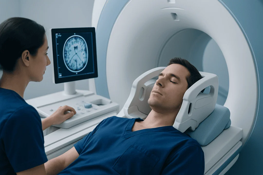
During MRI, the body lies inside a scanner that uses magnetic signals rather than ionizing radiation. The scanner collects images in several directions and sequences, allowing radiologists to distinguish fluid, soft tissue, blood products and other tissue characteristics. For craniopharyngioma, images focused on the pituitary and sellar region are commonly obtained.
MRI with gadolinium contrast is often recommended. The contrast agent is given through a small intravenous line and can make the solid parts and walls of cysts easier to evaluate. Clinicians review kidney health, prior contrast reactions and pregnancy status when deciding whether contrast is appropriate.
People may need a craniopharyngioma MRI when they have visual changes, persistent headaches, hormonal symptoms, delayed or early puberty, excessive thirst and urination, or a known mass in the pituitary region. The scan is also appropriate for adults and children who need long-term monitoring after treatment.
- Tell the MRI team about pacemakers, cochlear implants, aneurysm clips, neurostimulators or other implanted devices.
- Report metal fragments, prior surgery, kidney disease, allergies and pregnancy or possible pregnancy.
- Claustrophobia can often be managed with reassurance, an open-style scanner where available, or clinician-prescribed medication when suitable.
Preparing for and having a craniopharyngioma MRI

Most people can eat, drink and take regular medicines before a brain MRI. The imaging center will provide individual instructions, especially if contrast or sedation is planned. It is helpful to bring information about implants and previous imaging, as prior scans may allow more accurate comparison.
Before the scan, the person removes jewelry, watches, hearing aids, removable dental devices and clothing with metal fasteners. A technologist checks safety information and may place an intravenous line if contrast will be used. The person then lies on a padded table, with a lightweight head coil positioned around the head to improve image quality.
The table moves into the scanner, and the person must remain as still as possible while sequences are recorded. The machine makes loud tapping or knocking sounds, so earplugs or headphones are provided. Staff can see, hear and speak with the person throughout the examination, and a call button is available if assistance is needed.
A typical examination lasts about 30 to 60 minutes, although timing varies depending on the images required. Children and people unable to remain still may need sedation or anesthesia, which requires additional preparation and monitoring by an experienced team.
What are the MRI findings of craniopharyngioma?
Craniopharyngiomas are typically located in or above the sellar region, close to the pituitary gland. On MRI, they often appear as a well-defined mass with both solid and cystic components. The appearance can vary considerably because cyst fluid may contain protein, cholesterol or blood products, each of which can alter MRI signal patterns.
After contrast administration, solid portions of the tumor and the rim of a cyst may enhance. MRI also shows whether the mass contacts or compresses the optic chiasm, pituitary stalk, hypothalamus, third ventricle or nearby arteries. These details are especially important when planning surgery and considering radiation treatment.
Calcification is common in some forms of craniopharyngioma, particularly in children, but CT is generally more sensitive than MRI for detecting it. Radiologists interpret MRI alongside symptoms, hormone testing, eye examinations and, when needed, CT findings. Imaging features can suggest the diagnosis, but tissue assessment may be required in selected cases.
What imaging is used for craniopharyngioma?
MRI with and without contrast is the primary imaging method for craniopharyngioma. It provides the best assessment of the tumor’s soft-tissue features and its relationship to the pituitary gland, optic nerves and hypothalamus. It is also the preferred tool for follow-up because it does not use radiation.
CT of the head may be added when clinicians need a clearer view of calcification or bone anatomy. CT can complement MRI rather than replace it, particularly before surgery. In some situations, specialized MRI sequences, vascular imaging or repeat imaging may be needed to answer a specific planning question.
Imaging is usually combined with formal visual-field testing and blood tests that assess pituitary hormone function. Children may also need evaluation of growth and puberty. This coordinated approach helps identify complications that may not be visible on a scan alone.
Results, benefits, limitations and possible risks
A radiologist interprets the MRI and sends a report to the referring clinician, who explains the findings in the context of the person’s symptoms and medical history. Results may be available within a few days, although urgent findings are communicated promptly. The report commonly describes the tumor’s location, dimensions, cystic and solid components, enhancement pattern, and impact on nearby structures.
The main benefit of MRI is its detailed, radiation-free view of the brain and pituitary region. It supports diagnosis, guides treatment decisions and provides a reliable baseline for future comparison. It can also help the team decide whether observation, surgery, cyst drainage, radiation therapy or a combination of approaches may be appropriate.
MRI is generally very safe. Some people find the noise or enclosed space uncomfortable. Gadolinium contrast reactions are uncommon and are usually mild, but the imaging team should know about previous reactions and severe kidney disease. Sedation adds its own risks and requires recovery monitoring, so it is used only when necessary.
An MRI does not by itself determine every aspect of treatment. The scan may not fully distinguish craniopharyngioma from every other pituitary-region lesion, and it cannot replace endocrine and visual assessment. A multidisciplinary review is often valuable when results are complex.
How serious is a craniopharyngioma?
Craniopharyngioma is usually classified as a benign, or noncancerous, tumor, meaning it does not typically spread to distant parts of the body. However, it can still be serious because it grows in a small, sensitive area near structures that control vision, hormones, fluid balance and many functions regulated by the hypothalamus.
Its impact differs from person to person. Some tumors are found after gradual headaches or visual changes, while others cause hormone-related symptoms such as fatigue, growth changes in children, changes in menstrual cycles, low libido, excessive thirst or frequent urination. Cysts can sometimes change size and contribute to symptoms.
With specialist treatment and follow-up, many people can manage the condition over the long term. Care often involves neurosurgery, endocrinology, ophthalmology, neuroradiology and radiation oncology. Decisions aim to control the tumor while protecting vision, hormone function and quality of life as much as possible.
Treatment planning, surgery recovery and follow-up
Treatment is individualized according to age, symptoms, tumor anatomy, prior treatment and personal priorities. Options may include careful monitoring for selected stable tumors, surgery to remove or reduce the tumor, drainage or treatment of cysts, radiation therapy, and lifelong hormone replacement when pituitary function is affected. MRI findings help the team weigh the potential benefits and limitations of each option.
Surgery may be performed through the nose using an endoscopic approach or through an opening in the skull, depending on the tumor’s position and extension. The goal is not always complete removal; in some cases, preserving critical structures and using additional treatment is safer than pursuing extensive surgery.
How long does it take to recover from craniopharyngioma surgery? Recovery varies widely with the surgical approach, tumor size, complications and hormone or hypothalamic effects. Hospital recovery may take several days to longer in complex cases, while strength and adjustment to hormone treatment can continue for weeks to months. Follow-up visits commonly include MRI, visual testing and endocrine review.
Acibadem International’s multidisciplinary specialists and JCI-accredited hospitals diagnose and treat craniopharyngioma for international patients, coordinating neurosurgical, endocrine and visual care where needed.
When to seek medical care
Prompt medical assessment is important for new or worsening vision loss, double vision, severe or persistent headaches, vomiting with headache, marked drowsiness, confusion, or sudden changes in thirst and urination. These symptoms can have many causes, but they should not be ignored, especially in a person with a known pituitary-region tumor.
Anyone diagnosed with craniopharyngioma should maintain planned follow-up with their specialist team, even if they feel well. Contact the care team sooner if headaches, visual symptoms, fatigue, unusual weight change, menstrual changes, sexual-function changes, excessive thirst or frequent urination develop or worsen.
Emergency care is appropriate for sudden major vision changes, severe headache with confusion or fainting, repeated vomiting, or symptoms suggesting a severe disturbance in fluid balance. Early evaluation can help clinicians identify and treat urgent complications safely.
Frequently asked questions
Is an MRI painful for craniopharyngioma?
No. MRI does not involve injections into the tumor or painful radiation exposure. The examination can be noisy and requires lying still, and an intravenous line may be used if contrast is needed.
Do I need contrast for a craniopharyngioma MRI?
Contrast-enhanced MRI is often useful because it can better define solid tumor tissue and cyst walls. The radiology and clinical teams decide whether contrast is appropriate based on the clinical question and individual health factors.
Can an MRI diagnose craniopharyngioma on its own?
MRI can strongly suggest craniopharyngioma by showing its location and characteristic features. However, doctors also consider symptoms, hormone tests, eye examinations, CT findings when needed, and sometimes tissue analysis.
Can craniopharyngioma return after treatment?
Yes, craniopharyngioma can recur or cysts can enlarge after treatment, including after apparently successful surgery. This is why long-term MRI surveillance and specialist follow-up are important.
How soon can a person return to normal activities after the MRI?
Most people can return to their usual activities immediately after a standard MRI. If sedation was used, they may need observation afterward and should follow instructions about rest, driving and having an accompanying adult.
Why are hormone and eye tests needed alongside MRI?
A craniopharyngioma can affect the pituitary gland and optic pathways even when symptoms are subtle. Hormone testing and formal visual assessment provide important information that MRI alone cannot measure.
References
- World Health Organization
- National Cancer Institute
- RadiologyInfo.org
- Endocrine Society
- Children's Oncology Group
This article is for general information only and is not a substitute for professional medical advice. Please consult a qualified doctor about your individual situation.
Oncology care in Turkey — second opinion and treatment plan
JCI-accredited · board-certified surgeons · reply within 24h
Add us as a Preferred Source to see more of our trusted health content across Google Search, AI Overviews and Discover.
Check your numbers in seconds
BMI, calories, due date, blood pressure and 30+ more clinical calculators — free, instant, doctor-reviewed ranges.