

Horseshoe Kidney Ultrasound: Preparation, Procedure and Results

Ultrasound uses sound waves to create images of the kidneys and urinary tract without radiation. A horseshoe kidney is a congenital difference in which the kidneys are joined, usually at their lower poles.
Key Takeaways
- Ultrasound uses sound waves to create images of the kidneys and urinary tract without radiation.
- A horseshoe kidney is a congenital difference in which the kidneys are joined, usually at their lower poles.
- Preparation varies by clinic, but fasting and arriving with a comfortably full bladder may be requested.
- The scan can identify kidney position, dilation of the urine-collecting system, many stones and some structural changes.
- Results may lead to further imaging or urine and blood tests when more detail is needed.
A horseshoe kidney ultrasound is a noninvasive imaging test that helps clinicians confirm the position and structure of a horseshoe kidney and look for related concerns such as urinary blockage, stones or swelling. It does not use radiation, usually takes less than an hour, and most people can return to normal activities immediately afterward.
Horseshoe Kidney Ultrasound: What It Shows
A horseshoe kidney ultrasound is an imaging examination used to view kidneys that are joined together, typically by a bridge of tissue called an isthmus. It is often the first test used when a clinician suspects this congenital condition, when it was identified previously, or when symptoms such as recurrent urinary infections, flank discomfort, blood in the urine or kidney stones need assessment.
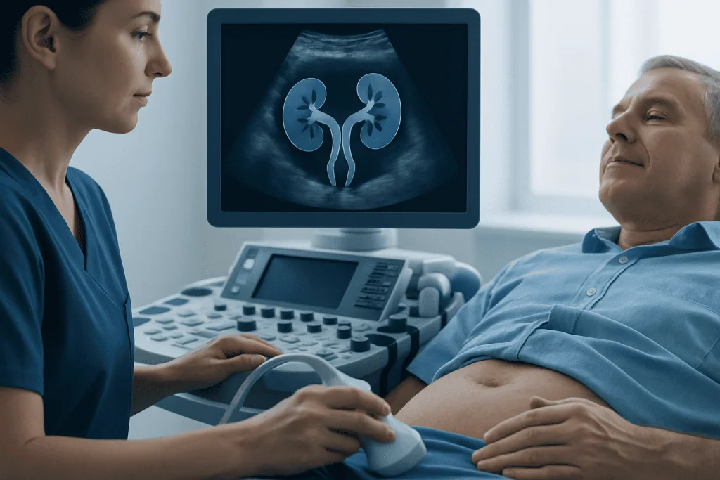
During the scan, a trained sonographer moves a small handheld device over the abdomen. The device sends high-frequency sound waves into the body and receives returning echoes, which are converted into live images. Because the test uses no ionizing radiation and usually does not require injections, it is suitable for many adults and children, including people who need repeat monitoring.
The examination can show the kidneys’ location, overall size and shape, the tissue joining them, and whether the urine-collecting areas appear enlarged. It may also identify cysts, some stones, or changes that suggest impaired urine drainage. However, ultrasound cannot answer every question, so a clinician may recommend CT, MRI or other tests if images are unclear or a complication is suspected.
How Horseshoe Kidney Affects the Urinary System
A horseshoe kidney develops before birth. Instead of separating fully as they rise into their usual position in the upper abdomen, the two kidneys remain connected and often sit lower than typical kidneys. Many people have no symptoms and discover the condition incidentally during imaging for another reason.
The different position and rotation of the kidneys can affect the course of the ureters, the tubes that carry urine to the bladder. This may make urine drainage less efficient in some people. Not everyone with a horseshoe kidney develops health problems, but clinicians consider its anatomy when investigating urinary symptoms or interpreting imaging findings.
Ultrasound is especially useful for assessing whether urine is backing up inside a kidney, a finding known as hydronephrosis. It can also provide a baseline image for future comparison. For broader information about the condition and its possible effects, see horseshoe kidney.
Who May Need a Horseshoe Kidney Ultrasound

A clinician may request this scan after a prenatal, childhood or adult imaging study suggests fused kidneys. It may also be used to investigate repeated urinary tract infections, suspected kidney stones, unexplained blood in the urine, persistent side or abdominal pain, or abnormal kidney-related blood or urine test results.
People already known to have a horseshoe kidney may have an ultrasound when new symptoms appear or as part of follow-up recommended by their urologist or nephrologist. The timing and frequency of monitoring are individualized. Many people do not need regular scans unless they have symptoms, associated urinary tract changes or a history of complications.
Ultrasound is often appropriate during pregnancy, in children, and for people who should avoid radiation or contrast material where possible. Its limitations include reduced image quality in some body types or when bowel gas obscures structures. In these situations, the care team may use another imaging method to obtain more detail.
Do I Need to Do Anything to Prepare for a Kidney Ultrasound?
Preparation instructions depend on the reason for the test and the imaging department’s protocol. Patients should follow the instructions provided with their appointment, as some renal ultrasounds require a period without food while others do not. Medications are usually continued as prescribed unless the clinician gives different advice.
In many cases, patients are asked to arrive with a comfortably full bladder. A full bladder can improve views of the bladder and the lower urinary tract and may help assess whether urine drains after voiding. Patients should ask the imaging center whether they need to avoid urinating before the appointment.
It is helpful to bring previous imaging reports, a medication list and relevant medical information. Loose, two-piece clothing can make the examination more convenient, although a gown may be provided. Patients should tell the team about pregnancy, recent surgery, a catheter or any difficulty lying flat so the scan can be adapted for comfort and safety.
How Much Water to Drink Prior to a Kidney Ultrasound?
The amount of water needed varies between facilities and according to whether the bladder will be evaluated. A common instruction is to drink several glasses of plain water in the hour before the scan and avoid emptying the bladder, but the exact volume and timing should come from the imaging center.
People with fluid restrictions, such as those with certain heart, kidney or liver conditions, should not increase fluid intake without checking with their clinician. Those who find a full bladder painful or difficult to maintain should inform the sonographer on arrival; the team can often adjust the sequence of images or offer guidance.
Water is generally preferred because carbonated drinks may increase bowel gas and make imaging more difficult. If the appointment instructions differ from general advice, the appointment instructions should always take priority.
What Not to Eat Before a Kidney Ultrasound?
Some centers ask patients not to eat for several hours before an abdominal or kidney ultrasound. This can reduce gas in the intestines, which may block sound waves and limit the view of the kidneys. Other centers allow a normal light meal, particularly when the scan focuses on the kidneys and bladder alone.
If fasting is requested, patients may be advised to avoid foods and drinks that commonly increase gas, such as fizzy drinks, large fatty meals and certain gas-producing foods. They should not change diabetes medication, insulin, or other prescribed treatment without individualized instructions from their healthcare professional.
Because protocols differ, patients should contact the radiology department if they have not received clear directions. Being accurately prepared can reduce the need for repeat images and supports the most useful assessment possible.
What Happens During and After the Procedure?
At the appointment, the patient usually lies on an examination bed, first on the back and sometimes on either side. A clear water-based gel is placed on the skin of the abdomen or back. The sonographer gently moves the transducer over the area and may ask the patient to take a deep breath, hold it briefly, or change position to improve the images.
The procedure is painless, although mild pressure may be felt where the probe is placed. It typically takes about 20 to 45 minutes, depending on the detail needed and whether the bladder or nearby structures are also assessed. In some examinations, images are taken before and after the patient urinates to check how well the bladder empties.
There is no recovery period. The gel is wiped away, and patients can eat, drink, drive and resume usual activities straight afterward unless they have other appointments or medical instructions. A radiologist interprets the images and sends a report to the referring clinician, who explains what the findings mean in the context of symptoms and other tests.
Benefits include a radiation-free, accessible evaluation of kidney anatomy and potential blockage. Risks are minimal and mainly relate to temporary discomfort from pressure or a full bladder. Ultrasound does not cause kidney damage, but it may not detect every stone or precisely define every abnormality, so additional imaging is sometimes necessary.
Results, Common Complications and When to Seek Medical Care
A report may describe the horseshoe kidney’s position, the appearance of the joining tissue, kidney size, stones, cysts and whether there is hydronephrosis. A normal or stable scan can be reassuring, but results should be discussed with the clinician who ordered the test. They may recommend urine tests, kidney function blood tests, or more detailed imaging when findings require clarification.
What is the most common complication of horseshoe kidney? Urinary drainage problems and urinary tract infections are among the most frequent clinically relevant concerns. Kidney stones are also more likely in some people because of altered urine flow. These complications are not inevitable, and prompt evaluation can help protect kidney health when symptoms develop.
Medical care should be sought promptly for fever with urinary symptoms, severe or worsening flank or abdominal pain, vomiting that prevents fluid intake, visible blood in the urine, difficulty passing urine, or symptoms of a urinary infection. Urgent assessment is particularly important when pain is severe or is accompanied by fever, chills or feeling very unwell, as these may indicate infection with obstruction.
Management depends on the specific finding and may range from observation and infection treatment to procedures for stones or urinary blockage. Kidney stone treatment and urology assessment may be considered when stones are found. Acibadem International’s multidisciplinary specialists and JCI-accredited hospitals diagnose and treat kidney and urinary tract conditions for international patients.
Frequently asked questions
Can an ultrasound diagnose a horseshoe kidney?
Ultrasound can often identify the joined kidneys and their lower-than-usual position. It may also show the connecting tissue and detect related changes such as swelling of the urine-collecting system. CT or MRI may be needed if the anatomy is not clearly seen or more detailed evaluation is required.
Is a horseshoe kidney ultrasound painful?
The scan is generally painless and does not involve needles, radiation or anesthesia. Some people feel mild pressure from the transducer, especially if an area is tender. A full bladder may feel uncomfortable temporarily, but the patient can usually empty it when instructed.
How long does a renal ultrasound take?
Most kidney ultrasounds take around 20 to 45 minutes. The timing can vary based on whether both kidneys, the bladder, urine flow or additional abdominal structures need to be examined. Patients should allow extra time for registration and preparation.
Can ultrasound show kidney stones in a horseshoe kidney?
Ultrasound can identify many kidney stones, particularly larger stones, and can show signs of urine blockage. Very small stones or stones in certain locations may be difficult to see. A clinician may recommend CT if symptoms strongly suggest a stone but the ultrasound is inconclusive.
Do people with a horseshoe kidney always need treatment?
No. Many people with a horseshoe kidney have no symptoms and require no treatment. Care is usually directed at complications, such as urinary infections, stones or urinary obstruction, rather than at the fused kidney itself.
When will I receive my ultrasound results?
A radiologist usually reviews the images and sends a report to the clinician who requested the examination. Timing varies by facility and urgency. The referring clinician can explain the results, whether follow-up is needed and what the findings mean for the individual patient.
References
- National Institute of Diabetes and Digestive and Kidney Diseases
- Radiological Society of North America
- American Urological Association
- National Kidney Foundation
This article is for general information only and is not a substitute for professional medical advice. Please consult a qualified doctor about your individual situation.
Ultrasound in Turkey — costs, top hospitals & a free quote
JCI-accredited · board-certified surgeons · reply within 24h
Add us as a Preferred Source to see more of our trusted health content across Google Search, AI Overviews and Discover.

Check-up & Preventive Medicine
Comprehensive health screenings designed for early detection and prevention.
2 specialists in this unitMore from the Health Library



Related Specialists

Dr. Salih Gülten
Kidney Transplant Center
Dr. Murat Apaydın
Pulmonary Medicine
Dr. Shahad Abdulhadı Abdulrahman Al Amoudı
Inpatient Clinic Physicians
Prof. Dr. Ülkem Çakır
Nephrology