Can You See Tendonitis in an X-Ray: Preparation, Procedure and Results

X-rays do not usually show tendon inflammation or small tendon tears directly. They can identify fractures, arthritis, bone spurs, calcium deposits and joint changes that may contribute to pain.
Key Takeaways
- X-rays do not usually show tendon inflammation or small tendon tears directly.
- They can identify fractures, arthritis, bone spurs, calcium deposits and joint changes that may contribute to pain.
- A clinical examination is central to diagnosing tendonitis; ultrasound or MRI may be used when the diagnosis is uncertain or tendon damage is suspected.
- X-rays are quick, painless and use a small amount of ionizing radiation.
- Many cases improve with activity modification, rehabilitation and a gradual return to loading, but persistent symptoms need medical assessment.
Can you see tendonitis in an X-ray? In most cases, no: tendons are soft tissues and are not shown clearly on standard X-rays. However, an X-ray can help doctors look for bone-related causes of pain and decide whether ultrasound or MRI is needed.
Can You See Tendonitis in an X-Ray?
Can you see tendonitis in an X-ray? Usually, tendonitis cannot be seen directly on a standard X-ray because tendons are soft tissues and X-rays primarily show bones. An X-ray may still be useful because it can reveal other explanations for pain, such as a fracture, arthritis, a bone spur, joint misalignment or calcium deposits near a tendon.
Tendonitis, also called tendinopathy in many longer-lasting cases, describes pain and changes affecting the strong bands of tissue that connect muscle to bone. It commonly affects the shoulder, elbow, wrist, hip, knee, ankle and foot. Diagnosis often begins with a discussion of symptoms and a focused physical examination; imaging is selected when it will help answer a specific clinical question.
If a clinician suspects tendon thickening, a tear, inflammation around the tendon or another soft-tissue problem, ultrasound or magnetic resonance imaging (MRI) may offer more information than an X-ray. Imaging findings are considered alongside symptoms, activity level and examination results rather than used alone.
Why an X-Ray May Still Be Part of the Assessment
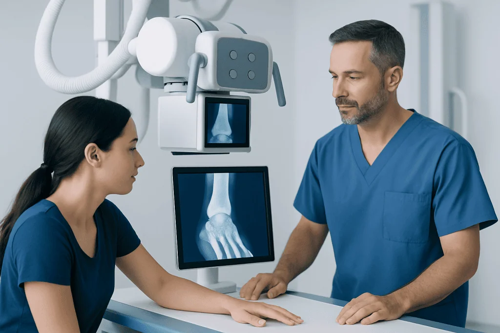
An X-ray produces images using a small amount of radiation that passes through the body. Dense structures such as bone absorb more radiation and appear clearly on the image, while tendons, muscles and many other soft tissues are much less distinct. For this reason, an X-ray cannot reliably confirm uncomplicated tendonitis.
Its main benefit is helping rule out or identify conditions that can cause similar symptoms. Depending on the painful area, an X-ray may show a previous or recent bone injury, osteoarthritis, a stress-related bone change, calcification near a tendon, altered joint alignment or a bony prominence that may irritate nearby tissues.
Calcific tendinopathy is one exception in which X-rays can be particularly informative. Calcium deposits within or around a tendon may be visible, especially in the shoulder. Their presence does not automatically explain every symptom, but it can help shape the next steps in care.
- Benefits: X-rays are widely available, fast, painless and useful for evaluating bones and joints.
- Limitations: They do not provide detailed pictures of tendon fibers, subtle inflammation or many partial tears.
- What happens next: Further imaging is considered if symptoms, examination findings or the X-ray result indicate it is needed.
How Do Doctors Tell If You Have Tendonitis?

Doctors usually diagnose tendonitis by combining a person’s history with a physical examination. They ask when pain began, which activities provoke it, whether there was a sudden injury, and whether there are symptoms such as swelling, weakness, stiffness, clicking or loss of function. Repetitive movement, a recent increase in training or work demands, and pain in a specific location can provide useful clues.
During the examination, the clinician may gently press along the tendon, assess joint movement, test muscle strength and ask the person to perform movements that load the affected area. Local tenderness and pain triggered by a particular resisted movement can support the diagnosis. The assessment also looks for signs that suggest a different condition, including nerve symptoms, joint disease or a significant tear.
Imaging is not always required when symptoms and examination findings are typical. It is more likely to be recommended after a traumatic injury, when pain does not improve as expected, when diagnosis is unclear, or when weakness or a tendon tear is a concern. A specialist may also assess related movement patterns and recommend physical therapy and rehabilitation to support safe recovery.
What Is Often Mistaken for Tendonitis?
Several conditions can cause pain near a tendon and may be mistaken for tendonitis. These include muscle strain, ligament injury, bursitis, joint arthritis, stress fracture, nerve compression and pain referred from the neck, back or another nearby area. The location of pain alone is not enough to distinguish among these possibilities.
For example, shoulder pain may arise from rotator cuff tendon problems, bursitis, arthritis or pain referred from the neck. Pain on the outside of the elbow may reflect tendon overload but can also occur with nerve irritation or joint problems. Heel pain may result from Achilles tendon disease, plantar fasciitis or a bone-related condition. Accurate diagnosis matters because the most helpful treatment varies by cause.
A sudden pop, extensive bruising, marked weakness, inability to bear weight or difficulty using a limb may indicate an injury more serious than routine tendon irritation. A clinician can determine whether imaging, urgent treatment or a referral to an orthopedic specialist is appropriate. Related conditions affecting joints and mobility may also be evaluated through osteoarthritis assessment when symptoms and examination findings point in that direction.
What Test Shows Tendon Damage?
Ultrasound and MRI are the imaging tests most commonly used to examine tendon damage. Ultrasound uses sound waves and can show tendon thickening, fluid around a tendon, calcifications and some partial or complete tears. It can also assess the tendon while the person moves the joint, making it useful for certain shoulder, elbow, wrist, knee, ankle and foot concerns.
MRI uses magnetic fields and radio waves rather than ionizing radiation. It provides detailed images of tendons, muscles, ligaments, cartilage, bone marrow and surrounding soft tissue. MRI may be considered when a deeper structure needs assessment, the extent of an injury is uncertain, symptoms persist despite appropriate care, or surgery is being considered.
Neither test is routinely necessary for every person with suspected tendonitis. Scans can show age- or activity-related changes that do not necessarily cause symptoms, so results should always be interpreted by a qualified clinician in context. For significant tears or complex injuries, orthopedic surgery may be discussed only when non-surgical care is unlikely to restore function or when repair is needed.
X-Ray Preparation, Procedure, Results and Safety
Most people need little preparation for a tendon-area X-ray. They may be asked to remove jewelry, watches, belts or clothing with metal near the body part being examined, and to tell the radiology team about pregnancy or the possibility of pregnancy. It is important to mention recent injuries, previous surgery, implants and the exact location of symptoms.
During the procedure, a radiographer positions the affected body part on or near the X-ray detector and may take images from more than one angle. The person needs to stay still for a few seconds while each image is taken. The examination commonly takes only a short time, although positioning can be uncomfortable if the area is very painful.
There is no recovery period after a routine X-ray. Normal activities can generally be resumed immediately unless a clinician has advised otherwise because of the underlying injury. A radiologist reviews the images, and the referring doctor explains what the findings mean in relation to the person’s symptoms and whether any further tests are appropriate.
X-rays involve low levels of ionizing radiation, and imaging teams use the lowest practical exposure needed to obtain diagnostic images. The benefit of an appropriately requested X-ray generally outweighs its small risk. X-rays do not cause tendonitis and do not make an existing tendon problem worse.
Does Tendonitis Ever Go Away?
Yes, many people recover from tendon pain with the right approach, although improvement is often gradual rather than immediate. The timeline depends on the tendon involved, how long symptoms have been present, the degree of tissue change, underlying health conditions and whether the tendon continues to be overloaded. Symptoms that have lasted many months may require a more structured rehabilitation plan.
Early care often includes reducing or modifying the activity that repeatedly provokes pain, while avoiding complete inactivity for longer than necessary. Guided, progressive exercise can help restore tendon capacity and surrounding muscle strength. Ice or heat may provide short-term comfort for some people, while pain medicines should be used only according to advice from a doctor or pharmacist, particularly for people with kidney, stomach, heart or bleeding risks.
Returning to sport, exercise or manual work is usually safest when it is gradual and based on improving strength and function, not only on the absence of pain. A rehabilitation professional can tailor loading exercises and address technique, footwear, equipment, workplace setup or training progression. At Acibadem International, multidisciplinary specialists in JCI-accredited hospitals can assess tendon conditions and coordinate care for international patients.
When to Seek Medical Care
Medical assessment is advisable for tendon pain that is severe, follows an injury, limits normal movement, causes persistent weakness or does not begin improving with sensible activity modification. A doctor can confirm whether the symptoms are likely tendon-related and determine whether an X-ray, ultrasound, MRI or another investigation is appropriate.
Prompt medical care is important after a sudden pop or snapping sensation, visible deformity, major swelling or bruising, inability to use the affected limb, inability to bear weight, or new numbness or tingling. These signs can occur with a tendon rupture, fracture, nerve problem or other injury that needs timely evaluation.
Urgent assessment is also needed if pain is accompanied by fever, a hot and markedly swollen joint, spreading redness, an open wound or feeling generally unwell. These symptoms are not typical of simple tendonitis and may need prompt treatment. People with diabetes, immune suppression or circulation problems should seek advice earlier for new foot, ankle or wound-related symptoms.
Frequently asked questions
Can an X-ray confirm tendonitis?
No, an X-ray usually cannot confirm tendonitis because tendons are not shown in detail on standard X-ray images. It can help rule out fractures, arthritis, bone spurs or calcium deposits that may be causing or contributing to pain. Diagnosis is usually based on symptoms and examination, with ultrasound or MRI used when needed.
Should I have an X-ray or an MRI for tendon pain?
The appropriate test depends on the suspected cause and the body area involved. An X-ray is often used first when a bone or joint problem needs to be excluded, while MRI provides more detail about tendons and other soft tissues. A clinician can recommend imaging based on the examination and the duration and severity of symptoms.
Can ultrasound detect a torn tendon?
Ultrasound can often identify tendon thickening, inflammation around a tendon, calcification and many partial or complete tears. It has the advantage of showing movement in real time. However, MRI may be preferred for deeper structures or when a more detailed assessment is required.
Why does my tendon hurt if my X-ray is normal?
A normal X-ray does not rule out tendonitis, because uncomplicated tendon problems may not be visible on it. Pain may also come from muscles, ligaments, bursae, nerves or other soft tissues. Further clinical assessment or soft-tissue imaging may be helpful if symptoms persist.
How long does tendonitis take to heal?
Recovery varies widely and can take weeks to months, particularly when symptoms have been present for a long time. Reducing aggravating activity and following a progressive rehabilitation plan can support recovery. Persistent pain, weakness or loss of function should be reviewed by a healthcare professional.
Is it safe to exercise with tendonitis?
Complete rest is not always necessary, but painful activities usually need to be modified while the tendon recovers. Controlled strengthening and gradual loading are often part of rehabilitation, ideally guided by a clinician or physiotherapist. Sudden increases in activity or pushing through worsening pain can delay improvement.
References
- American College of Radiology
- American Academy of Orthopaedic Surgeons
- National Institute of Arthritis and Musculoskeletal and Skin Diseases
- Radiological Society of North America
- National Health Service
This article is for general information only and is not a substitute for professional medical advice. Please consult a qualified doctor about your individual situation.
Joint and spine care in Turkey — expert assessment & treatment
JCI-accredited · board-certified surgeons · reply within 24h
Add us as a Preferred Source to see more of our trusted health content across Google Search, AI Overviews and Discover.