Lymphography Procedure: An Evidence-Based Patient Guide
Lymphography and lymphangiography generally describe imaging of the lymphatic system; terminology may vary by center. Modern lymphatic imaging may use contrast material with X-ray, fluoroscopy, CT, MRI, or nuclear medicine techniques such as lymphoscintigraphy.
Key Takeaways
- Lymphography and lymphangiography generally describe imaging of the lymphatic system; terminology may vary by center.
- Modern lymphatic imaging may use contrast material with X-ray, fluoroscopy, CT, MRI, or nuclear medicine techniques such as lymphoscintigraphy.
- The test is usually considered when symptoms or previous imaging suggest a lymphatic flow problem, including persistent limb swelling or fluid leakage.
- Preparation, imaging method, recovery time, and risks depend on the exact technique and whether an intervention is performed at the same time.
- Patients should report allergies, pregnancy, kidney problems, blood-thinning medicines, and previous reactions to contrast before the procedure.
A lymphography procedure is a specialized imaging examination used to show how lymph fluid travels through lymphatic vessels and lymph nodes. It can help clinicians investigate swelling, lymphatic leaks, vessel blockages, and selected abnormalities, while also supporting treatment planning.
Overview: what is a lymphography procedure?
A lymphography procedure is an imaging test that helps doctors examine the lymphatic system: the network of vessels, nodes, and organs that returns fluid to the bloodstream and supports immune function. By showing the route and flow of lymph fluid, it can help identify where fluid is leaking, collecting, slowed, or blocked.
The terms lymphography and lymphangiography are often used interchangeably. In practical use, lymphangiography procedure radiology may refer specifically to imaging after contrast material is placed into lymphatic vessels or nodes, often with real-time X-ray guidance. The exact test chosen is individualized and may be diagnostic only or may be combined with a minimally invasive treatment.
Lymphatic imaging is not a routine test for every swollen limb. It is usually requested when the clinical history, examination, ultrasound, CT, MRI, or prior tests suggest a lymphatic disorder, such as suspected lymphedema, an injury-related leak, or an abnormal lymphatic pathway. It can complement assessment for lymphedema when a specialist needs more detailed information about lymph drainage.
How lymphatic imaging works
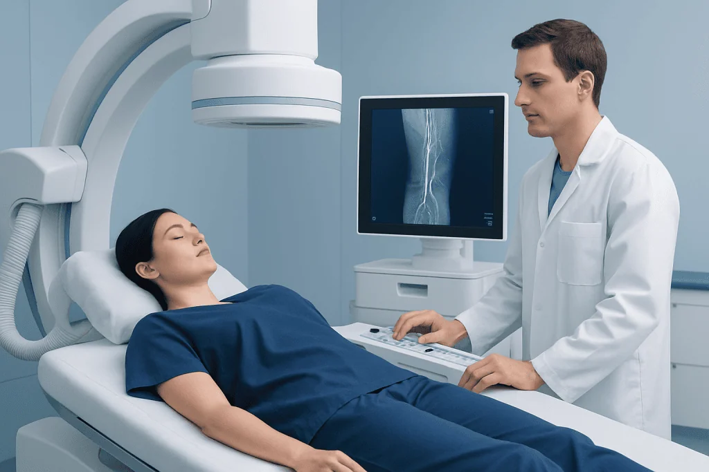
In conventional oil-based lymphangiography, a radiologist introduces a contrast agent into selected lymphatic channels, commonly in the feet, or injects it into a lymph node under ultrasound guidance. Fluoroscopy and X-ray images track the contrast as it travels through lymphatic vessels and nodes. Delayed X-rays or CT images may be needed because lymph flow is slow.
Several other methods can be used. Intranodal lymphangiography places contrast directly into a groin lymph node and is often used to investigate leaks in the abdomen, pelvis, chest, or groin. Dynamic contrast-enhanced MR lymphangiography uses MRI and contrast in selected situations, while indocyanine green lymphography may map superficial lymphatic vessels, particularly in the limbs.
Lymphography vs lymphangiography is therefore usually a difference in wording rather than a completely different test. Lymphoscintigraphy is distinct: it is a nuclear medicine examination in which a very small amount of radiotracer is injected near the area of concern and a gamma camera follows lymphatic drainage. Lymphoscintigraphy procedure guidelines, including injection sites and imaging timing, vary according to the body area and clinical question.
The imaging findings may show normal drainage, delayed transport, reflux, narrowed or interrupted vessels, enlarged nodes, abnormal connections, or the location of a leak. A radiologist interprets the pictures together with symptoms, medical history, examination findings, and other imaging results.
Who may be a candidate?

A clinician may recommend a lymphography procedure when a detailed map of lymphatic flow could change diagnosis or treatment. Common reasons include unexplained or persistent swelling of an arm, leg, genital area, or other body region; suspected primary or secondary lymphedema; fluid collections after surgery or trauma; and suspected chyle leak into the chest or abdomen.
It may also be considered after cancer treatment, lymph node surgery, radiation therapy, transplant surgery, or major pelvic, abdominal, chest, or vascular procedures when a lymphatic complication is suspected. The purpose is not simply to confirm swelling; it is to identify whether a lymphatic cause is likely and, when possible, pinpoint the involved vessel or leak.
Not everyone needs invasive contrast lymphangiography. Doctors may first use physical examination, blood tests, ultrasound, CT, MRI, or lymphoscintigraphy. The imaging team will consider the expected benefit, the availability of alternative tests, kidney function, prior contrast reactions, pregnancy status, infection at a possible injection site, and the person’s ability to lie still for imaging.
Patients should share a full medication and allergy list. In particular, they should mention anticoagulants or antiplatelet medicines, diabetes medicines, kidney disease, thyroid disease, breathing disorders, and any previous reaction to iodinated contrast or anesthesia. The care team will advise whether any medicines need adjustment; patients should not stop prescribed medicines unless instructed.
What happens during the procedure?
The lymphangiography protocol depends on the indication, the body area being examined, and the type of imaging. Before the procedure, the team confirms consent, reviews medical history, checks relevant tests, and explains how to prepare. Some patients may be asked not to eat for a set period, especially if sedation or a possible intervention is planned; others may follow their usual diet. Clear instructions should come from the treating facility.
For intranodal lymphangiography, the patient usually lies on an imaging table. The skin over the groin is cleaned, local anesthetic is used, and ultrasound helps the radiologist place a fine needle into a lymph node. Contrast is injected slowly while fluoroscopy records its movement. Depending on the findings, imaging can continue immediately or be repeated later with X-ray or CT.
For pedal lymphangiography, small lymphatic channels in the foot may be identified and accessed after local anesthetic. This technique can take longer and is used less often in some centers than intranodal approaches. For lymphoscintigraphy, injections are generally placed just under the skin, followed by images taken over time; it does not use the same contrast approach as conventional lymphangiography.
Most diagnostic examinations use local anesthetic, and some patients receive medicine to help them relax. If the study identifies a leak or suitable target, the interventional radiology team may discuss treatment during the same session or at a later date. This can include lymphatic embolization, a minimally invasive method intended to close selected abnormal lymphatic channels. Whether it is appropriate depends on the imaging findings and the person’s overall health.
Benefits, limitations, and possible risks
The principal benefit of a lymphography procedure is detailed, functional information about lymphatic anatomy and drainage. Locating a leak, obstruction, or abnormal pathway may avoid uncertainty and can help a multidisciplinary team choose observation, compression-based care, surgery, or image-guided treatment more precisely.
Like all medical tests, it has limitations. Lymph flow may be very slow, abnormal vessels can be difficult to access, and results may not explain every case of swelling. A normal or inconclusive study does not always exclude a lymphatic problem, so doctors interpret the results alongside other clinical information.
Possible short-term effects include tenderness, bruising, mild bleeding, or temporary discomfort at an injection site. Contrast-related effects can include nausea or allergic-type reactions, although serious reactions are uncommon. Depending on the contrast agent and technique, there can also be risks of infection, damage to a lymphatic vessel, contrast moving outside the intended pathway, or complications related to sedation.
Rare but important complications are technique-specific and are discussed during consent. Oil-based contrast can persist in the body for some time and may cause temporary image changes; in unusual circumstances, it can travel to unintended areas. Promptly reporting chest pain, shortness of breath, severe rash, fever, worsening pain, or marked swelling after the procedure allows the team to assess for a complication without delay.
Recovery and understanding the results
Recovery after a diagnostic lymphography procedure is often straightforward. Patients are usually observed for a period after the examination, particularly if they received sedation or had an interventional treatment. The puncture site may remain mildly sore or bruised for a few days, and activity instructions depend on the access site and procedure performed.
When sedation is used, a responsible adult may need to take the patient home, and driving, alcohol, and important decisions should be avoided for the period advised by the care team. Drinking fluids may be encouraged unless a clinician has provided fluid restrictions. Wound-care instructions should be followed carefully, including when bathing and when to remove any dressing.
The radiologist prepares a report that is sent to the referring clinician. Results may be available quickly for urgent inpatient concerns, while more complex imaging may require additional review. The next step could include monitoring, lymphedema therapy, referral to surgery, or an interventional radiology discussion. For people with confirmed chronic swelling, lymphedema treatment often includes individualized skin care, exercise guidance, compression strategies, and specialist follow-up.
Billing terminology is separate from medical decision-making. A lymphangiography procedure CPT code may differ according to the imaging technique, body area, contrast administration, image guidance, and whether a therapeutic intervention was carried out. The hospital coding or insurance team can clarify the code used for an individual case.
When to seek medical care
Medical assessment is appropriate for new, persistent, or worsening swelling, especially when it affects one limb more than the other, occurs after surgery or cancer therapy, or is accompanied by skin tightness, heaviness, recurrent fluid collections, or leakage from a wound. Early assessment can help distinguish lymphatic swelling from venous, heart, kidney, liver, medication-related, or inflammatory causes.
Urgent medical care is needed for sudden one-sided swelling with pain, redness, warmth, or breathing symptoms, because these can have causes that require prompt evaluation. Fever, rapidly spreading redness, severe pain, or drainage from an injection or surgical site should also be assessed without delay.
Patients scheduled for lymphatic imaging should contact their care team before arrival if they develop fever, active skin infection near the planned access site, a new pregnancy concern, or a new reaction to contrast material. Acibadem International’s multidisciplinary specialists and JCI-accredited hospitals can evaluate lymphatic conditions and coordinate imaging and treatment plans for international patients.
Frequently asked questions
Is lymphography painful?
Most people feel a brief sting from the local anesthetic and may notice pressure or mild discomfort during injection. The team works to keep the procedure as comfortable as possible and can discuss sedation options when appropriate. Tenderness or bruising at the access site may occur afterward.
How long does a lymphography procedure take?
Timing varies substantially with the technique and the speed of lymphatic flow. The injection and initial imaging may take an hour or longer, and delayed X-ray or CT images may be scheduled later. If treatment is performed during the same visit, the overall appointment will usually be longer.
What is the difference between lymphography and lymphoscintigraphy?
Lymphography or lymphangiography generally uses contrast material and radiologic imaging to visualize lymphatic vessels and nodes in detail. Lymphoscintigraphy uses a small amount of radioactive tracer and a gamma camera to assess lymph drainage. The most suitable test depends on the clinical question and the part of the body involved.
Can lymphangiography treat a lymphatic leak?
The imaging portion is primarily used to locate and characterize a leak or abnormal lymphatic pathway. In selected cases, the same or a later procedure may include image-guided treatment, such as embolization. A specialist determines whether this is safe and likely to be helpful based on the imaging findings.
Do patients need to fast before lymphography?
Some patients may need to fast, particularly if sedation, anesthesia, or a possible intervention is planned. Requirements differ among facilities and procedure types. Patients should follow the instructions provided by their imaging or interventional radiology team.
How soon can normal activities resume after lymphography?
Many people can return to light daily activities within a day after a diagnostic procedure, but individual advice may differ. Rest, driving restrictions after sedation, and care of the puncture site should follow the treating team’s instructions. More extensive procedures or embolization may require a longer recovery period.
References
- Radiological Society of North America
- Society of Interventional Radiology
- International Society of Lymphology
- National Cancer Institute
- American College of Radiology
This article is for general information only and is not a substitute for professional medical advice. Please consult a qualified doctor about your individual situation.
Explore treatments in Turkey — costs, top hospitals & a free quote
JCI-accredited · board-certified surgeons · reply within 24h
Add us as a Preferred Source to see more of our trusted health content across Google Search, AI Overviews and Discover.