Ommaya Shunt MRI Safety: Preparation, Procedure and Results

An Ommaya reservoir is a surgically placed device that provides access to cerebrospinal fluid or allows medicines to be delivered into the fluid around the brain and spinal cord. Many modern Ommaya reservoirs can be scanned with MRI, including some at 3 Tesla, but MRI conditions depend on the exact device and any associated shunt hardware.
Key Takeaways
- An Ommaya reservoir is a surgically placed device that provides access to cerebrospinal fluid or allows medicines to be delivered into the fluid around the brain and spinal cord.
- Many modern Ommaya reservoirs can be scanned with MRI, including some at 3 Tesla, but MRI conditions depend on the exact device and any associated shunt hardware.
- A patient should bring implant information when available and inform the MRI team and treating neurosurgeon before the scan.
- Ommaya placement can be very effective when carefully planned, but outcomes depend on the underlying condition and correct catheter positioning.
- Infection, bleeding, catheter blockage and device malfunction are important possible complications that require medical assessment.
Ommaya shunt MRI safety is generally good when the reservoir and any connected shunt components are confirmed as MRI-compatible or MRI-conditional. Patients should always tell the imaging team about their device so its model, materials and scan conditions can be checked before an MRI.
Overview: Ommaya Shunt MRI Safety
Ommaya shunt MRI safety is generally good when the exact implanted device has been identified and the MRI team follows the manufacturer’s instructions. Many Ommaya reservoirs are made from materials that are MRI-conditional, meaning MRI can be performed under specified conditions rather than being automatically suitable for every scanner or setting.
An Ommaya reservoir is not always the same as a conventional shunt. It is a small dome-shaped chamber placed beneath the scalp and connected to a catheter that enters a fluid-filled space in the brain. It may be used to collect cerebrospinal fluid (CSF), monitor aspects of care, or give medication directly into CSF when this is clinically appropriate.
Before an MRI, patients should tell the radiology team that they have an Ommaya reservoir, whether or not they think it is visible externally. The team may review operative records, implant cards or prior imaging to confirm the device model and determine whether other implanted components, such as a programmable valve, require additional precautions.
What Is an Ommaya Shunt?
An Ommaya reservoir is a neurosurgical access device named after the physician who developed it. It consists of a silicone reservoir under the scalp and a thin catheter placed into a brain ventricle, which is a natural cavity containing CSF. A clinician can access the reservoir with a specially designed needle through the skin when sampling fluid or administering prescribed treatment.
The term “Ommaya shunt” is sometimes used informally, although an Ommaya reservoir does not necessarily drain fluid in the way a ventriculoperitoneal shunt does. Its main purpose is repeated, controlled access to CSF without needing repeated lumbar punctures or repeated direct procedures involving the brain.
It may be considered for selected people who need intrathecal or intraventricular medicines, frequent CSF testing, or treatment related to certain cancers or infections affecting the central nervous system. The decision is individualized and involves the treating specialist, often including neurosurgery, oncology, neurology, radiology and infectious disease teams.
Are Ommaya Reservoir MRI Safe?

For many people, the answer is yes: an MRI can be performed safely with an Ommaya reservoir when the device’s MRI labeling and scan conditions are verified. However, no patient should assume that every reservoir, catheter or related shunt component has identical MRI requirements. The radiology department should make the final safety check before the examination.
Questions about ommaya MRI safety often arise because MRI uses a powerful magnetic field and radiofrequency energy. Some implants can move, heat up, create image distortion or have settings affected by the magnetic field. Standard Ommaya reservoirs are commonly made with nonmetallic or minimally metallic materials, but connected hardware and older devices may differ.
For ommaya reservoir MRI safety at 3T, the same principle applies: 3 Tesla scanning may be possible for some device models but should be performed only within the manufacturer’s stated conditions. If the device type cannot be confirmed, the MRI safety team may recommend another imaging method or seek more information before proceeding.
- Tell the MRI staff about the reservoir when booking and again on arrival.
- Bring an implant card, surgical report or device details if these are available.
- Report any additional implants, including a shunt valve, clips, stimulators or hearing devices.
- Ask whether any device setting needs checking after the MRI, particularly if a programmable shunt valve is present.
How MRI Preparation and the Scan Usually Work
Preparation begins before the appointment. The imaging team completes an MRI safety questionnaire and may contact the neurosurgeon or review records from the reservoir placement. If there is uncertainty about the implant, a plain X-ray or previous scan may sometimes help identify related shunt hardware, although this does not replace device-specific safety information.
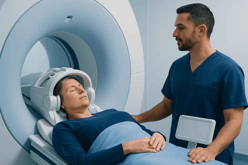
During the MRI, the patient lies on a padded table that moves into the scanner. A head coil may be used for brain imaging. The examination is painless, but the scanner produces loud tapping or knocking sounds, so hearing protection is provided. Staff remain in contact through an intercom, and the patient can alert them if they feel uncomfortable.
Contrast material may be recommended for some MRI examinations to provide more detail. This is separate from the Ommaya reservoir itself. The care team will ask about kidney disease, past contrast reactions, pregnancy and other relevant health information before deciding whether contrast is appropriate.
After scanning, most patients can return to usual activities immediately unless sedation was used or their medical condition requires observation. If a programmable device is part of the system, the care team may arrange a prompt setting check after MRI. The imaging report is then interpreted alongside symptoms, examination findings and other tests.
Candidacy, Benefits and Success of Ommaya Reservoir Placement
An Ommaya reservoir may be suitable when repeated access to CSF is expected to support diagnosis or treatment. For example, it can reduce the need for multiple lumbar punctures in some circumstances and can allow certain medicines to reach CSF directly. It is not needed for every person with a neurologic condition, cancer or infection.
Suitability depends on the reason for treatment, brain anatomy, clotting status, current infection, previous surgery and the person’s overall health. Before placement, the neurosurgical team reviews brain imaging and explains why the reservoir is being considered, what alternatives exist and how it may fit within the wider treatment plan.
What is the success rate of Ommaya reservoir placement? There is no single success rate that applies to everyone. Technical success means the catheter and reservoir are placed in the intended position and can be used safely; clinical benefit depends largely on the condition being treated, the medication or testing plan, and whether complications occur. Modern image-guided neurosurgical techniques help clinicians confirm placement, but individual outcomes should be discussed with the operating neurosurgeon.
When cancer-related treatment is being considered, the reservoir may be part of coordinated care that includes systemic therapy, radiation therapy or other approaches. Its benefit is assessed over time through symptoms, CSF results and imaging, rather than by the device alone.
What Are the Risks of Ommaya Reservoir Use?
What are the risks of Ommaya reservoir use? Like all implanted medical devices, an Ommaya reservoir has potential complications. These include infection, bleeding during or after placement, catheter misplacement, obstruction, leakage, skin problems over the reservoir and device malfunction. Rarely, irritation or injury to brain tissue may occur.
Risks can also arise when the reservoir is accessed. Careful sterile technique is important because introducing bacteria into the reservoir or CSF can lead to a serious infection. The clinical team will use an appropriate procedure and may postpone access if there are concerns about skin infection or other signs of illness.
Some people experience headaches, nausea, scalp tenderness or swelling after surgery. These symptoms may be temporary, but new or worsening symptoms should be assessed. The care team may use examination, CSF testing and imaging to evaluate whether the reservoir is working as intended.
Potential benefits must be balanced against these risks. For an appropriate candidate, reliable CSF access can make ongoing care more practical and may avoid repeated invasive procedures. The treating team should explain the expected benefit for the individual’s specific diagnosis before placement or use.
Recovery, Everyday Care and When to Seek Medical Care
Recovery after placement varies with the person’s health and the reason for surgery. Some people stay in hospital for monitoring, while others may have a shorter recovery depending on their overall treatment needs. The scalp incision generally needs time to heal, and the surgeon provides instructions about washing, dressings, activity and follow-up appointments.
Patients should avoid pressing repeatedly on the reservoir unless their clinical team has specifically instructed them to do so. They should attend scheduled follow-up visits and keep a record of the implant details. Before future MRI, surgery or procedures, they should inform all relevant healthcare professionals about the reservoir and any other implanted devices.
When to seek medical care: Prompt medical advice is important for fever, increasing headache, neck stiffness, confusion, persistent vomiting, seizure, new weakness, unusual drowsiness, redness or drainage around the scalp incision, or swelling and increasing pain over the reservoir. These symptoms do not always mean there is a device complication, but they need timely assessment.
Acibadem International’s multidisciplinary specialists and JCI-accredited hospitals support international patients who need evaluation and treatment involving neurosurgical devices. Care is coordinated with the relevant neurosurgery, radiology, oncology and neurology teams according to the person’s diagnosis and treatment goals.
Frequently asked questions
Can a person with an Ommaya reservoir have an MRI?
Many people with an Ommaya reservoir can have an MRI, but the device must be checked before the scan. The MRI team needs to confirm the reservoir model, any connected hardware and the manufacturer’s MRI conditions. Patients should always tell the imaging department about the implant in advance.
Is an Ommaya reservoir safe in a 3 Tesla MRI scanner?
Some Ommaya reservoirs may be MRI-conditional at 3 Tesla, while conditions can vary by manufacturer and device version. A 3T MRI should only proceed after the radiology safety team confirms that the specific implanted system is suitable. Any additional shunt valve or implanted hardware may have separate requirements.
Will an MRI damage an Ommaya reservoir?
When the scan is performed within the approved conditions for the identified device, MRI is not expected to damage the reservoir. The main concern is confirming the full implanted system, especially if there are additional components. If device information is unavailable, the team may need further assessment before scanning.
Does an Ommaya reservoir need to be checked after MRI?
A standard reservoir may not require a special check after MRI, but the treating team may recommend follow-up depending on the device and clinical situation. If a programmable shunt valve is also present, its setting may need to be verified after exposure to the magnetic field. The radiology and neurosurgery teams can advise on the appropriate plan.
How long does recovery take after Ommaya reservoir placement?
Initial surgical recovery commonly involves monitoring for wound healing, neurological symptoms and signs of infection. The exact timeline varies according to the person’s overall health, the reason for the device and any other treatments being received. The surgeon will give individualized guidance about returning to normal activities and when the reservoir can be used.
Can an Ommaya reservoir be removed?
Yes, an Ommaya reservoir can be removed if it is no longer needed or if there is a complication such as infection or malfunction. Removal requires assessment by a neurosurgeon, who considers the reason for the device, current symptoms and any ongoing need for CSF access. It should not be removed without a clear clinical reason.
References
- U.S. Food and Drug Administration
- Radiological Society of North America
- American Association of Neurological Surgeons
- National Cancer Institute
- MedlinePlus
This article is for general information only and is not a substitute for professional medical advice. Please consult a qualified doctor about your individual situation.
Explore treatments in Turkey — costs, top hospitals & a free quote
JCI-accredited · board-certified surgeons · reply within 24h
Add us as a Preferred Source to see more of our trusted health content across Google Search, AI Overviews and Discover.