Eye CT Scan: Preparation, Procedure and Results
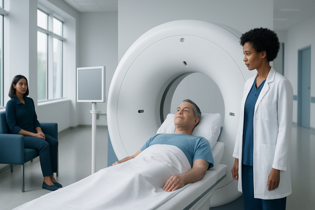
An eye CT scan is a quick, noninvasive imaging examination of the orbit and nearby facial structures. It can help identify fractures, bleeding, foreign bodies, sinus-related problems, masses and changes around the optic nerve.
Key Takeaways
- An eye CT scan is a quick, noninvasive imaging examination of the orbit and nearby facial structures.
- It can help identify fractures, bleeding, foreign bodies, sinus-related problems, masses and changes around the optic nerve.
- Some scans use contrast dye, which may require temporary fasting and additional safety checks.
- Most people can return to usual activities immediately after a non-contrast scan.
- Urgent results are usually communicated promptly when the scan shows findings that need rapid care.
An eye CT scan, also called an orbital CT scan, creates detailed cross-sectional images of the eye sockets, surrounding bones, sinuses and nearby soft tissues. It is commonly used after facial injury or when doctors need to investigate symptoms such as eye pain, swelling, double vision or a suspected problem behind the eye.
Eye CT Scan Overview
An eye CT scan is a computed tomography examination focused on the orbits, which are the bony sockets that contain the eyes. The scan uses a series of X-ray images and computer processing to create detailed views of the eye sockets, eye muscles, bones, sinuses, optic nerve region and tissues behind the eyes. It is different from a standard eye examination because it can show structures that cannot be seen directly through the front of the eye.
Doctors may request this test after an injury to the face or eye, or to investigate unexplained swelling, pain, double vision, reduced eye movement or changes in the position of an eye. It may also help assess conditions affecting the tissues around the eye, including thyroid eye disease, sinus disease or suspected infection. An eye CT scan is usually performed in a radiology department and often takes only a few minutes of actual scanning time.
CT uses ionizing radiation, so clinicians recommend it when its diagnostic value outweighs the small associated radiation exposure. In many situations, especially after trauma, CT provides faster and clearer information about bone injuries and certain urgent orbital problems than other imaging methods.
How an Eye CT Scan Works and Who May Need One
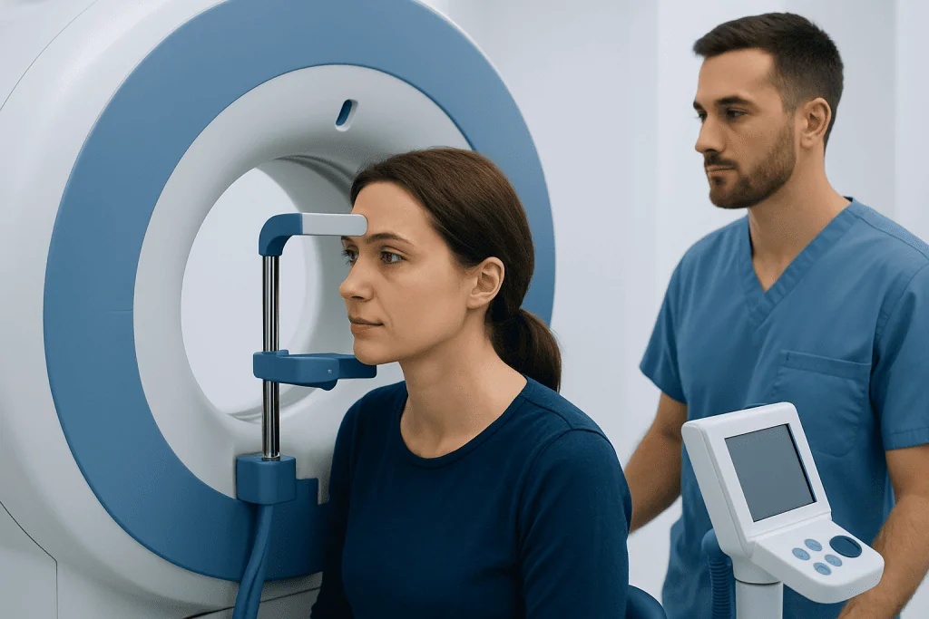
A CT scanner is a large ring-shaped machine. During the examination, an X-ray tube rotates around the head while detectors collect images from multiple angles. A computer combines these images into thin slices that a radiologist can review in several planes. This allows detailed assessment of delicate structures in and around the eye.
An eye CT scan may be recommended for a person with a suspected orbital fracture, injury caused by a sharp or high-speed object, facial trauma, eye protrusion, severe swelling or restricted eye movement. It may also be used to look for sinus inflammation extending toward the orbit, calcifications, certain tumors, abscesses or other causes of symptoms behind the eye.
CT is particularly useful for evaluating bone, air, acute bleeding and many foreign bodies. However, an MRI scan may be more appropriate for selected concerns involving soft tissues, the optic nerve or brain, especially when the situation is not urgent. The referring clinician will choose the imaging test based on the symptoms, examination findings and medical history.
Preparation and Candidacy

Most people can have an eye CT scan safely. Before the appointment, the imaging team should be told about pregnancy or a possible pregnancy, previous reactions to contrast dye, kidney disease, asthma, thyroid conditions and all regular medicines. These details help the team decide whether contrast is needed and whether any additional precautions are appropriate.
Some eye CT scans are performed without contrast dye. If contrast is planned, the hospital may ask the person not to eat for a specified period before the scan. Instructions vary by hospital and the reason for imaging, so the appointment guidance should always take priority. Drinking clear fluids may be allowed in many cases, but patients should confirm this with the imaging department.
Metal objects around the head can affect image quality. Patients may be asked to remove glasses, earrings, hearing aids, removable dental devices, hair clips and other items before entering the scanner room. They should wear comfortable clothing and bring relevant previous scans or medical records if requested.
Children and people who find it difficult to remain still may need additional support. Sedation is uncommon for a routine eye CT scan, but it may be considered in selected circumstances under appropriate medical supervision.
Eye CT Scan Procedure: Step by Step
On arrival, a radiology staff member confirms the person’s identity, reviews the reason for the scan and checks important safety information. If contrast is required, an intravenous line may be placed in the arm or hand. The person then lies on their back on a narrow table, usually with the head supported to help prevent movement.
The table moves slowly through the opening of the CT scanner. The scanner does not usually enclose the whole body, and the technologist can communicate with the patient throughout the test. The patient may be asked to keep their eyes still, look straight ahead or briefly hold their breath, depending on the images required.
If contrast dye is injected, some people notice a brief warm sensation, a metallic taste or a feeling of needing to urinate. These sensations typically pass quickly. The images themselves are painless, and the scanning portion often lasts less than 10 minutes, although the overall appointment can take longer.
After the images are obtained, a radiologist interprets them and sends a report to the requesting doctor. In cases of injury, the CT findings may guide urgent care, including assessment by ophthalmology, ear-nose-throat, maxillofacial or neurosurgical specialists when needed.
Benefits, Risks and Recovery Timeline
The main benefit of an eye CT scan is that it can provide rapid, detailed information about the orbit and surrounding facial structures. It can identify fractures that may trap an eye muscle, bleeding, swelling, sinus-related complications and many foreign bodies. This information can help doctors make timely decisions about observation, medication, specialist review or surgery.
CT scans expose the body to radiation. The amount is carefully controlled, and imaging teams use the lowest practical dose needed to obtain useful images. Patients should still avoid unnecessary repeat scans and share any relevant prior imaging with their clinician. Contrast dye, when used, can rarely cause an allergic-like reaction or affect kidney function in people with significant kidney impairment.
Recovery is generally immediate after a scan without contrast. Most people can eat, drink, drive and resume normal activities unless their doctor gives different instructions. If contrast was used, patients are commonly advised to drink fluids unless they have a medical reason to limit intake. Those who received sedation require a responsible adult and temporary restrictions on driving and important decisions.
Seek urgent medical advice after contrast if there is trouble breathing, swelling of the face or throat, widespread rash, faintness or severe persistent symptoms. These reactions are uncommon, and imaging departments are prepared to manage them promptly.
Is There Anything I Shouldn't Do Before a CT Scan?
Before an eye CT scan, a person should not ignore the preparation instructions provided by the imaging department. If contrast dye is planned, they may be asked not to eat for several hours beforehand. They should not assume fasting is needed for every CT scan, however, because many non-contrast eye scans require no dietary restriction.
Patients should not wear or bring unnecessary metal items around the head and face into the scanning area. It is also important not to withhold information about pregnancy, kidney disease, allergies, prior contrast reactions or medicines. This information allows the radiology team to make the examination as safe and useful as possible.
Regular medicines can often be taken as usual, but patients should ask in advance if they have diabetes, kidney disease or take medicines that require special planning around contrast. The care team can provide individualized instructions rather than asking the patient to change medication without advice.
What Happens If You Eat 2 Hours Before a CT Scan?
Eating two hours before a CT scan is usually not a problem for many non-contrast eye CT scans. However, if contrast dye, sedation or a particular type of examination is planned, the department may have fasting requirements. The safest step is to call the imaging center and explain what was eaten and when.
The scan may still proceed, or the appointment may be adjusted depending on the planned protocol and the person’s health needs. Patients should not skip an urgent scan after an injury simply because they have eaten; the clinical team can determine the safest approach. For people with diabetes or other conditions requiring regular meals, fasting instructions should always be individualized.
If a scan is scheduled because of a sudden eye injury, severe symptoms or a possible emergency, clinical urgency generally takes priority. The treating team will manage food intake, contrast and any other precautions as needed.
Is There Anything You Shouldn't Do After a CT Scan?
After a CT scan performed without contrast or sedation, there are usually no special restrictions. A person can generally return to work, eat normally and continue usual activities. If symptoms that prompted the scan are ongoing, such as pain or changes in vision, they should follow the advice of the clinician who arranged the test rather than relying only on the scan appointment instructions.
After contrast dye, patients should follow the department’s guidance about fluids and monitoring for delayed reactions. They should seek medical help for symptoms such as hives, worsening rash, breathing difficulty, facial swelling or severe vomiting. People with kidney disease may receive additional advice based on their individual situation.
After sedation, patients should not drive, operate machinery, drink alcohol or make important legal or financial decisions until the effects have fully worn off, typically for the period specified by their care team. A routine eye CT scan itself does not impair vision or make someone radioactive.
Frequently asked questions
Does an eye CT scan hurt?
The scan itself is painless. The person needs to lie still while the table moves through the CT scanner. If contrast dye is used, there may be a brief warm feeling or metallic taste during the injection.
How long does an eye CT scan take?
The actual image acquisition often takes only a few minutes. Allowing time for registration, safety checks, positioning and possible contrast injection, the visit may take around 20 to 60 minutes. Timing varies by facility and clinical situation.
Can an eye CT scan show an orbital fracture?
Yes. CT is commonly used to detect fractures of the eye socket and nearby facial bones. It can also show whether a fracture affects nearby muscles, soft tissues or sinuses.
How soon do doctors receive CT scan results if serious?
When an eye CT scan is performed in an emergency setting or shows a potentially urgent finding, images are commonly reviewed quickly and the treating team is informed promptly. The exact timing depends on the hospital, the reason for the scan and the availability of radiology specialists. A complete written report may follow after the initial urgent communication.
Can I drive after an eye CT scan?
Most people can drive after a routine CT scan without sedation, provided their vision and overall condition allow it. Driving is not appropriate after sedation, and it may not be safe if the person has an eye injury, double vision or other symptoms affecting sight. The care team can give specific advice.
Is MRI better than CT for eye problems?
Neither test is always better; they provide different information. CT is often preferred for fractures, acute trauma, bone detail and some foreign bodies, while MRI may better evaluate selected soft-tissue, optic nerve or brain concerns. A doctor chooses the test that best fits the clinical question.
References
- Radiological Society of North America
- American College of Radiology
- National Institute of Biomedical Imaging and Bioengineering
- U.S. Food and Drug Administration
- American Academy of Ophthalmology
This article is for general information only and is not a substitute for professional medical advice. Please consult a qualified doctor about your individual situation.
Explore treatments in Turkey — costs, top hospitals & a free quote
JCI-accredited · board-certified surgeons · reply within 24h
Add us as a Preferred Source to see more of our trusted health content across Google Search, AI Overviews and Discover.

More from the Health Library



Related Specialists

Prof. Dr. Lütfi Özkan
Radiation Oncology
Prof. Dr. Ahmet Alponat
General Surgery
Prof. Dr. Aziz Yazar
Medical Oncology
Prof. Dr. Meral Bayramoğlu
Physical Medicine & Rehabilitation