MRI Diffusion Weighted Imaging Stroke: Procedure, Recovery and Results
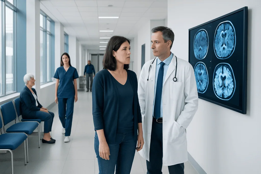
Diffusion-weighted MRI is highly sensitive for many recent ischemic strokes because it detects changes in water movement within injured brain tissue. A stroke MRI protocol may include several MRI sequences and can take roughly 20 to 60 minutes, depending on the clinical situation and scanner.
Key Takeaways
- Diffusion-weighted MRI is highly sensitive for many recent ischemic strokes because it detects changes in water movement within injured brain tissue.
- A stroke MRI protocol may include several MRI sequences and can take roughly 20 to 60 minutes, depending on the clinical situation and scanner.
- A normal early DWI scan does not always exclude stroke, particularly with very small, posterior circulation or transient events.
- MRI does not use ionizing radiation, but screening for implanted devices, metal fragments and kidney problems is important before contrast is considered.
- Stroke recovery varies widely; early rehabilitation, risk-factor management and follow-up care can support the best possible recovery.
MRI diffusion weighted imaging (DWI) is an MRI sequence used in suspected stroke to identify areas of recent brain injury, often within minutes to hours of reduced blood flow. It supports urgent clinical decisions, but results are interpreted alongside symptoms, examination findings and other imaging tests.
Overview: What MRI Diffusion Weighted Imaging Shows in Stroke
MRI diffusion weighted imaging stroke assessment is a specialized MRI approach used when clinicians suspect a recent stroke. Diffusion-weighted imaging, usually called DWI, detects changes in the microscopic movement of water molecules. When blood flow to part of the brain is suddenly reduced or blocked, as in an ischemic stroke, brain cells can swell and restrict water movement; this often appears as a bright area on DWI images.
DWI can help identify the location, approximate extent and likely timing of recent brain injury. It is especially useful when symptoms began recently, when computed tomography (CT) does not yet show a clear abnormality, or when doctors need more detail about the affected brain tissue. It is one part of stroke evaluation rather than a stand-alone diagnosis: symptoms, neurological examination, blood tests, CT or blood-vessel imaging, and heart assessment may also be needed.
Stroke is a medical emergency. Brain imaging should not delay urgent treatment when a person has sudden stroke symptoms. Depending on local stroke pathways, a rapid CT scan may be performed first to rule out bleeding, while MRI is used immediately or soon afterward when it can safely add important information. For broader information about the condition, see stroke.
How Diffusion-Weighted MRI Works
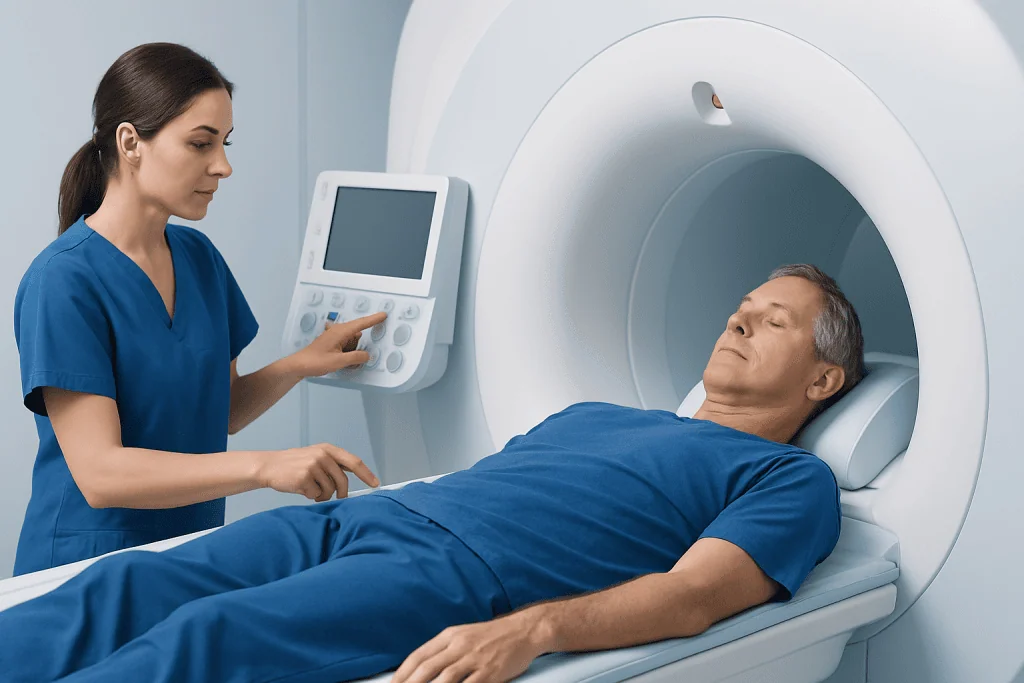
An MRI scanner uses a strong magnetic field and radiofrequency waves to create detailed pictures of the brain. DWI applies additional magnetic gradients that make the images sensitive to water motion. Healthy tissue and newly injured tissue handle water movement differently, allowing radiologists to identify areas of restricted diffusion.
Radiologists usually review DWI alongside an apparent diffusion coefficient (ADC) map. A true area of restricted diffusion generally appears bright on DWI and dark on the ADC map. This comparison helps distinguish a recent ischemic injury from certain imaging effects that can look bright on DWI without true restriction.
A complete stroke MRI protocol may also include fluid-attenuated inversion recovery (FLAIR), T2- and T1-weighted images, susceptibility-weighted imaging for blood products, and magnetic resonance angiography (MRA) to evaluate blood vessels. The pattern of findings may help clinicians determine whether a stroke is recent, whether bleeding is present, and which blood vessel territory may be involved.
Candidacy and Step-by-Step Procedure
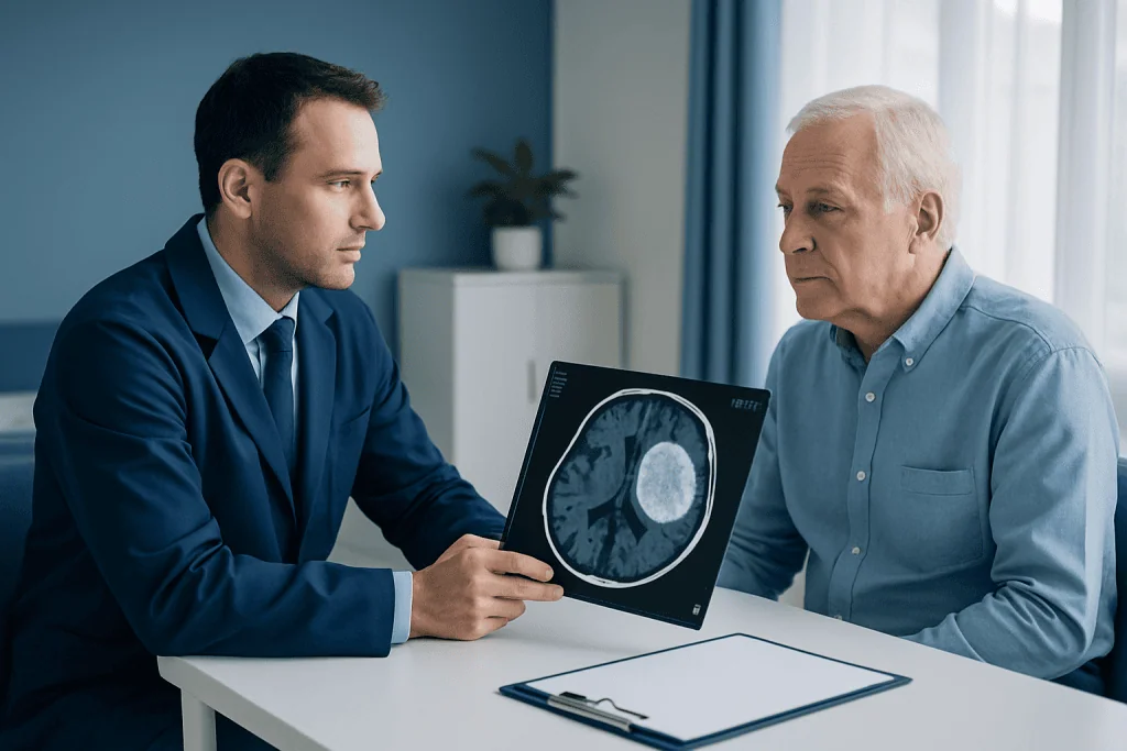
DWI is commonly considered for people with sudden weakness or numbness on one side, facial drooping, speech or language difficulty, vision changes, loss of balance, severe unexplained dizziness, or other symptoms that may represent stroke. It may also be used after a transient ischemic attack (TIA), sometimes called a “mini-stroke,” even if symptoms have resolved. The treating team decides which scan is appropriate based on symptom onset, clinical stability and the urgency of treatment.
Before the scan, staff ask about pacemakers, implanted stimulators, cochlear implants, clips used in brain surgery, metal fragments, medication pumps and prior operations. Many modern devices can be scanned under specific conditions, but every implant must be checked. Patients should also mention pregnancy, claustrophobia and kidney disease, particularly if contrast material might be considered. DWI itself generally does not require contrast.
During the examination, the person lies on a padded table with the head positioned in a special imaging coil. The table moves into the MRI scanner, and it is important to stay as still as possible. The machine makes loud tapping or knocking sounds, so ear protection is provided. Staff can communicate with the patient throughout the scan, and monitoring may be used for people who are unwell.
For patients with confirmed stroke, imaging findings guide urgent medical care and longer-term planning, which may include stroke treatment and rehabilitation. The appropriate treatment depends on stroke type, timing, cause and individual health needs.
How Long Does the MRI Stroke Protocol Take?
A focused brain MRI with diffusion-weighted images may be completed in a relatively short time, but a full MRI stroke protocol commonly takes about 20 to 60 minutes. The exact duration depends on the scanner, whether blood-vessel imaging or contrast sequences are included, how easily the person can remain still, and whether monitoring is required.
In an emergency, the clinical team prioritizes the fastest safe imaging method that will answer the immediate question. CT is often used first because it is widely available and rapidly identifies brain bleeding. MRI may follow when it can clarify whether there is early ischemic injury, assess the brainstem or posterior circulation, or evaluate uncertain symptoms.
Results are often reviewed urgently by a radiologist and stroke team. The report may describe a “focus of restricted diffusion,” its location and size, and whether there is evidence of hemorrhage or older changes. A result should always be discussed with the treating clinician, because the clinical meaning depends on the person’s symptoms and examination.
Benefits, Limitations and Safety Considerations
The main benefit of DWI is its sensitivity for many acute ischemic strokes. It can reveal small or early areas of injury that may not be obvious on other tests, helping clinicians confirm a suspected diagnosis, identify the brain region involved and guide further investigation. MRI also avoids ionizing radiation.
However, DWI is not perfect. A scan can be negative very early after symptoms begin, with very small strokes, or with some strokes affecting the brainstem or posterior circulation. Conversely, restricted diffusion can occasionally occur in conditions other than stroke, such as seizures, migraine-related changes, inflammation, infection or metabolic disturbances. Follow-up imaging may be recommended if symptoms strongly suggest stroke despite an initially normal scan.
MRI is generally safe when proper screening is completed. The most common challenges are noise, the enclosed space and the need to lie still. Sedation may occasionally be considered for severe claustrophobia or inability to remain still, but this is individualized and requires appropriate monitoring. Gadolinium contrast, if needed for another part of the assessment, has separate considerations and is used only when its likely benefit outweighs potential risks.
Recovery, Follow-Up and Everyday Care After Stroke
There is no single recovery timeline after stroke. Some people improve quickly over days or weeks, while others need longer-term support. Recovery depends on the location and size of the injury, the person’s age and general health, complications, rehabilitation access and the cause of the stroke. DWI findings contribute useful information, but they cannot predict an individual outcome on their own.
Follow-up commonly addresses the cause of stroke and ways to reduce the chance of another event. This may involve management of blood pressure, diabetes, cholesterol, heart rhythm disorders and sleep apnea, along with medication review, smoking cessation and individualized nutrition and activity advice. Physical therapy, occupational therapy and speech-language therapy can address movement, daily activities, communication, swallowing and thinking skills.
Families and caregivers can help by supporting medication routines, attending follow-up appointments when appropriate, and watching for changes in mood, cognition, swallowing or mobility. Sudden new neurological symptoms should never be assumed to be part of recovery. Acibadem International’s multidisciplinary specialists and JCI-accredited hospitals diagnose and treat stroke for international patients, including coordinated assessment and rehabilitation planning.
What Are Good Signs After a Stroke?
Good signs after a stroke can include gradually improving strength, clearer speech, better attention, improved swallowing, increased ability to sit, stand or walk safely, and greater independence with daily tasks. Early return of movement or speech can be encouraging, but progress is not always steady. Fatigue, slower thinking and temporary setbacks may occur during recovery.
Meaningful improvement can continue for months and, for some people, longer. The rehabilitation team measures progress with practical goals rather than comparing one person’s recovery with another’s. Regular therapy, prevention of complications and treatment of underlying risk factors are important parts of continued recovery.
What Is the Average Life Expectancy After a Frontal Lobe Stroke?
There is no reliable single average life expectancy for a person after a frontal lobe stroke. Outcome and longevity vary greatly according to stroke severity, the amount of brain affected, age, other medical conditions, the cause of stroke, treatment received and success in preventing future strokes.
Frontal lobe strokes may affect movement, planning, attention, behavior, language or emotional regulation, depending on the exact area involved. Many of these changes can improve with medical follow-up and rehabilitation. Rather than relying on a general estimate, clinicians can discuss an individual outlook after reviewing imaging, recovery progress and health risks over time.
What Not to Do After Brain Stroke?
After a brain stroke, a person should not stop prescribed medicines, miss follow-up appointments, smoke, use recreational drugs, or make major changes to diet, exercise or supplements without discussing them with the clinical team. It is also important not to drive, return to hazardous work, or resume activities with a high fall risk until cleared by an appropriate professional.
Alcohol use should be discussed with the treating clinician because it may interact with medicines, increase fall risk and affect blood pressure or recovery. People should not ignore depression, anxiety, sleep problems, swallowing difficulty or new cognitive changes; these are common, treatable concerns that deserve medical attention. Family members should avoid taking over every task if the person can safely practice skills, as supervised independence supports rehabilitation.
When to Seek Medical Care
Emergency care is needed immediately for sudden facial drooping, arm or leg weakness or numbness, speech difficulty, confusion, new vision loss, severe balance problems, sudden severe headache, or loss of consciousness. Even if symptoms improve within minutes, urgent assessment is essential because a TIA can be a warning sign of a future stroke.
After a known stroke, seek urgent medical care for any new or worsening neurological symptoms, repeated vomiting, seizures, severe headache, chest pain, shortness of breath, falls with head injury, or marked changes in alertness. Calling local emergency services is safer than driving the person to hospital, as emergency teams can begin assessment during transport.
Non-urgent follow-up is appropriate for persistent fatigue, mood changes, memory concerns, difficulty with daily activities, medication side effects or questions about return to work, travel and exercise. Early communication with the stroke and rehabilitation team can help address these concerns safely.
Frequently asked questions
Can diffusion-weighted MRI detect a stroke immediately?
DWI can detect many ischemic strokes very early, sometimes within minutes to hours after reduced blood flow begins. However, an initially normal DWI scan does not completely rule out stroke, especially with very small or posterior circulation strokes. Doctors interpret the scan together with symptoms, examination and other tests.
Does diffusion-weighted MRI require contrast dye?
No. Diffusion-weighted imaging itself does not require contrast material. Other MRI sequences may sometimes use contrast if clinicians need additional information, but this is decided individually.
What does restricted diffusion on an MRI mean?
Restricted diffusion means water molecules are moving less freely in a part of the brain than expected. In the appropriate clinical setting, it commonly indicates recent ischemic stroke, but it can also occur in other conditions. A radiologist and treating clinician determine what it means in the full clinical context.
Can an MRI tell how old a stroke is?
MRI patterns can help estimate whether a brain injury is recent, subacute or old, particularly when DWI and FLAIR images are reviewed together. It cannot always assign an exact time of onset. The person’s symptom history remains important.
Is MRI safe after a stroke?
MRI is generally safe after stroke when the person is stable and has been screened for implanted devices and metal. The choice between CT and MRI depends on urgency, availability and the clinical question. Emergency stroke treatment should not be delayed while waiting for an MRI.
What happens if the MRI is negative but stroke symptoms occurred?
A negative MRI may lead clinicians to consider a TIA, a very small or early stroke, or a condition that can mimic stroke. Further observation, repeat imaging, blood-vessel tests, heart rhythm monitoring and risk-factor assessment may be recommended. Follow-up remains important because transient symptoms can still signal future stroke risk.
References
- American Stroke Association
- American College of Radiology
- National Institute of Neurological Disorders and Stroke
- European Stroke Organisation
- World Health Organization
This article is for general information only and is not a substitute for professional medical advice. Please consult a qualified doctor about your individual situation.
Explore treatments in Turkey — costs, top hospitals & a free quote
JCI-accredited · board-certified surgeons · reply within 24h
Add us as a Preferred Source to see more of our trusted health content across Google Search, AI Overviews and Discover.