Can MRI Detect Polyps in Colon: Preparation, Procedure and Results
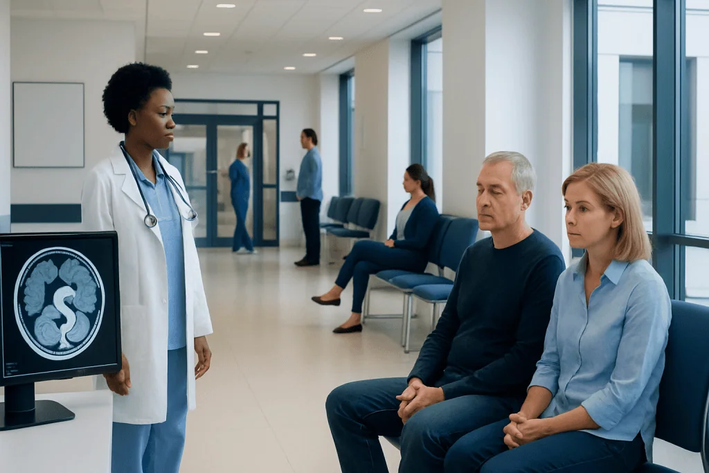
MRI may detect larger colon abnormalities but can miss small polyps. Colonoscopy is generally the most accurate test for detecting and removing colon polyps.
Key Takeaways
- MRI may detect larger colon abnormalities but can miss small polyps.
- Colonoscopy is generally the most accurate test for detecting and removing colon polyps.
- MRI is particularly useful for assessing some known or suspected colorectal cancers, including local spread and nearby structures.
- A positive stool-based screening test or concerning symptoms usually needs follow-up with colonoscopy.
- Most colorectal cancers develop from certain precancerous polyps over time, making screening and polyp removal important.
MRI can sometimes show larger colon polyps, masses, bowel-wall changes or signs that cancer may have spread, but it is not usually the preferred test for routine polyp screening. Colonoscopy remains the standard because it can closely examine the colon, take biopsies and remove most polyps during the same procedure.
Can MRI Detect Polyps in Colon?
Can MRI detect polyps in colon? MRI may show larger polyps, a sizable mass, bowel-wall thickening or changes around the colon, but it is not the usual first-choice test for finding colon polyps. Small polyps can be difficult to see on standard MRI, and MRI cannot remove a polyp or collect a tissue sample.
For most people, colonoscopy is the preferred examination when the goal is to identify and prevent colorectal cancer by finding and removing polyps. MRI has an important but different role: it may help doctors evaluate certain suspected or confirmed bowel cancers, define how far a tumor extends, or assess areas outside the bowel wall.
Polyps are growths that arise from the lining of the colon or rectum. Many are harmless, but some types can become cancerous gradually over years. Finding them early gives clinicians an opportunity to remove them before cancer develops. For information about the wider condition, see colon cancer.
How MRI Examines the Colon
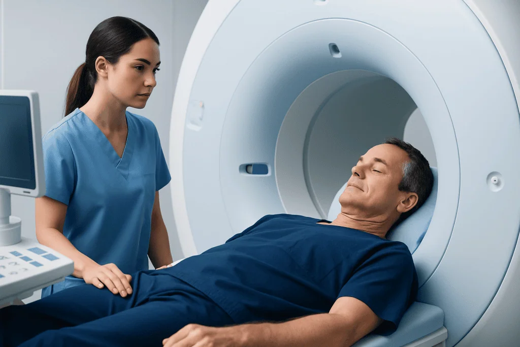
MRI uses a strong magnetic field and radio waves to create detailed images without ionizing radiation. During an abdominal or pelvic MRI, a person lies on a movable table that passes through the scanner. The images can show soft tissues, the bowel wall, lymph nodes, blood vessels and organs around the colon.
Routine MRI is not designed specifically to inspect the inner lining of the colon in the way a colonoscope can. The colon naturally moves and may contain stool, gas and fluid, all of which can affect image quality. Very small or flat polyps may not create enough contrast from the surrounding bowel lining to be reliably visible.
In selected settings, specialized MRI techniques may be used to assess the rectum or other parts of the bowel. For example, pelvic MRI is commonly valuable after a rectal cancer diagnosis because it can help the care team assess the tumor’s relationship to nearby tissues and plan treatment. It complements, rather than replaces, endoscopy and biopsy.
MRI may involve contrast material given through a vein to make particular tissues easier to distinguish. The radiology team reviews kidney health, allergy history, implanted devices and pregnancy status beforehand to determine whether MRI and contrast are appropriate.
Would colon cancer show up on a MRI scan?
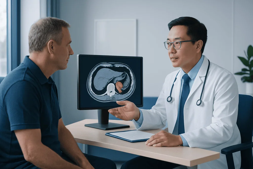
Would colon cancer show up on a MRI scan? A larger colon or rectal cancer may appear on MRI as a mass, irregular bowel-wall thickening or an area that narrows the bowel. MRI can also help identify findings outside the bowel, such as involvement of nearby organs, enlarged lymph nodes or, in some circumstances, disease in other parts of the body.
However, an MRI result alone usually cannot confirm that an abnormality is cancer. Diagnosis generally requires direct visualization with endoscopy and a biopsy, in which a small tissue sample is examined by a pathologist. A normal MRI also does not reliably rule out early cancer or small precancerous polyps.
MRI is especially established in the assessment of rectal cancer, where detailed pelvic images can guide decisions about surgery, radiation therapy or chemotherapy. The exact imaging plan depends on the suspected location and stage of disease, as well as a person’s symptoms and previous test results.
Symptoms such as rectal bleeding, persistent change in bowel habits, unexplained iron-deficiency anemia, abdominal pain or unintentional weight loss should be medically assessed. These symptoms have many possible causes, but they should not be evaluated with imaging alone when colonoscopy or another direct bowel assessment is indicated.
Is an MRI as good as a colonoscopy?
Is an MRI as good as a colonoscopy? For finding and treating colon polyps, MRI is generally not as effective as colonoscopy. Colonoscopy allows the specialist to inspect the colon lining directly, identify small and flat lesions, take biopsies and remove many polyps immediately. This combination makes it both a diagnostic and preventive procedure.
MRI may be considered when a person cannot undergo colonoscopy, when an examination is incomplete, or when clinicians need additional information about a known or suspected cancer. Other noninvasive options may also be considered in selected patients, including stool-based screening tests or CT colonography. Each option has strengths and limitations, and an abnormal result commonly still requires colonoscopy.
Colonoscopy preparation is important because a clean colon improves the ability to detect small lesions. If polyps are found, their size, number and microscopic type help determine when the next surveillance examination may be needed. Polyp removal is often performed through colonoscopy during the same visit.
The best screening method is individualized. A doctor considers age, family history, prior polyp findings, symptoms, medical conditions, medication use and local screening guidance before recommending a test.
How to detect colon polyps without colonoscopy?
How to detect colon polyps without colonoscopy? No non-colonoscopy method detects every polyp, but several tests can help identify people who may need further assessment. Stool tests can look for hidden blood or altered DNA associated with some colorectal cancers and advanced polyps. They are convenient screening tools, but they do not visualize or remove polyps.
CT colonography, sometimes called virtual colonoscopy, uses CT imaging to create pictures of the colon after bowel preparation and inflation of the bowel with gas. It can identify some larger polyps, but it uses radiation and any significant finding needs follow-up colonoscopy for biopsy or removal. It may be useful for certain people when standard colonoscopy is unsuitable or incomplete.
Capsule endoscopy is used for selected digestive conditions, but it is not a routine replacement for colonoscopy in colon-polyp screening. MRI is likewise not routinely used as a stand-alone screening test for polyps. A specialist can explain which tests are available and appropriate in the person’s healthcare setting.
A negative noninvasive test does not mean that new symptoms should be ignored. If bleeding, anemia, persistent bowel changes or other warning signs develop, prompt clinical review is important even if a previous screening test was normal.
What percent of colon cancer starts as polyps?
What percent of colon cancer starts as polyps? Most colorectal cancers are believed to develop from precancerous polyps, particularly adenomatous polyps and certain serrated polyps. Not every polyp becomes cancer, and the likelihood varies according to the polyp’s type, size, microscopic features and number.
Because the transformation from a precancerous polyp to cancer often takes years, screening can reduce risk by detecting and removing lesions before they progress. Hyperplastic polyps, especially small ones in the lower colon, are often low risk, but a pathology report is needed to classify a removed polyp accurately.
Some colorectal cancers develop through pathways that may not follow the typical polyp-to-cancer sequence, and some polyps can be harder to recognize. This is why appropriate screening intervals and careful follow-up after polyp removal matter. A clinician can explain what an individual pathology result means for future surveillance.
- Risk may be higher with increasing age, a personal history of polyps or colorectal cancer, or a strong family history.
- Inflammatory bowel disease and inherited cancer syndromes may require earlier or more frequent surveillance.
- Healthy lifestyle habits can support overall colorectal health but do not replace recommended screening.
MRI Preparation, Procedure, Recovery, Risks and Benefits
Preparation and candidacy: The required preparation depends on why MRI is being performed. Some abdominal or pelvic MRI scans require avoiding food for a period beforehand, and some may involve bowel preparation. The imaging center provides specific instructions. People should tell the team about pacemakers, cochlear implants, metal fragments, clips, joint replacements, kidney disease, pregnancy, claustrophobia and previous contrast reactions.
Step by step: On arrival, the person changes into suitable clothing and removes metal objects. An intravenous line may be placed if contrast is planned. They lie still on the scanner table while images are obtained; the machine makes loud tapping sounds, so hearing protection is provided. The scan commonly takes between about 30 and 60 minutes, depending on the body area and imaging protocol.
Recovery timeline: Most people can resume normal activities immediately after an MRI. If contrast is used, the team may advise drinking fluids unless there is a medical reason to limit intake. A radiologist interprets the images, and the ordering clinician discusses the results and whether endoscopy, laboratory testing or further imaging is needed.
Benefits and risks: MRI does not use radiation and provides excellent soft-tissue detail. Limitations include reduced ability to identify small colon polyps, the need to remain still and possible discomfort in an enclosed space. Contrast reactions are uncommon, but they can occur; very rarely, certain contrast agents require special caution in people with severe kidney impairment.
When to Seek Medical Care
Medical review is advisable for rectal bleeding, black stools, persistent change in bowel habits, ongoing abdominal discomfort, unexplained fatigue, iron-deficiency anemia or unintentional weight loss. These symptoms do not necessarily mean cancer, but timely assessment can identify treatable causes and determine whether colonoscopy or another test is needed.
People with a first-degree relative who has had colorectal cancer or advanced polyps, a history of inflammatory bowel disease, or a known inherited cancer syndrome should discuss individualized screening with a doctor. Screening may need to begin earlier or occur more often than for average-risk adults.
If a screening test is positive, follow-up should not be delayed. A stool test, CT colonography or MRI result may provide useful information, but colonoscopy is often the next step when clinicians need to inspect the lining closely, obtain tissue or remove a lesion.
Acibadem International’s multidisciplinary specialists and JCI-accredited hospitals can evaluate digestive symptoms, coordinate imaging and provide endoscopic care for international patients. The appropriate test and follow-up plan should always be decided with a qualified healthcare professional.
Frequently asked questions
Can an MRI miss colon polyps?
Yes. MRI can miss small, flat or early polyps because it does not directly inspect the inner lining of the colon. It is therefore not commonly used as the main test for routine colon-polyp screening.
Can MRI replace colonoscopy for colorectal cancer screening?
MRI generally cannot replace colonoscopy for most people who need colon-polyp assessment. Colonoscopy is more effective for direct visualization, biopsy and removal of polyps, while MRI is more often used to assess the extent of known or suspected disease.
Why might a doctor order an MRI after colonoscopy?
A doctor may order MRI to obtain detailed images of tissues around a suspected or confirmed cancer, particularly in the rectum. It can support staging and treatment planning, while colonoscopy and biopsy establish what the lesion is.
Do I need bowel preparation before a colon MRI?
Some MRI examinations require fasting, bowel preparation or medication to reduce bowel movement, while others do not. The imaging department will provide instructions based on the type of scan and the reason it has been requested.
Can stool tests find colon polyps?
Some stool tests may detect blood or DNA changes linked with certain advanced polyps and colorectal cancers. They do not directly see polyps, and a positive result generally requires colonoscopy for confirmation and possible removal.
Are colon polyps always cancerous?
No. Many colon polyps never become cancer, but some types have the potential to change over time. Removing and examining polyps helps determine their type and reduces the chance that precancerous lesions will progress.
References
- World Health Organization
- American Cancer Society
- National Cancer Institute
- European Society of Gastrointestinal Endoscopy
- American College of Radiology
This article is for general information only and is not a substitute for professional medical advice. Please consult a qualified doctor about your individual situation.
Persistent digestive symptoms? Get evaluated in Turkey
JCI-accredited · board-certified surgeons · reply within 24h
Add us as a Preferred Source to see more of our trusted health content across Google Search, AI Overviews and Discover.