Xray of the Bladder: Preparation, Procedure and Results

A bladder X-ray commonly involves contrast dye placed through a urinary catheter before images are taken. The medical term for an X-ray examination of the bladder is cystography; a study performed while urinating is called a voiding cystourethrogram.
Key Takeaways
- A bladder X-ray commonly involves contrast dye placed through a urinary catheter before images are taken.
- The medical term for an X-ray examination of the bladder is cystography; a study performed while urinating is called a voiding cystourethrogram.
- The examination may help identify bladder injury, urinary reflux, structural changes, stones, or problems with bladder emptying.
- Preparation is usually straightforward, but patients should report pregnancy, contrast reactions, infections, and relevant medicines beforehand.
- Mild urinary discomfort after catheter removal can occur, but severe pain, fever, or inability to urinate needs medical attention.
An xray of the bladder is usually a contrast imaging examination called a cystogram. It helps clinicians assess the bladder’s shape, capacity, emptying, and possible problems such as leakage, stones, obstruction, or urine flowing backward toward the kidneys.
Overview: What Is an Xray of the Bladder?
An xray of the bladder is an imaging test that usually uses a contrast liquid to make the bladder visible on X-ray images. The contrast is most often placed directly into the bladder through a thin catheter, allowing the radiology team to assess its outline, filling, and emptying. This is different from a routine abdominal X-ray, which may show some stones but does not show the bladder lining and function in the same detail.
The X-ray examination of the bladder is called cystography, and the images are called a cystogram. The term sometimes appears in study materials as “xray of the bladder quizlet,” but in clinical practice the specific name depends on how the test is performed. An X-ray of the bladder and urethra during urination is called a voiding cystourethrogram (VCUG), also known as a micturating cystourethrogram.
These tests are used selectively, often when a clinician needs information that ultrasound, CT, or other imaging has not provided. They can be particularly useful after pelvic trauma, during evaluation of recurrent urinary problems in selected patients, or when bladder leakage or urinary reflux is suspected.
How the Examination Works and Who May Need It
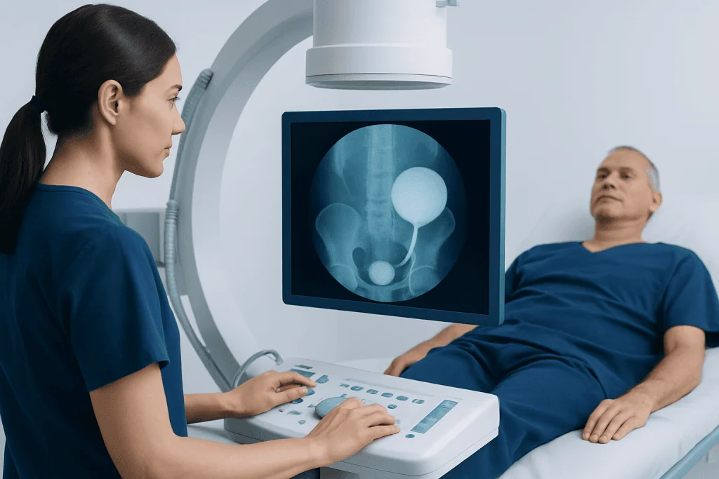
During cystography, contrast material fills the bladder and absorbs X-rays differently than surrounding tissues. This creates a clear outline of the bladder on a series of images. In a VCUG, images are also taken as the person urinates, which helps show the urethra and whether urine travels backward from the bladder toward the ureters and kidneys.
A doctor may recommend this test for people with a suspected bladder injury, unexplained urine leakage, difficulty emptying the bladder, or concern for an abnormal connection between the bladder and another organ. In children, a VCUG may be considered when recurrent urinary tract infections or other findings suggest vesicoureteral reflux, a condition in which urine flows backward toward the kidneys.
The examination is not required for every urinary symptom. Depending on the clinical question, a clinician may first recommend urine testing, ultrasound, CT imaging, MRI, or cystoscopy. For people being assessed for urinary symptoms or structural concerns, related evaluation and care may include urinary tract infection assessment and kidney stone evaluation when these conditions are suspected.
People should tell the care team if they might be pregnant, have had a previous reaction to contrast material, have a current urinary infection, recently had urinary surgery, or take medicines that may affect bleeding or kidney function. The team can explain whether the test should proceed, be adjusted, or be postponed.
Why Would a Doctor Order a Bladder Scan?

The phrase “bladder scan” can mean different tests. In many settings, it refers to a quick ultrasound measurement of urine left in the bladder after urination. However, people may also use it loosely to describe a bladder X-ray or cystogram. A clinician will clarify which test is being ordered and what question it is intended to answer.
A doctor may order a bladder X-ray when there is concern about a leak after pelvic injury or bladder surgery, particularly if urine could be escaping outside the bladder. It may also be used to investigate suspected reflux, structural abnormalities, certain fistulas, diverticula, or difficulties with the outlet of the bladder and urethra.
An X-ray study of the bladder and urethra can also provide functional information during urination. This may help identify narrowing, obstruction, or poor coordination in the lower urinary tract. Findings are interpreted alongside symptoms, physical examination, urine tests, and other imaging rather than in isolation.
For persistent symptoms such as weak urine flow, straining, incomplete emptying, or recurrent infections, the next steps may involve urological assessment. Urology care can bring together diagnostic imaging, laboratory testing, endoscopic evaluation, and individualized management when appropriate.
What Can Show Up on a Bladder Scan?
A cystogram can show the bladder’s size, shape, contour, and ability to hold contrast. It may reveal contrast leaking outside the bladder, which can indicate a tear or injury. It can also show outpouchings in the bladder wall, called diverticula, or an abnormal connection between the bladder and nearby structures.
When images are taken during urination, a VCUG can show the urethra and the way the bladder empties. It may identify urine moving backward into one or both ureters, known as vesicoureteral reflux. It may also suggest a narrowing or obstruction, although further tests are often needed to confirm the cause and guide treatment.
Some abnormalities may not be visible on a bladder X-ray. Small tumors, inflammation of the bladder lining, and certain stones may require other tests, such as cystoscopy, ultrasound, or CT. A normal result is reassuring but does not always exclude every cause of urinary symptoms.
The radiologist prepares a report describing the findings and sends it to the referring clinician. The clinician explains what the result means in the person’s wider health context and whether follow-up tests or treatment are necessary.
Preparation and Step-by-Step Procedure
Preparation instructions vary by hospital and by the reason for the examination. Most people can eat and drink normally unless they receive different directions. The care team may request a urine sample beforehand, especially if infection is a concern. Patients should bring a list of medicines and tell staff about allergies, pregnancy, recent procedures, and urinary symptoms.
At the imaging department, the person changes into a gown and lies on an X-ray table. A clinician gently cleanses the genital area and inserts a lubricated catheter into the urethra and bladder. Contrast material is then allowed to flow into the bladder through the catheter. Filling can create pressure and a strong urge to urinate, but it should not cause severe pain.
X-ray images are taken from different positions while the bladder is filled. For a voiding cystourethrogram, the catheter is removed or adjusted according to the protocol, and additional images are taken while the person urinates into a private collection device. This portion is important because it shows the urethra and bladder emptying in real time.
After the images are complete, the person can usually dress and return home the same day. The imaging team aims to maintain dignity and privacy throughout the test. Children may need additional reassurance, caregiver support, or age-appropriate preparation to help them cooperate comfortably.
How Long Does a Bladder Scan Take?
A simple ultrasound bladder scan often takes only a few minutes. A bladder X-ray examination, including catheter placement, bladder filling, positioning, and image review, commonly takes about 30 to 60 minutes. The exact time depends on the type of study, the number of images needed, and whether a voiding phase is included.
The actual X-ray exposures are brief. Much of the appointment is used for preparation, explanation, safe placement of the catheter, and positioning. If a patient has trouble emptying the bladder or requires additional images, the appointment may take longer.
Results are not always available immediately because a radiologist needs to review the images carefully. In urgent situations, such as suspected traumatic bladder injury, the treating team may receive preliminary information promptly. For non-urgent examinations, the referring clinician generally discusses the finalized report at a follow-up visit or through the agreed communication method.
Recovery, Benefits, and Possible Risks
Most people return to normal activities immediately after a cystogram or VCUG. Drinking water after the examination may help reduce temporary stinging or irritation when urinating. Mild discomfort, urinary urgency, or a small amount of pink-tinged urine can occur after catheterization and usually settles quickly.
The main benefit is that the examination can provide direct information about bladder integrity and urinary flow that may not be available from other tests. This can help clinicians make timely decisions after injury, clarify whether reflux is present, or plan further urological care when a structural issue is suspected.
Risks are generally low but include temporary urethral discomfort, a urinary tract infection, and a rare reaction to contrast material. The test uses a small amount of ionizing radiation; clinicians weigh this against the expected benefit and use appropriate imaging protocols. Pregnant patients or those who may be pregnant should inform the team before the examination.
Contact a healthcare professional if fever, chills, worsening burning on urination, heavy bleeding, severe lower abdominal pain, or inability to pass urine develops after the test. These symptoms are uncommon, but prompt assessment is important if they occur.
When to Seek Medical Care
Urgent medical assessment is important after major pelvic trauma, especially when there is lower abdominal pain, blood in the urine, inability to urinate, or a visibly swollen lower abdomen. These symptoms can have several causes, including injury to the urinary tract, and should not be managed at home.
A non-urgent medical appointment is appropriate for repeated urinary tract infections, ongoing pain with urination, persistent blood in the urine, recurrent leakage, weak urine stream, or the feeling that the bladder does not empty fully. These symptoms do not necessarily mean a serious condition, but they deserve evaluation.
Acibadem International’s multidisciplinary specialists and JCI-accredited hospitals diagnose and treat urinary conditions for international patients. When imaging identifies a problem requiring further care, a urologist can discuss suitable options, including uro-oncology evaluation when a cancer-related concern needs specialist assessment.
Frequently asked questions
What is the medical term for an X-ray of the bladder?
The medical term is cystography, and the resulting images are called a cystogram. If X-ray images are taken while the person urinates, the test is called a voiding cystourethrogram, or VCUG.
Is a bladder X-ray painful?
The X-ray itself is painless. Some people feel temporary discomfort during catheter insertion and pressure as the bladder fills with contrast material. The care team uses lubrication and explains each step to make the procedure as comfortable as possible.
Do I need to fast before a cystogram?
Many people do not need to fast for a cystogram, but instructions can differ depending on the reason for the test and the hospital protocol. The imaging department should provide specific preparation guidance before the appointment.
Can a cystogram detect a urinary tract infection?
A cystogram is not the main test used to diagnose a urinary tract infection. Urine testing is usually used for that purpose, while cystography may be considered if recurrent infections raise concern about reflux or a structural urinary problem.
What happens if the cystogram result is abnormal?
An abnormal result may lead to further testing, follow-up imaging, or consultation with a urologist, depending on the finding. Treatment is guided by the underlying cause, symptoms, and the person’s overall health rather than the X-ray result alone.
How much radiation is used in a bladder X-ray?
Cystography uses ionizing radiation, but imaging teams follow protocols designed to keep exposure as low as reasonably achievable while obtaining useful images. The expected diagnostic benefit is considered before the test is recommended, particularly for children and people who may be pregnant.
References
- Radiological Society of North America
- American College of Radiology
- National Institute of Diabetes and Digestive and Kidney Diseases
- National Health Service
- European Association of Urology
This article is for general information only and is not a substitute for professional medical advice. Please consult a qualified doctor about your individual situation.
Explore treatments in Turkey — costs, top hospitals & a free quote
JCI-accredited · board-certified surgeons · reply within 24h
Add us as a Preferred Source to see more of our trusted health content across Google Search, AI Overviews and Discover.