Enchondroma Xray: Preparation, Procedure and Results

An X-ray is commonly the first test used when an enchondroma is suspected or found incidentally. Most enchondromas are benign and do not cause symptoms or require surgery.
Key Takeaways
- An X-ray is commonly the first test used when an enchondroma is suspected or found incidentally.
- Most enchondromas are benign and do not cause symptoms or require surgery.
- Pain, growth after skeletal maturity, or unusual X-ray features may require further assessment.
- MRI or CT may be used when X-ray findings do not fully explain symptoms or need clarification.
- Regular follow-up is individualized and should be guided by an orthopedic specialist or musculoskeletal radiologist.
An enchondroma X-ray is usually the first imaging test used to assess a cartilage-based growth within a bone. The examination is quick, painless and can often show features that help clinicians decide whether observation, additional imaging or specialist assessment is appropriate.
Enchondroma X-ray: what it shows
An enchondroma X-ray is an imaging examination of a bone that may contain an enchondroma, a growth made of cartilage cells inside the bone marrow space. Enchondromas are commonly benign and may be discovered by chance when an X-ray is performed after an injury, for joint pain, or for another reason. They are often seen in the small bones of the hands and feet, but can also occur in long bones such as the femur, humerus, or tibia.
On an X-ray, an enchondroma may appear as a well-defined area within the bone with a characteristic pattern of calcification. The image also helps show whether the surrounding bone cortex is intact, expanded, thinned, or otherwise changed. These details, together with a person’s age, symptoms, and the lesion’s location, help the clinician judge whether the appearance is typical of a benign enchondroma.
An X-ray alone may not provide every answer. If the lesion has features that are not clearly typical, or if there is persistent pain that cannot be explained by another condition, the clinician may recommend MRI, CT, repeat X-rays over time, or referral to an orthopedic tumor specialist. The purpose is to reach an accurate diagnosis while avoiding unnecessary procedures.
How the X-ray works and who may need one
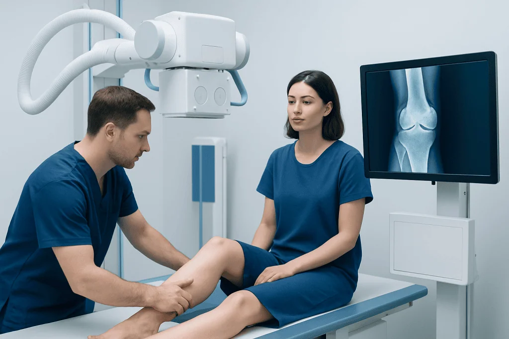
X-rays use a small, controlled amount of radiation to create images of dense structures, especially bones. Different tissues absorb X-rays differently, allowing the radiology team to see the outline of the bone and changes within it. For suspected enchondroma, images are usually taken from two or more angles so the lesion can be assessed in three dimensions.
An enchondroma X-ray may be appropriate for a person with unexplained bone pain, localized swelling, a fracture after a minor injury, or an incidental finding on previous imaging. It may also be requested to monitor a known lesion when a clinician considers interval imaging useful. Many people with an enchondroma have no symptoms, and not every incidental finding needs frequent follow-up.
Pregnancy should be discussed with the imaging team before any X-ray. Extremity X-rays generally involve a low radiation dose, but the team can consider whether imaging should be postponed, modified, or replaced with another test. People should also mention any previous scans, surgeries, injuries, or known bone conditions, as these can help the radiologist interpret the images accurately.
Preparing for an enchondroma X-ray and what happens during it

Preparation for a standard enchondroma X-ray is usually simple. Fasting, injections, contrast dye, and sedation are not normally needed. The patient may be asked to remove jewelry, watches, clothing with metal fasteners, or other items near the body part being examined, since metal can obscure the image.
During the examination, a radiographer positions the affected hand, foot, arm, leg, or other body part on or beside the X-ray detector. The patient may be asked to hold still for a few seconds while each image is taken. Several views are commonly obtained, and the radiographer may gently adjust the limb to obtain the clearest angles.
The test itself is painless and often takes about 10 to 20 minutes, depending on the body area and number of images required. Holding a sore limb in position may cause brief discomfort, especially after an injury, but the radiographer can assist with positioning and should be told if pain makes it difficult to remain still.
After the images are acquired, a radiologist reviews them and provides a report to the referring clinician. The report may describe the lesion’s size, location, borders, calcification pattern, and any effect on the surrounding bone. The clinician then explains what these findings mean in the context of the patient’s symptoms and medical history.
Understanding enchondroma X-ray results
A typical enchondroma often appears as a centrally located lesion within the bone, sometimes with small areas of calcification that can resemble rings, arcs, or stippling. In the hands and feet, it may cause gradual expansion of the bone without destroying its outer layer. A stable, well-defined appearance is generally reassuring, particularly when there are no concerning symptoms.
The report may also identify findings that need closer review, such as marked cortical destruction, a soft-tissue mass, rapid change compared with prior imaging, or other atypical features. These findings do not automatically mean cancer is present. They do mean that further assessment, often with MRI and specialist review, may be important to distinguish among possible cartilage-related bone lesions.
If the X-ray suggests a fracture through an enchondroma, treatment focuses first on the fracture and then on evaluating the underlying lesion. In selected cases, a specialist may discuss surgical removal of the lesion, known as curettage, sometimes followed by filling the space with graft material or another bone substitute. Decisions are based on the bone involved, fracture risk, symptoms, and imaging appearance.
For people needing coordinated assessment, Acibadem International’s multidisciplinary specialists in orthopedics, radiology, and oncology can evaluate bone lesions at JCI-accredited hospitals for international patients. A qualified clinician should interpret imaging results rather than relying on an X-ray report alone.
Benefits, limitations, risks and recovery timeline
The main benefit of an enchondroma X-ray is that it is widely available, fast, noninvasive, and particularly useful for examining bone structure. It can identify many typical enchondromas and establish a baseline image for comparison if follow-up is needed. It also helps identify fractures or weakening of the bone that may affect activity advice and treatment planning.
An X-ray has limitations. It cannot always distinguish a benign enchondroma from every other cartilage-forming lesion, especially when the lesion is in a long bone or symptoms do not match the imaging appearance. MRI provides more detail about cartilage, bone marrow, and nearby soft tissues, while CT can better define mineralization and cortical bone in some situations.
The radiation exposure from a routine extremity X-ray is low. There are no recovery restrictions after the examination, and people can usually return to work, school, driving, and normal daily activities immediately. If a painful injury was the reason for imaging, activity recommendations should follow the clinician’s advice rather than the X-ray procedure itself.
Further testing or treatment is not required for every enchondroma. When observation is chosen, the care team may recommend follow-up based on the lesion’s characteristics and symptoms. New or worsening pain should be reported rather than waiting until a scheduled review.
When should I be concerned about an enchondroma?
An enchondroma deserves medical review if there is persistent or increasing pain near the lesion, pain at rest or during the night, new swelling, reduced movement, or an unexplained fracture. These symptoms can have many causes, including arthritis, tendon problems, injury, or a fracture, but they should not be assumed to be caused by an enchondroma without evaluation.
Concern may also be appropriate when a lesion appears to enlarge after a person has reached skeletal maturity, when a new lesion is found in a long bone, or when an imaging report describes atypical features. Comparing current images with previous X-rays, if available, can be very helpful. A clinician may recommend MRI or referral to an orthopedic specialist with experience in bone tumors.
People with multiple enchondromas or a known condition that affects cartilage growth may need more individualized surveillance. The appropriate monitoring plan depends on the type, location, and behavior of the lesions, as well as the person’s symptoms and family history.
How painful is an enchondroma?
Most enchondromas are not painful. They are commonly found incidentally, meaning that the X-ray was ordered for another reason and the person did not know the lesion was present. When pain occurs, it is important to assess whether the discomfort truly comes from the enchondroma or from a more common nearby problem, such as an injury, joint disease, or soft-tissue irritation.
An enchondroma may cause discomfort if it weakens a bone, causes expansion of a small bone in the hand or foot, or is associated with a fracture. Pain can also occur when a lesion is in a location exposed to repeated pressure or movement. The severity varies and cannot be predicted from size alone.
Persistent, progressive, or unexplained pain should be discussed with a doctor, particularly if it occurs at rest or at night. The clinician may review the X-ray, examine the area, and arrange additional imaging when needed. Self-treatment with pain medicines should not replace assessment of new or worsening bone pain.
Can an enchondroma become cancerous? Can it increase in size?
A solitary enchondroma is usually benign, and transformation into a malignant cartilage tumor is considered uncommon. However, cartilage tumors can sometimes be difficult to classify from a single image, and some concerning lesions may resemble enchondromas initially. This is why symptoms, lesion location, imaging characteristics, and changes over time all matter.
Enchondromas can increase in size while a child or adolescent is growing because the bones are still developing. In adults, apparent growth should be assessed carefully, particularly if it is accompanied by pain or changes in the bone’s outer cortex. Differences between imaging techniques or X-ray positioning can sometimes make a lesion seem different in size, so direct comparison by an experienced radiologist is valuable.
Most people do not need preventive treatment for an asymptomatic, typical enchondroma. Maintaining general bone health, avoiding high-impact activity only if advised because of fracture risk, and attending recommended reviews are practical steps. Prompt reassessment is appropriate if symptoms change or a new injury occurs near the affected bone.
Frequently asked questions
What does an enchondroma look like on an X-ray?
An enchondroma often appears as a well-defined area within a bone, sometimes with small calcifications in a ring-like or arc-like pattern. It may gently expand the bone, especially in the fingers or toes, while leaving the outer bone layer intact. A radiologist interprets these features together with the lesion location and a person’s symptoms.
Do I need to prepare for an enchondroma X-ray?
Usually, no special preparation is needed. The patient may need to remove jewelry or metal objects near the area being imaged and should tell the team about pregnancy or possible pregnancy. Food, drink, and regular medicines can generally continue unless another test is planned at the same visit.
When should I be concerned about an enchondroma?
Medical review is important for persistent or worsening pain, pain at rest or at night, swelling, reduced function, or a fracture after minor trauma. It is also appropriate when imaging suggests growth or atypical changes. These signs do not confirm cancer, but they may indicate a need for more detailed assessment.
How painful is an enchondroma?
Many enchondromas cause no pain at all. If pain occurs, it may relate to a fracture, pressure within a small bone, or another condition near the lesion. Persistent pain should be assessed because pain alone cannot determine the nature of a cartilage lesion.
Can an enchondroma become cancerous?
A single enchondroma is usually benign, and malignant transformation is uncommon. Still, clinicians evaluate symptoms and imaging features carefully because some cartilage tumors can have overlapping appearances. Further imaging or specialist review may be advised when findings are unclear or concerning.
Can an enchondroma increase in size?
An enchondroma may appear to grow while bones are developing in childhood and adolescence. Growth or new changes in adulthood should be evaluated, especially when accompanied by pain. Comparing X-rays taken over time helps determine whether a meaningful change has occurred.
References
- American Academy of Orthopaedic Surgeons
- American College of Radiology
- National Cancer Institute
- OrthoInfo
- Radiopaedia
This article is for general information only and is not a substitute for professional medical advice. Please consult a qualified doctor about your individual situation.
Joint and spine care in Turkey — expert assessment & treatment
JCI-accredited · board-certified surgeons · reply within 24h
Add us as a Preferred Source to see more of our trusted health content across Google Search, AI Overviews and Discover.

More from the Health Library



Related Specialists

Assoc. Prof. Dr. Emrah Erdoğan
Cardiology
Dr. Evrim Çeliker Yapıcı
Neonatal Intensive Care Unit
Dr. Hayati Yavuz
Internal Medicine
Dr. Sinan Aksu
Orthopedic Surgery & Traumatology