How to Prepare for a Screening Mammogram: How It Works, Results and What to Expect
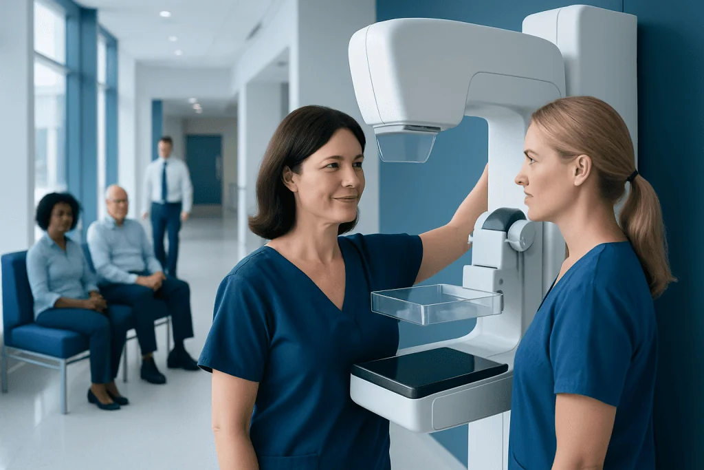
Do not apply deodorant, antiperspirant, powder, lotion, perfume, or glitter to the chest or underarms on the day of the examination. Tell the imaging team about breast symptoms, implants, pregnancy or possible pregnancy, breastfeeding, prior procedures, and previous imaging.
Key Takeaways
- Do not apply deodorant, antiperspirant, powder, lotion, perfume, or glitter to the chest or underarms on the day of the examination.
- Tell the imaging team about breast symptoms, implants, pregnancy or possible pregnancy, breastfeeding, prior procedures, and previous imaging.
- Most screening mammograms take about 20 to 30 minutes, with breast compression lasting only a few seconds for each image.
- A callback for additional imaging is common and often reflects the need for clearer views, not a cancer diagnosis.
- Regular screening recommendations vary with age, personal risk, family history, and local guidance, so an individual plan should be discussed with a clinician.
To prepare for a screening mammogram, schedule it when breast tenderness is least likely, avoid deodorant and similar products on the day, wear a two-piece outfit, and bring previous breast imaging if it was done elsewhere. A mammogram is a brief X-ray examination that can identify breast changes before they can be felt, although an abnormal result does not by itself mean cancer.
Overview: preparing for a screening mammogram
A screening mammogram is a breast X-ray performed when a person has no new breast symptoms. It is designed to look for early changes, including small cancers or calcifications, that may not be noticeable during a breast examination. The best preparation is simple: make the appointment at a time when the breasts are less tender if possible, avoid products on the skin of the chest and underarms on the day, and provide the imaging center with relevant health and prior imaging information.
Screening mammography is different from diagnostic mammography. Diagnostic imaging is used when there is a concern such as a new lump, nipple discharge, skin change, or focal breast pain. People with symptoms should not wait for their next routine screening appointment; they should contact a clinician so the most appropriate breast assessment can be arranged.
Recommendations for when to start and how often to have screening differ among professional organizations and countries. Age, breast density, family history, inherited gene changes, previous chest radiation, and personal history of breast disease can all affect the plan. A clinician can help decide which screening approach is appropriate for the individual.
How screening mammography works and who may need it
Mammography uses low-dose X-rays to create images of breast tissue. During the examination, each breast is gently but firmly compressed between two plates for a few seconds. Compression spreads the tissue, reduces movement, and improves image quality while allowing the lowest practical radiation exposure. Standard screening usually includes more than one view of each breast.
Many centers use digital mammography, and some use digital breast tomosynthesis, often called 3D mammography. Tomosynthesis takes images from multiple angles to create thin image slices of the breast. It may improve visibility in some situations, particularly where overlapping tissue makes interpretation more difficult, but the most suitable technique depends on local availability and an individual’s circumstances.
Routine screening is generally intended for people at average risk who do not have breast symptoms. Those at higher risk may need earlier or additional screening, such as breast MRI, based on individualized medical advice. People with breast implants can usually have mammograms, but they should notify the center when booking because additional views may be needed.
Prior mammograms are particularly valuable because radiologists compare images over time. Comparison can help distinguish a stable, benign-looking finding from a new change that needs closer assessment. If previous studies were done at another center, ask how they can be transferred before the appointment.
How to prepare the day before and on the day

When possible, some people prefer to schedule screening for the week after their menstrual period, when the breasts may be less tender. This is optional, and it should not delay an important screening examination. At booking, mention any breast implants, breast surgery, hormone use, previous biopsies, family history of breast or ovarian cancer, breastfeeding, and possible pregnancy.
What should you not do the night before a mammogram? There are usually no special dietary restrictions, and most people can eat, drink, exercise, and take regular medicines as usual. The main practical step is to avoid applying products that may remain on the skin, such as deodorant, antiperspirant, talcum powder, body glitter, lotions, creams, or perfume around the breasts and underarms. These products can sometimes create image artifacts that resemble calcifications.
Should I shower before my mammogram? Yes. Showering or bathing is fine and does not affect the examination. Afterward, it is best not to apply deodorant, antiperspirant, powder, perfume, lotion, or similar products to the breasts, chest, or underarms until after the images have been taken.
Should I shave my armpits before a mammogram? Shaving is not required for mammography. A person may shave if that is part of their usual routine, but it is sensible to avoid shaving immediately before the appointment if it causes irritation or small cuts. The imaging team is accustomed to all normal body hair and skin variations.
- Wear a two-piece outfit so only the top clothing needs to be removed.
- Bring photo identification, referral details if required, and information about prior breast imaging.
- Tell the technologist about any new symptom, implant, prior breast operation, or difficulty standing or raising the arms.
- Consider asking the booking team whether results are sent directly to the patient, a referring clinician, or both.
What happens during the procedure and afterward
At the appointment, a radiology technologist will explain the process and ask relevant safety questions. The person removes clothing and jewelry from the waist up and is given a gown. The technologist positions one breast at a time on the mammography unit, carefully adjusting posture to include as much breast tissue as possible.
The compression plates close briefly while each image is taken. The pressure can feel uncomfortable, especially if the breasts are sensitive, but it should last only a short time. Speaking up is helpful if the position is painful, if there is limited shoulder movement, or if anxiety makes it difficult to continue; the technologist can make reasonable adjustments while still aiming for clear images.
There is no recovery period after a screening mammogram. Most people return to usual activities immediately. Mild temporary breast soreness, skin redness, or pressure marks can occur and generally settle quickly. Normal skin care products can be used after the appointment unless the imaging center gives different instructions.
The main benefit of screening is the chance to identify breast cancer at an earlier stage, sometimes before symptoms appear. Mammograms do not find every cancer, particularly in dense breast tissue, and they can also lead to false-positive findings that require more imaging or occasionally a biopsy. These limitations are important reasons to continue reporting new breast symptoms even after a recent normal mammogram.
Results, callbacks, and the limits of mammography
A radiologist interprets the images and sends a report to the appropriate clinician or directly to the patient, depending on the local process. Results may be available the same day or may take several days. A normal screening result means no concerning changes were seen on that examination; it does not guarantee that cancer is absent or that future screening is unnecessary.
Can a radiologist tell if you have breast cancer from a mammogram? A mammogram can show findings that are suspicious for cancer, but it usually cannot confirm a diagnosis on its own. If an area needs further evaluation, the next step may be additional mammographic views, breast ultrasound, MRI in selected circumstances, or a biopsy. A biopsy, in which a small tissue sample is examined in a laboratory, is the definitive way to diagnose most breast cancers.
A callback after screening can feel worrying, but it is often due to overlapping tissue, a technical issue, or a finding that needs a closer look. Most people recalled for additional imaging do not receive a cancer diagnosis. Comparing prior images and completing recommended follow-up promptly helps the care team reach the clearest conclusion.
If a biopsy is recommended after abnormal imaging, clinicians may discuss breast biopsy options and what the findings could mean. If cancer is diagnosed, care is planned by a multidisciplinary team and may include surgery, radiation therapy, systemic treatment, or a combination, tailored to the type and stage of disease.
When to seek medical care
A screening mammogram is not the right pathway for a new breast concern. A person should arrange medical assessment promptly if they notice a new lump or thickened area, persistent change in breast shape or size, dimpling or redness of the skin, a new inverted nipple, bloody or spontaneous nipple discharge, or swelling near the collarbone or underarm. These changes are often caused by non-cancerous conditions, but they still deserve evaluation.
Persistent pain in one specific area of the breast, especially when accompanied by a lump or skin or nipple change, should also be discussed with a clinician. Breast symptoms do not always require emergency care, but rapid or severe inflammation, significant swelling, fever, or feeling unwell should be assessed without delay because infection or another urgent problem may need treatment.
People with a strong family history of breast or ovarian cancer, a known inherited cancer-related gene change, past chest radiation, or a previous high-risk breast lesion should ask about a personalized screening plan. This may differ from routine population screening and can involve additional imaging or genetic counseling.
Planning ongoing breast health care
Keeping a personal record of screening dates, imaging locations, prior biopsies, operations, and family history can make future appointments more efficient. It is also helpful to know what the breasts normally look and feel like, so a new or persistent change is easier to recognize. Breast awareness supports screening but does not replace scheduled mammography when it is recommended.
Breast density is a common imaging description that refers to the proportion of fibrous and glandular tissue compared with fatty tissue. Dense breasts are normal, but they can make mammograms harder to interpret and are associated with a modestly increased breast cancer risk. A clinician can explain whether breast density was reported and whether additional screening is appropriate for the person’s overall risk profile.
For international patients, Acibadem International’s multidisciplinary specialists and JCI-accredited hospitals can coordinate breast imaging, diagnostic assessment, and treatment planning when needed. Decisions about screening and follow-up should always be made with a qualified healthcare professional who understands the patient’s personal history and local screening recommendations.
Frequently asked questions
How long does a screening mammogram take?
The imaging portion commonly takes about 20 to 30 minutes, although appointment timing varies by center. Compression is applied for only a few seconds for each image. Allow extra time for registration, changing, and any questions for the imaging staff.
Can I eat or drink before a screening mammogram?
Yes. Screening mammography does not usually require fasting, so people can eat and drink normally unless they have been given separate instructions for another test. Regular medicines can generally be taken as prescribed.
Why should deodorant and powder be avoided before a mammogram?
Some deodorants, antiperspirants, powders, lotions, and cosmetic products contain particles that can appear on an X-ray image. These may mimic tiny calcifications and make interpretation less clear. Showering is fine; simply avoid applying these products to the breasts, chest, and underarms until after the examination.
Is mammogram compression necessary?
Yes, compression is an important part of obtaining useful mammogram images. It spreads breast tissue for a clearer view, reduces motion, and helps keep radiation exposure as low as reasonably achievable. The pressure is brief, and the technologist can explain each step and respond to discomfort.
What happens if my mammogram result is abnormal?
An abnormal or incomplete result usually means that more information is needed, not that cancer has been diagnosed. The care team may recommend extra mammogram views, ultrasound, MRI in selected cases, or a biopsy. Completing follow-up promptly allows the radiologist and clinician to clarify the finding.
Can I have a mammogram if I have breast implants?
Yes, most people with breast implants can have mammography. It is important to tell the imaging center when making the appointment because specialized positioning and additional images may be required. The technologist will use techniques intended to visualize as much breast tissue as possible while taking care around the implant.
References
- World Health Organization
- American Cancer Society
- U.S. Food and Drug Administration
- American College of Radiology
- National Cancer Institute
This article is for general information only and is not a substitute for professional medical advice. Please consult a qualified doctor about your individual situation.
Oncology care in Turkey — second opinion and treatment plan
JCI-accredited · board-certified surgeons · reply within 24h
Add us as a Preferred Source to see more of our trusted health content across Google Search, AI Overviews and Discover.