

How CT, Ultrasound and Urine Tests Shape a Kidney Stone Treatment Plan
Key Takeaways
- Non-contrast CT detects nearly every stone type, including uric acid stones that are invisible on a plain X-ray, which is why X-rays are now rarely used for diagnosis.
- In a randomized trial of 2,759 emergency patients, starting with ultrasound instead of CT led to no more missed serious diagnoses at six months and far less cumulative radiation.
- Fever alongside flank pain converts a routine stone into an emergency, because an infected kidney behind a blockage needs urgent drainage regardless of stone size.
- The radiology report's two key numbers are stone diameter in millimeters and its position along the ureter; together they predict whether it will pass on its own.
- Only laboratory analysis of the passed or removed stone reliably tells you its composition; CT density offers a hint, not an answer.
- A 24-hour urine collection after the crisis identifies the specific chemistry behind stone formation and is the basis for a personalized prevention plan.
A non-contrast CT scan is the most accurate single test for finding kidney stones and measuring their size and position, but it is not always the right first test. Ultrasound can safely start the work-up for many stable adults and is preferred in pregnancy, while urinalysis, blood tests and stone analysis reveal infection and chemistry that no image shows. Together these results guide whether a stone is watched, helped to pass or treated with a procedure.
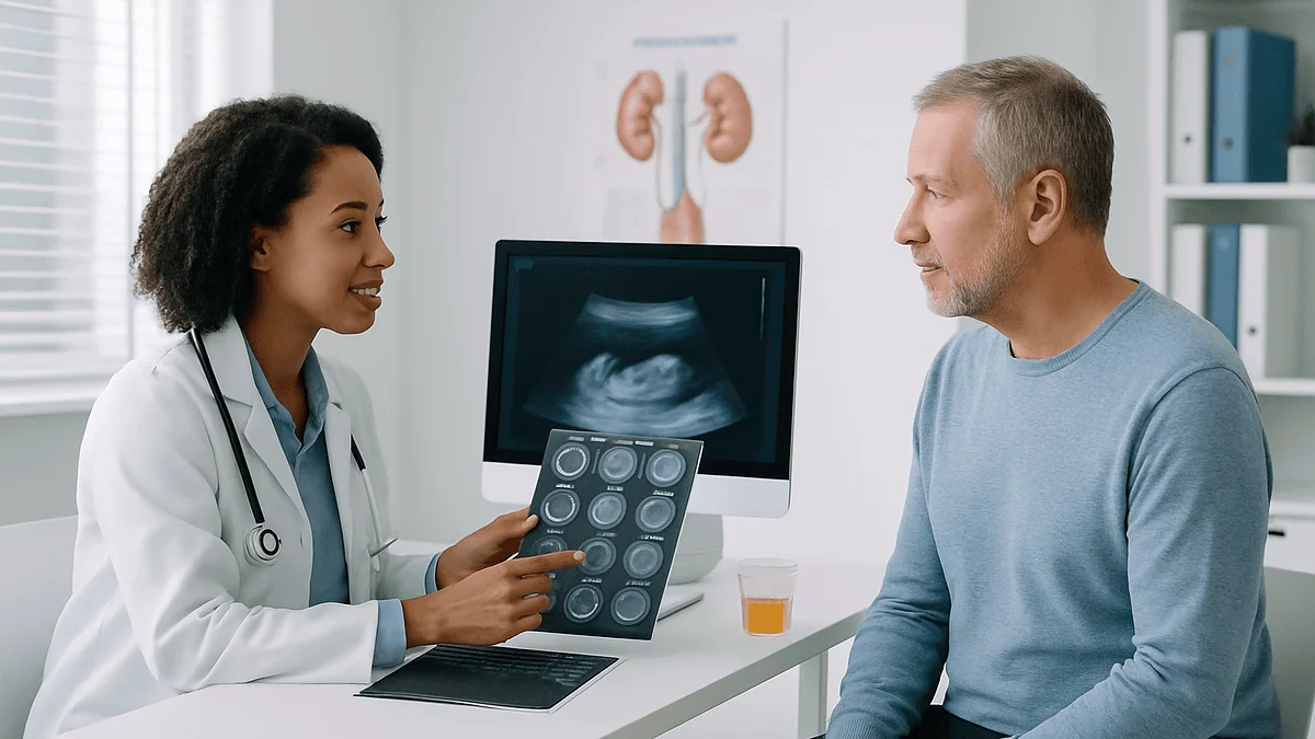
At two in the morning the pain arrives without introduction: a deep, twisting ache under one rib that no position relieves, spreading toward the groin in waves. By the time the triage nurse asks about blood in the urine, the person in the chair has already guessed. Then comes the sentence that turns a bad night into a decision tree: the doctor wants a CT scan for kidney stones, and possibly an ultrasound and a urine sample too.
Most people assume the scan is the whole story. It is not. The image answers where and how big; the urine answers whether infection is hiding behind the blockage and why the stone formed at all. Each result closes some doors and opens others.
This explainer walks through what each test actually shows, when doctors choose one over another, and how the numbers on a radiology report translate into waiting, medicine or a procedure, with the evidence laid out honestly.
Why a CT scan for kidney stones is usually the first test doctors order
Non-contrast CT is a scan that builds cross-sectional images of the abdomen from many X-ray angles without injecting dye. For suspected kidney stones it has become the default in most emergency departments for a simple reason: it finds stones of nearly every chemical type, including ones too small or too faint to appear on a plain X-ray. Mayo Clinic notes that this kind of CT can reveal even tiny stones, which is why the older abdominal X-ray is used far less often now.
The scan does more than confirm a stone exists. It measures the stone’s diameter, pinpoints where it sits along the ureter (the narrow tube carrying urine from kidney to bladder), and shows whether urine is backing up and swelling the kidney, a finding called hydronephrosis. Those three facts, size, position and blockage, are the spine of every treatment decision that follows. A stone the width of a grain of rice sitting near the bladder is a very different problem from one the size of a pea lodged at the top of the ureter.
There is a cost, and it is not money. CT uses ionizing radiation, and people who form stones tend to form them again, so scans can add up over a lifetime. That tension is the reason this article exists. The honest question is rarely whether CT is a good test; it plainly is. The better question is whether this particular person, at this particular moment, needs it. Sometimes the answer is yes within the hour. Sometimes an ultrasound and a urine sample will do the job with no radiation at all.
What actually happens during a CT scan for kidney stones, and how long it takes
Expect an anticlimax. You lie on a padded table, usually on your back with arms raised above your head, and the table glides through a ring-shaped opening while the machine hums. Nobody is sealed inside a tunnel; the ring is open at both ends. A technologist may ask you to hold your breath for a few seconds so the images are not blurred by a moving diaphragm.
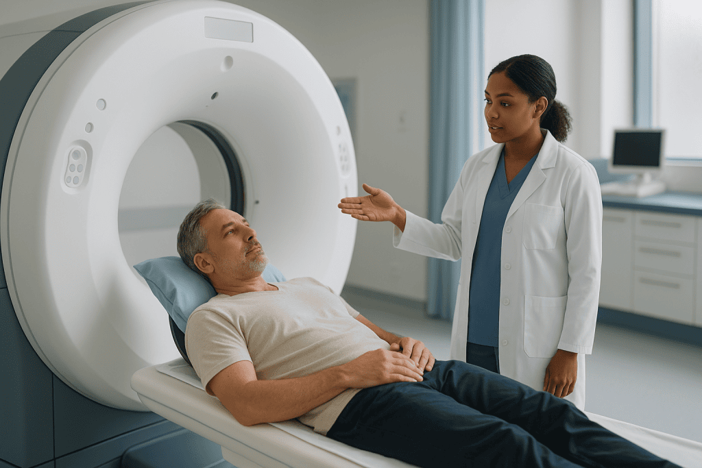
The scanning itself lasts only minutes. According to Johns Hopkins Medicine, a CT of the kidney performed without contrast generally needs little preparation, and most of the appointment goes to checking in, changing and positioning rather than to the scan. In an emergency department, the wait for the radiologist’s written report often takes longer than the scan did.
For stone hunting, doctors usually order the scan without contrast dye. Dye is an iodine-based liquid injected into a vein that makes blood vessels and the urine-collecting system glow white on the image. That glow can hide a stone, which is also white, so the plain scan comes first. Many hospitals now use a low-dose protocol, which lowers the X-ray energy delivered while still showing stones clearly; radiologists accept slightly grainier pictures in exchange for less radiation.
Afterward you walk out and resume your day. There is no recovery, no sedation and no restriction on driving. What you take home is a report describing each stone’s location, its longest measurement in millimeters, the degree of any hydronephrosis, and any unrelated findings the radiologist happened to notice along the way.
Does a CT scan show kidney stones every time? What it can miss or mistake
Almost always, yes. Calcium oxalate, calcium phosphate, uric acid, struvite and cystine stones all appear as bright white specks on CT because they are denser than the tissue around them. The rare exception is a stone made of crystallized medicine: certain older HIV protease inhibitors can form stones so soft that CT sees them poorly, and diagnosis then leans on symptoms and the medication history.
Being visible, though, is not the same as being unmistakable. The pelvis is dotted with phleboliths, harmless little calcifications inside veins that look uncannily like a stone sitting in the lower ureter. Radiologists tell them apart by tracing the ureter’s path and looking for a rim of soft tissue around a true stone. Hardened arterial plaque, calcified lymph nodes and fragments of previous stones can also sow doubt.
The reverse error matters more. Flank pain that turns out not to be a stone can be appendicitis, diverticulitis, a twisted ovary, an ectopic pregnancy, gallbladder inflammation, a kidney infection without a stone, or, rarely and most dangerously, a leaking aortic aneurysm. This is where CT earns its reputation: it images the whole abdomen, so it tends to catch alternative explanations at the same time. In the large randomized trial comparing ultrasound and CT as first tests, fewer than one percent of patients in any group had a serious alternative diagnosis missed in the first month, a low number that reflects careful follow-up rather than luck.
A stone that has already passed is another source of confusion. By the time the scan happens, the ureter may be empty and only swelling remains. The report then says no stone was seen, which can be perfectly true and still consistent with the story the patient told.
Kidney stone ultrasound vs CT: when sound waves are the better first test
Ultrasound uses high-frequency sound waves bounced off internal organs to build a live picture; no radiation is involved. For kidneys it does two things well: it shows stones sitting inside the kidney itself, and it shows hydronephrosis, the telltale swelling of a blocked kidney. What it does poorly is see stones traveling down the ureter, where bowel gas and depth get in the way. Mayo Clinic describes ultrasound as a noninvasive, quick and easy option that may nonetheless miss small stones.
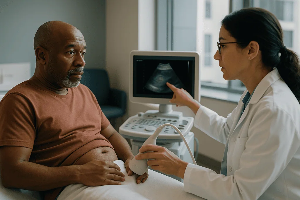
The best evidence on which test to start with comes from a randomized trial published in the New England Journal of Medicine that enrolled 2,759 emergency department patients across 15 centers. Participants were assigned to bedside ultrasound, radiology-department ultrasound, or CT as the first imaging test. Six months later there was no difference in serious missed diagnoses, return visits or complications among the groups. The ultrasound-first groups received markedly less cumulative radiation. Roughly four in ten of those who started with ultrasound went on to have a CT during the same visit because the ultrasound left questions open.
Read that carefully, because it is often misquoted. The trial did not show that ultrasound is as accurate as CT at spotting a stone; CT is more sensitive. It showed that starting with ultrasound, and moving to CT only when needed, is a safe strategy. The people who benefit most are those with a typical story, stable vital signs, no fever and a history of previous stones, particularly younger adults and anyone who is pregnant.
The practical lesson is not that ultrasound is good and CT is bad. It is that the order of tests is a choice, and a thoughtful clinician makes it based on the person in front of them.
Who usually gets a CT scan right away, and who is asked to wait
Emergency clinicians carry a mental shortlist for going straight to CT. Fever alongside flank pain tops it, because a stone blocking an infected kidney is a surgical emergency, and CT quickly shows both the stone and the extent of blockage. A single functioning kidney, a kidney transplant or known kidney disease also moves someone to the front of the line: there is no spare organ to absorb a mistake. So does uncertainty. When the pain is atypical, when the person is older and vascular disease is possible, or when the abdomen is tender in ways a stone should not cause, the whole-abdomen view of CT is worth its radiation.
At the other end are people who can reasonably be asked to wait, or to skip CT at first. Someone younger than about fifty with classic colicky flank pain, blood on the urine dipstick, previous stones, a normal temperature and a kidney that looks only mildly swollen on ultrasound fits a well-recognized pattern. Emergency medicine and urology guidance increasingly supports ultrasound first for this group, with CT reserved for pain that will not settle, symptoms that change, or a stone that has not passed within a few weeks.
Pregnancy sits in its own category. Ultrasound is the first-line test throughout pregnancy, and CT is generally avoided unless the answer cannot be obtained any other way. Magnetic resonance imaging without contrast sometimes serves as a middle step.
Children follow a similar logic. Because young tissues are more sensitive to radiation and children have more years ahead in which to accumulate exposure, pediatric protocols begin with ultrasound and reach for low-dose CT only when the result would change management.
What urine and blood tests reveal that no scan can
A scan shows shape. Urine shows chemistry, and chemistry decides whether the stone comes back. The first urine test, done within minutes on a dipstick and then under a microscope, looks for blood, which most stones cause as they scrape the lining of the urinary tract; for white blood cells and nitrites, which suggest infection; and for the urine’s acidity. Crystals under the microscope can hint at stone type before any stone is retrieved. A urine culture, in which a sample is grown in the laboratory for a day or two, confirms or rules out bacteria.
The infection question changes plans fastest. A stone with a clean urinalysis can often be watched at home. A stone with infected urine trapped behind it cannot, because that urine can spill bacteria into the bloodstream. One dipstick result can be the difference between a prescription and an urgent procedure to relieve the blockage.
Blood tests add a second layer. Creatinine measures how well the kidneys are filtering; a rise suggests the blockage is straining kidney function and raises urgency. A white cell count supports or weakens the suspicion of infection. Calcium and uric acid levels, drawn once the crisis has passed, screen for the metabolic conditions, such as an overactive parathyroid gland or gout, that quietly drive some people to form stone after stone.
Then there is the stone itself. Anyone expected to pass one is usually handed a small strainer and asked to catch it. Laboratory analysis of its composition is, according to the National Institute of Diabetes and Digestive and Kidney Diseases, one of the most useful pieces of information for planning prevention, because dietary and medication advice differ sharply between a calcium oxalate stone and a uric acid one.
The 24 hour urine test for kidney stones: why it matters after the emergency
Once the pain is gone and the stone is out, many people never hear from a doctor about it again. That is a missed opportunity. Cleveland Clinic and other sources commonly cite that roughly half of people who form one stone will form another within about a decade, and the tool for lowering that risk is a 24-hour urine collection.
The test is unglamorous. Every drop of urine over a full day goes into a container kept cool, sometimes on two separate days for accuracy. The laboratory measures total volume, calcium, oxalate, citrate, uric acid, sodium and pH. Each number maps onto a specific cause. Low volume means concentrated urine in which minerals crystallize more readily. High calcium or oxalate supplies the raw material. Low citrate removes a natural inhibitor that normally keeps calcium from clumping. High sodium drags calcium into the urine along with it.
Results shape a prevention plan that is individual rather than generic. One person leaves with the single instruction to drink enough to produce a target urine volume each day. Another learns that oxalate is the problem and receives specific dietary guidance. A third may be offered a medicine class that changes urine chemistry: thiazide diuretics to reduce urinary calcium, potassium citrate to raise citrate and pH, or a xanthine oxidase inhibitor to lower uric acid production. Whether, which and for how long are decisions for the prescribing clinician.
Not everyone needs the collection. Mayo Clinic frames it as most useful for people with recurrent stones, a first stone at a young age, a family history or a single kidney. Ask whether it applies to you, and if it does, ask before the next stone rather than after.
How stone size and location on imaging steer the treatment plan
Reports describe stones by their longest diameter in millimeters and by where they sit: in the kidney’s collecting system, at the top of the ureter, in its middle, or at the ureterovesical junction where ureter meets bladder. Together these two facts predict the odds of natural passage and shape the menu of options. The sizes below are landmarks used in urology guidance and by Mayo Clinic and Cleveland Clinic, not rigid cutoffs.
| Imaging finding | What it usually means | Options the team typically discusses |
|---|---|---|
| Stone under about 5 mm, lower ureter | Most likely to pass on its own | Fluids, pain control, sometimes an alpha-blocker, follow-up imaging |
| Stone 5 to 10 mm, mid or upper ureter | Passage possible but slower and less certain | Time-limited trial of passage, or shock wave lithotripsy or ureteroscopy |
| Stone over about 10 mm anywhere | Unlikely to pass unaided | Ureteroscopy, shock wave lithotripsy, or percutaneous surgery for very large stones |
| Any stone plus fever or infected urine | Emergency regardless of size | Urgent drainage with a stent or nephrostomy tube, antibiotics, stone treated later |
| Stone inside the kidney, not blocking, no pain | Often monitored | Periodic imaging and a prevention work-up |
Location matters as much as size because the ureter narrows at three points, and a stone that has reached the lowest one has already cleared the hardest obstacles. The grade of hydronephrosis adds nuance: mild swelling is expected and tolerated; severe swelling or thinning of kidney tissue pushes toward earlier intervention. The final call belongs to the urology team, who weigh these numbers against your pain, your other conditions and your own preferences.
Waiting for a stone to pass: what the following days and weeks usually look like
If imaging and urine results allow it, the most common plan is medical expulsive therapy, the clinical phrase for helping a stone pass on its own. You go home with instructions to drink enough fluid to keep urine pale, a pain plan that often starts with an anti-inflammatory class of painkiller if your kidneys and stomach allow it, and sometimes an alpha-blocker, a medicine originally developed for prostate symptoms that relaxes the smooth muscle of the ureter so the stone has a wider corridor. Whether an alpha-blocker is offered depends on stone size and location; the evidence suggests its benefit is largest for stones in the lower ureter that are more than a few millimeters across, and your prescriber will weigh that.
The first few days are the hardest. Pain comes in waves as the stone shifts and the ureter spasms around it, then eases between episodes. Blood in the urine, sometimes visible, is expected. You strain your urine to catch the stone for analysis.
Timelines are ranges, not promises. NHS guidance says most small stones pass within a few weeks, and many clinicians set a checkpoint around four to six weeks: if the stone has not appeared by then, or pain persists, repeat imaging is arranged. That repeat is frequently an ultrasound or a plain X-ray if the stone was clearly visible on the first CT, precisely to avoid stacking up radiation.
The waiting period ends in one of three ways. The stone passes, the stone stalls and a procedure is scheduled, or a red flag appears and the plan changes urgently. Knowing those three exits in advance makes the wait far less frightening.
When imaging points toward a procedure: shock waves, scopes and surgery
When a stone is too large, too high, too stubborn or too dangerous to wait for, the imaging report becomes a surgical map. Three approaches cover nearly all cases, and the choice among them turns on the stone’s size, position, density and the anatomy around it.
Shock wave lithotripsy sends focused pressure waves through the skin to shatter the stone into fragments small enough to pass. Mayo Clinic notes the procedure takes about 45 to 60 minutes and can cause moderate pain, so sedation or light anesthesia is used. It suits stones in the kidney or upper ureter that are not too dense; a CT measurement called Hounsfield units, a scale of how much X-ray a tissue absorbs, helps predict which stones will fragment and which will resist.
Ureteroscopy threads a thin telescope up through the urethra and bladder into the ureter, where the stone is either pulled out in a basket or broken up with a laser. It handles ureteral stones of most sizes and leaves no incision. A temporary stent, a soft plastic tube that keeps the ureter open while swelling settles, is often left in place for days to weeks and removed later.
Percutaneous nephrolithotomy is reserved for large stones, generally those bigger than about two centimeters or filling the kidney’s branches. A surgeon makes a small incision in the back and enters the kidney directly, and a hospital stay of a night or two is typical.
Each carries risks the team will describe in plain terms: bleeding, infection, injury to the ureter, leftover fragments and the discomfort of a stent. Alternatives, including continued observation for stones that are not blocking anything, remain part of that conversation.
Radiation, contrast dye and pregnancy: the safety questions people ask
A CT scan for kidney stones delivers a measurable dose of ionizing radiation, and pretending otherwise helps no one. A single scan carries a very small increase in lifetime cancer risk; the real concern is repetition, because stone formers may have many episodes over decades. Radiology and urology bodies have responded with low-dose stone protocols that preserve stone detection while cutting exposure well below standard abdominal CT, and with a habit of switching to ultrasound or plain X-ray for follow-up once a stone’s position is known.
You can help. Tell the ordering clinician how many CT scans you have had for stones in recent years. Ask whether a low-dose protocol is available. Ask whether follow-up can be done with ultrasound. None of these questions is rude, and good departments welcome them.
Contrast dye is rarely needed to diagnose a stone, but when it is used, typically to map the collecting system before surgery, two safety points apply. People with reduced kidney function face some risk of a temporary further decline after iodinated contrast, so creatinine is checked first. Anyone with a previous reaction to contrast should say so; allergic reactions are uncommon but real.
Pregnancy reorders everything. Ultrasound is the first test and can be repeated freely. If it cannot answer the question, non-contrast MRI is generally the next step. CT is held back for situations where the mother’s safety demands it and nothing else will do. Stones in pregnancy are managed conservatively when possible, with temporary drainage preferred over definitive stone surgery until after delivery, a decision made jointly by urology and obstetric teams.
What people often get wrong about kidney stone imaging
The first misunderstanding is that a plain X-ray settles the matter. Uric acid stones are invisible on ordinary X-rays and perfectly visible on CT, so a clear X-ray rules out very little. Mayo Clinic notes that simple abdominal X-rays are now used far less often for this reason.
The second is that ultrasound is a consolation prize. In pregnancy it is the primary tool, and in the large randomized trial described earlier it proved a safe starting point for most stable adults. Its weakness is the mid-ureter, not the whole urinary tract.
The third is that a bigger stone always means worse pain. Pain comes from blockage and the ureter’s spasm against it, not from diameter. A three-millimeter stone wedged at a narrow point can hurt more than a large stone resting quietly in the kidney that never obstructs anything.
The fourth is that once a stone passes, the matter is closed. Recurrence is common, and the metabolic work-up described above exists precisely because the chemistry that produced the first stone is usually still present.
The fifth is that a CT can tell you what your stone is made of. Density on the scan offers a hint, since uric acid stones tend to be less dense than calcium stones, but only laboratory analysis of the stone itself gives a reliable answer.
The sixth is the folk remedy: that cranberry juice, beer or a specific herbal drink dissolves or flushes stones. No mainstream evidence supports those claims. Fluid volume itself matters, which is why water is the recommendation, and cranberry products may actually raise urinary oxalate in some people.
Questions to ask your care team
A radiology report is written for other doctors, and the appointment that follows can move quickly. Bringing a short list of questions turns a monologue into a plan you understand. Consider asking, in your own words, some of the following.
- How big is the stone in millimeters, and exactly where is it sitting?
- Is there any sign of blockage or swelling of the kidney, and how severe?
- Did my urine test show any sign of infection, and does that change the plan?
- Is it reasonable to wait for this stone to pass, and for how long before we re-check?
- If we re-image, can that be done with ultrasound or X-ray rather than another CT?
- What symptoms should send me back to the emergency department tonight?
- Should I have a 24-hour urine collection or blood tests to find out why I formed this stone?
- If a procedure is recommended, what are the alternatives, including doing nothing for now?
Two further questions deserve a place. Ask how many CT scans you have had in total for stones, because you may need that number for future visits. Ask, too, what the stone was made of once it is analyzed, and what that means for your diet.
Write down the answers, or ask a companion to. People in pain remember remarkably little of what they are told, and the plan matters most on the days when the pain returns and the clinic is closed. Every decision about waiting, medicine or surgery rests with your treating team, but a well-informed patient makes that team’s job easier and the plan safer.
When to call your doctor
Most stones that are being watched at home pass without drama, but a handful of signs mean the plan has stopped working and you should not wait for the scheduled follow-up. Call your doctor promptly, or go to an emergency department, if any of the following appears.
- Fever, shaking chills or feeling generally unwell, which can signal infection trapped behind the stone
- Pain so severe that the prescribed plan does not control it, or pain that lasts for hours without any break
- Persistent vomiting that stops you keeping fluids down
- Passing very little urine, or none at all, for many hours
- Urine that is heavily bloody, cloudy or foul-smelling, or a burning that worsens rather than eases
- Pain that shifts to a new place or a new character, especially if you have a single kidney
- Confusion, a racing heartbeat, dizziness on standing or a sense of impending collapse
Fever is the one that clinicians worry about most. A blocked, infected kidney can tip into sepsis, a life-threatening whole-body reaction to infection, within hours, and NHS guidance is explicit that fever with kidney stone pain needs urgent assessment. The treatment in that situation is usually to drain the kidney first with a stent or a tube through the back, and to deal with the stone once the infection is under control.
Less urgently, arrange a routine appointment if several weeks have passed without the stone appearing in your strainer, if you notice the pain settling in a new pattern, or if you simply want to understand the prevention options. Your care team decides the next step, but they can only act on what you report, so report early and specifically.
Frequently asked questions
Is it worth getting a CT scan for kidney stones?
For many people, yes, because a non-contrast CT is the most accurate single test for confirming a stone, measuring it and spotting blockage or an alternative cause of pain. It is not automatically worth it for everyone: stable adults with a typical story and previous stones can often start with ultrasound, and pregnant patients should. The deciding factors are fever, kidney function, diagnostic uncertainty and how many scans you have already had.
How long does a CT scan take for kidney stones?
The scan itself lasts only a few minutes; you lie on a table that passes through an open ring while holding your breath briefly. Johns Hopkins Medicine notes that a non-contrast kidney CT needs little preparation, and most of the appointment is spent on check-in and positioning. In an emergency setting the radiologist’s report, not the scan, is usually what you wait for.
Does a CT scan show kidney stones of every type?
Almost every type. Calcium, uric acid, struvite and cystine stones are all dense enough to appear white on CT, which is a major advantage over plain X-rays that miss uric acid stones. The rare exceptions are stones formed from certain crystallized medicines, such as some older HIV protease inhibitors, which can be nearly invisible. A stone that has already passed will also not be seen, leaving only kidney swelling as evidence.
What can be mistaken for kidney stones on a CT scan?
Phleboliths, harmless calcified spots in pelvic veins, are the classic look-alike for a stone in the lower ureter. Calcified arterial plaque, lymph nodes and fragments of old stones can also mimic one. Radiologists distinguish them by tracing the ureter and looking for a soft-tissue rim around a true stone. The more important error runs the other way: appendicitis, ovarian problems, diverticulitis or an aortic aneurysm can present with stone-like pain.
What symptoms usually prompt doctors to order stone imaging?
Clinicians typically image when someone has sudden, severe pain in the side or back that comes in waves and may spread toward the groin, often with nausea, blood in the urine, or a need to urinate urgently or frequently. None of these confirms a stone on its own, and similar symptoms have other causes, which is exactly why imaging and urine testing are used rather than symptoms alone.
Kidney stone ultrasound vs CT: which is more accurate?
CT is more sensitive, particularly for small stones in the middle of the ureter where ultrasound struggles. Ultrasound, however, reliably shows kidney swelling and stones inside the kidney without radiation. A large randomized trial found that starting with ultrasound and adding CT only when needed produced the same safety outcomes over six months, with about four in ten ultrasound-first patients going on to CT.
Why is a non contrast CT used for kidney stones instead of one with dye?
Because stones and contrast dye both appear white, injected dye can hide the very thing the scan is looking for. A non-contrast scan shows stones as bright specks against darker tissue, avoids the small risk of allergic reaction, and spares people with reduced kidney function the strain of iodinated contrast. Dye is added later only when surgeons need a detailed map of the urine-collecting system.
What does a 24 hour urine test for kidney stones measure?
It measures total urine volume plus calcium, oxalate, citrate, uric acid, sodium and acidity over a full day, sometimes on two separate days. Each value points to a specific reason stones form, such as concentrated urine, too much calcium or oxalate, or too little protective citrate. Mayo Clinic describes it as most useful for people with repeated stones, an early first stone, a family history or a single kidney.
Can a CT scan tell what my kidney stone is made of?
Only roughly. CT measures density in Hounsfield units, and uric acid stones tend to be less dense than calcium stones, which offers a hint that can influence treatment choices such as shock wave lithotripsy. It cannot reliably identify composition. Catching the stone in a strainer and sending it for laboratory analysis remains the standard way to learn what it is made of and to plan prevention.
How often will I need repeat scans for kidney stones?
It depends on whether the stone has passed and whether new symptoms arise. During a trial of passage, many clinicians re-check around four to six weeks if the stone has not appeared, and they often use ultrasound or a plain X-ray rather than another CT once the stone’s position is known. Keeping a personal tally of past CT scans helps your team choose the lowest-radiation option each time.
References
- Kidney stones (NHS)
- Diagnosis of Kidney Stones (NIH National Institute of Diabetes and Digestive and Kidney Diseases)
- Kidney Stones (Cleveland Clinic)
This article is for general information only and is not a substitute for professional medical advice. Please consult a qualified doctor about your individual situation.

More from the Blog

What Happens in Testicular Torsion Surgery: Detorsion, Orchiopexy and Fixing Both Sides
Testicular torsion surgery is an emergency operation to untwist a spermatic cord that has cut off blood flow to a testicle. Under general anesthesia,…

Vascular, Hormonal or Nerve-Related: How the Cause of Erectile Dysfunction Guides Treatment
The causes of erectile dysfunction fall into three broad groups: vascular (narrowed or leaky penile blood vessels), hormonal (low testosterone, thyroid or prolactin problems)…

Vasectomy Recovery Timeline: Rest, Cold Packs and When Everyday Activity Usually Resumes
Most people rest for the first 24 hours after a vasectomy, use wrapped cold packs and snug supportive underwear for about two days, and…