Intraoperative Neuromonitoring: Nerve Protection During Complex Surgery
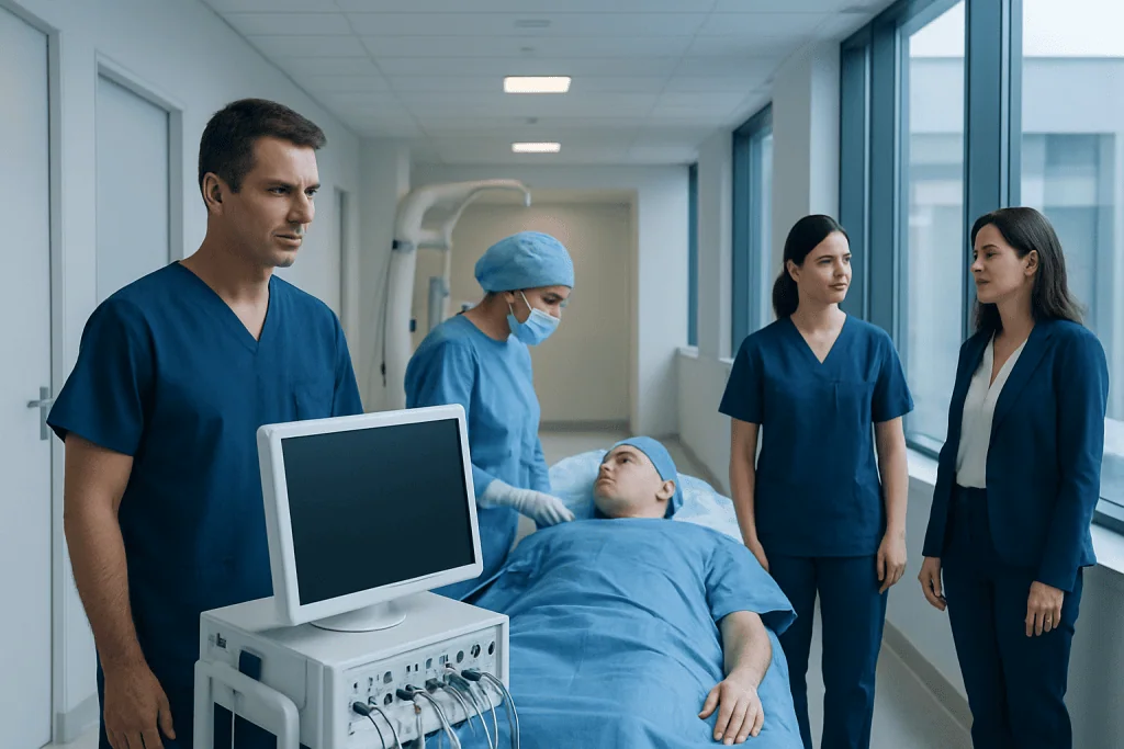
Intraoperative neuromonitoring, often called IONM, records electrical signals from nerves, muscles, the spinal cord, or the brain during surgery. It is commonly used in spine, brain, vascular, ENT, orthopedic, and some endocrine surgeries when important nerves may be at risk.
Key Takeaways
- Intraoperative neuromonitoring, often called IONM, records electrical signals from nerves, muscles, the spinal cord, or the brain during surgery.
- It is commonly used in spine, brain, vascular, ENT, orthopedic, and some endocrine surgeries when important nerves may be at risk.
- IONM does not replace the surgeon’s skill; it provides additional real-time information to guide surgical decisions.
- The technique is generally safe, but its usefulness depends on the procedure, the patient’s condition, anesthesia choices, and the monitoring methods used.
- Patients should ask their surgical team why IONM is recommended, what type will be used, and how results may influence the operation.
Intraoperative neuromonitoring is a specialized technology used during certain complex surgeries to track the function of nerves, the spinal cord, and parts of the brain in real time. It helps the surgical and anesthesia teams make timely decisions that support nerve protection and patient safety.
Overview
Intraoperative neuromonitoring, also known as IONM or intraoperative neurophysiological monitoring, is a technology used to observe the function of nerves and related structures during surgery. It records electrical activity from the nervous system while a procedure is taking place. The goal is to give the surgical team early warning if a nerve, the spinal cord, or certain brain pathways may be under stress.
Complex operations sometimes take place close to delicate nerves or neural pathways. These may include the spinal cord, facial nerve, recurrent laryngeal nerve, optic pathways, or nerves that control movement and sensation. Even when surgery is carefully planned, nerves can be affected by traction, pressure, changes in blood flow, positioning, temperature, or instruments placed near them.
IONM acts like an additional source of information in the operating room. It cannot prevent every complication and it does not replace expert surgical judgment. However, when used appropriately, it can help the team detect changes early and adjust the surgical approach, patient positioning, blood pressure management, or anesthesia plan to support nerve protection.
How Intraoperative Neuromonitoring Works
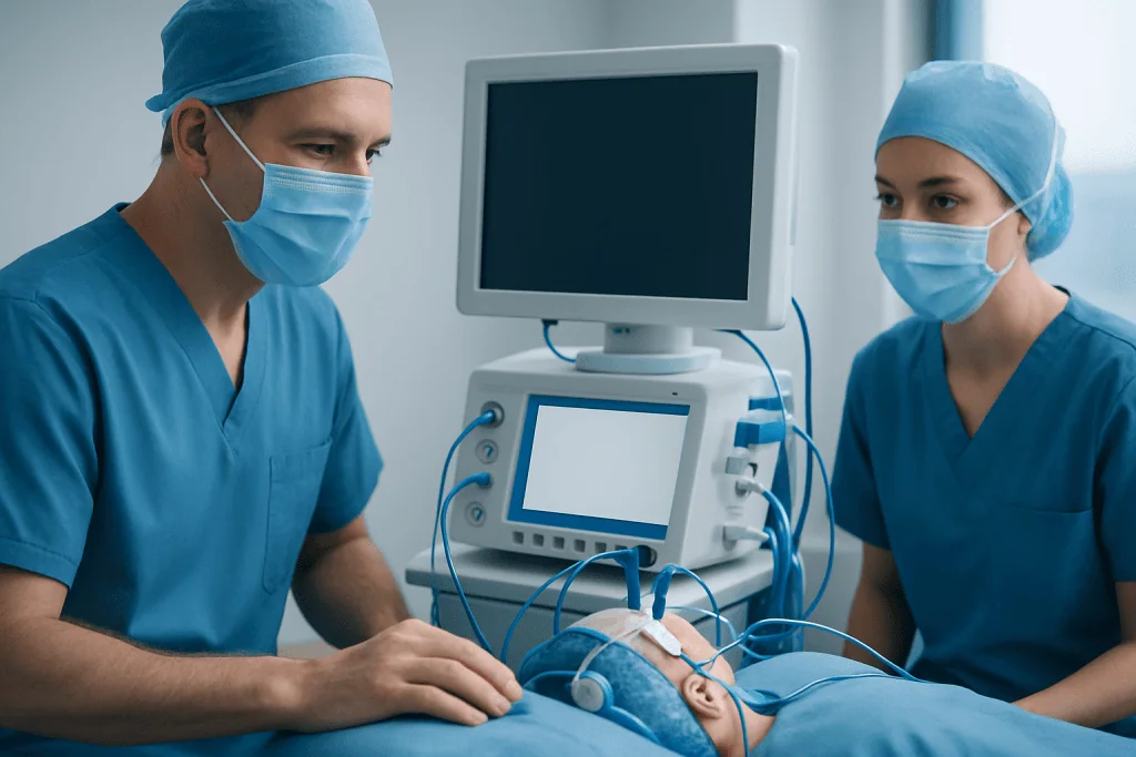
Nerves communicate through small electrical signals. IONM uses electrodes to stimulate and record these signals while a patient is under anesthesia. Depending on the operation, electrodes may be placed on the scalp, skin, muscles, vocal cord area, or directly near a nerve during surgery. The monitoring team then tracks signal strength, timing, and consistency throughout the procedure.
Several monitoring methods may be used alone or together. Somatosensory evoked potentials assess sensory pathways, often by stimulating a nerve in the arm or leg and recording how signals travel through the spinal cord and brain. Motor evoked potentials evaluate movement pathways by stimulating areas of the brain or spinal cord and recording muscle responses. Electromyography records muscle activity that may indicate irritation or stimulation of a nearby nerve.
Other specialized tests may be selected for specific procedures. Brainstem auditory evoked potentials can help monitor hearing pathways during selected skull base operations. Visual evoked potentials may be considered in certain surgeries near visual pathways, although they can be more sensitive to anesthesia and technical factors. For thyroid, parathyroid, and some neck surgeries, laryngeal nerve monitoring may help assess nerves involved in voice function.
During surgery, the neurophysiology team watches for meaningful changes in the signals. If a change occurs, it is communicated promptly to the surgeon and anesthesiologist. The team then evaluates possible causes, such as surgical manipulation, reduced blood pressure, body positioning, temperature, anesthetic effects, or equipment issues.
When IONM May Be Used
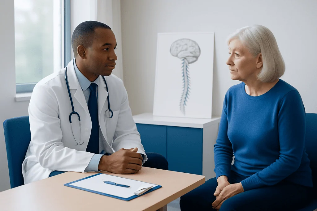
Intraoperative neuromonitoring is most often considered when surgery is close to important nerves or when temporary changes in nerve function would be difficult to detect while the patient is asleep. It is commonly used in many spine procedures, including scoliosis correction, spinal tumor surgery, complex deformity surgery, revision spine surgery, and operations involving the spinal cord or nerve roots.
IONM may also be used in brain and skull base surgery, especially when procedures are near areas involved in movement, sensation, hearing, facial movement, or swallowing. In vascular surgery, monitoring may help during selected operations where blood supply to the brain or spinal cord could be affected. In ENT and endocrine surgery, it may be used to identify and monitor nerves related to voice and swallowing.
Orthopedic procedures involving the pelvis, hip, or limbs may sometimes involve monitoring if major nerves are at risk. The decision is individualized. Not every surgery requires IONM, and not every patient benefits from the same type of monitoring. The surgical team weighs the location of the operation, the patient’s anatomy, previous surgeries, the complexity of the procedure, and the expected benefit of real-time nerve information.
Benefits and Limitations
The main benefit of intraoperative neuromonitoring is timely feedback. If a signal changes, the team can pause, check the surgical field, adjust instruments, improve blood pressure or oxygenation, reposition the patient, warm the patient, or modify the surgical plan. In some situations, these steps may allow signals to recover before permanent nerve injury occurs.
IONM can also help identify nerves during surgery. For example, stimulation may help confirm that a structure is a nerve and show which muscles it controls. This can be particularly useful in areas where anatomy is altered by tumors, scarring, congenital differences, or prior operations. Monitoring may also provide documentation of nerve pathway function at different stages of a procedure.
It is important to understand the limitations. IONM reduces uncertainty but does not eliminate risk. Some nerve injuries may occur without clear warning changes, and some signal changes may be temporary or related to anesthesia, temperature, blood pressure, or technical factors rather than direct nerve injury. The quality of monitoring depends on appropriate patient selection, equipment, anesthesia coordination, and experienced interpretation.
Patients should also know that IONM is not a single test. It is a set of techniques selected for the surgery being performed. A monitoring plan that is very useful for one operation may not be the best choice for another. This is why discussion between the surgeon, anesthesiologist, and neurophysiology team is essential before and during the procedure.
What Patients Can Expect Before and During Surgery
Before surgery, the medical team reviews the patient’s condition, diagnosis, imaging, neurological examination, and planned operation. Patients should inform the team about implanted devices, seizure history, heart rhythm conditions, skin allergies, nerve or muscle disorders, and any previous reactions to anesthesia. This information helps the team choose the safest and most suitable monitoring approach.
During the operation, the patient is under anesthesia and does not feel the monitoring. Small electrodes may be placed on the scalp, skin, arms, legs, hands, feet, face, throat area, or specific muscles, depending on the procedure. Some electrodes are placed after the patient is asleep and removed before leaving the operating room. The placement is done carefully and is part of the surgical safety workflow.
Anesthesia is closely coordinated with IONM because some anesthetic medicines can affect nerve signals. The anesthesiologist selects an approach that supports both patient comfort and reliable monitoring when possible. This may include specific combinations of medications and careful control of blood pressure, oxygen levels, temperature, and muscle relaxation.
After surgery, the surgeon examines nerve function as appropriate for the operation. This may include checking movement, sensation, voice quality, swallowing, facial movement, or limb strength. The IONM results are interpreted together with the surgical findings and the patient’s postoperative examination.
Safety, Risks, and Practical Considerations
Intraoperative neuromonitoring is generally considered safe when performed by trained professionals using appropriate equipment and protocols. The electrodes and stimulation levels are designed for surgical monitoring. Most patients do not have any lasting effects from the monitoring itself.
Minor temporary effects can occur. These may include small areas of skin irritation where electrodes were placed, mild soreness, or rarely a small superficial mark. Bite injury is a known but uncommon concern during some motor evoked potential monitoring because muscle responses can occur under anesthesia; protective bite blocks are used to reduce this risk. The team also considers any patient-specific factors that could affect safety.
Some conditions may require special planning. Patients with implanted electrical devices, certain cardiac conditions, epilepsy, significant neurological disease, or implanted neurostimulators should tell the surgical and anesthesia teams well before the operation. IONM can often still be performed, but the plan may need adjustment.
Practical factors also matter. Reliable monitoring requires communication in the operating room, accurate baseline signals, appropriate anesthesia, and rapid response to alerts. If baseline signals cannot be obtained because of severe pre-existing nerve damage or technical limitations, the usefulness of monitoring may be reduced. In those cases, the team may rely more heavily on anatomy, imaging guidance, surgical technique, and other safety measures.
Questions to Ask the Surgical Team
Patients are encouraged to ask clear questions before surgery. Helpful questions include: Why is intraoperative neuromonitoring recommended for this operation? Which nerves or pathways will be monitored? What type of monitoring will be used? How could the results change the surgical plan? Who will interpret the signals during the procedure?
It is also reasonable to ask how anesthesia will be coordinated with monitoring and whether any personal medical history could affect the plan. Patients should discuss implanted devices, neurological conditions, allergies to adhesive materials, and previous surgeries. These details help the team prepare and reduce avoidable interruptions on the day of surgery.
After surgery, patients should report any new weakness, numbness, voice change, swallowing difficulty, facial asymmetry, severe pain, or changes in bladder or bowel control, depending on the type of procedure performed. Some temporary symptoms can occur after complex surgery, but prompt assessment helps the team distinguish expected recovery from concerns that need attention.
For international patients seeking evaluation for complex procedures, Acibadem International provides access to multidisciplinary specialists and JCI-accredited hospitals where advanced diagnostic, surgical, anesthesia, and neuromonitoring teams can coordinate care. Patients should always receive individualized advice from their treating doctor based on their diagnosis and surgical plan.
Frequently asked questions
Is intraoperative neuromonitoring the same as anesthesia monitoring?
No. Anesthesia monitoring tracks vital signs such as heart rate, blood pressure, oxygen levels, and breathing. Intraoperative neuromonitoring focuses on nerve, spinal cord, brain pathway, or muscle signals. The two teams work closely together because anesthesia choices can affect the quality of nerve signals.
Does IONM guarantee that nerve injury will not happen?
No medical technology can guarantee that a complication will not occur. IONM provides real-time information that may help the surgical team detect and respond to potential nerve stress. It is one part of a broader safety strategy that includes surgical expertise, imaging, anesthesia care, and postoperative assessment.
Will the patient feel the electrical stimulation during surgery?
Patients are under anesthesia during intraoperative neuromonitoring, so they do not feel the stimulation or electrode placement. The monitoring is performed while the patient is asleep and is removed before or shortly after the procedure is completed. The anesthesia team remains responsible for comfort and safety throughout the operation.
Which surgeries most often use intraoperative neuromonitoring?
IONM is often used in complex spine surgery, brain and skull base surgery, selected vascular procedures, and operations near nerves that control movement, sensation, hearing, voice, or swallowing. It may also be used in some orthopedic, ENT, and endocrine surgeries. The need depends on the location and complexity of the operation.
Are there risks from the electrodes or monitoring equipment?
Most patients have no lasting effects from IONM. Minor skin irritation or temporary soreness can occur where electrodes are placed. For some types of monitoring, the team uses protective measures such as bite blocks to reduce the risk of injury from muscle contractions during stimulation.
Who performs and interprets intraoperative neuromonitoring?
IONM is performed by trained neurophysiology professionals using specialized equipment. Signal changes are interpreted in coordination with the surgeon and anesthesiologist, and in some settings a supervising physician with expertise in neurophysiology is involved. Effective communication among all team members is essential.
References
- American Society of Neurophysiological Monitoring
- American Clinical Neurophysiology Society
- International Society of Intraoperative Neurophysiology
- North American Spine Society
- American Association of Neurological Surgeons
This article is for general information only and is not a substitute for professional medical advice. Please consult a qualified doctor about your individual situation.
Joint and spine care in Turkey — expert assessment & treatment
JCI-accredited · board-certified surgeons · reply within 24h
Add us as a Preferred Source to see more of our trusted health content across Google Search, AI Overviews and Discover.
Check your numbers in seconds
BMI, calories, due date, blood pressure and 30+ more clinical calculators — free, instant, doctor-reviewed ranges.

More from the Health Library



Related Specialists

Dr. Volkan Ertuğrul
Anesthesiology
Assoc. Prof. Dr. Şevket Tolga Tombul
Urology
Dr. Mehmet İbrahim Cansunar
General Surgery
Prof. Dr. İsmail Çepni
Gynecology & Obstetrics