Stereotactic Radiosurgery: High-Precision Radiation for Brain and Spine Lesions

Stereotactic radiosurgery is not open surgery; it is a non-invasive or minimally invasive radiation treatment planned with advanced imaging. It may be used for certain benign and malignant tumors, brain metastases, vascular malformations, and selected functional disorders.
Key Takeaways
- Stereotactic radiosurgery is not open surgery; it is a non-invasive or minimally invasive radiation treatment planned with advanced imaging.
- It may be used for certain benign and malignant tumors, brain metastases, vascular malformations, and selected functional disorders.
- Treatment is usually completed in one session or a small number of sessions, depending on the size, location, and type of lesion.
- A multidisciplinary team reviews imaging, symptoms, previous treatments, and overall health before recommending SRS.
- Side effects are often temporary, but careful follow-up imaging is essential to monitor response and detect delayed changes.
Stereotactic radiosurgery is a highly focused radiation technique used to treat selected brain and spine lesions without making an incision. It delivers strong, carefully planned radiation beams to a defined target while limiting exposure to nearby healthy tissue.
Overview
Stereotactic radiosurgery, often abbreviated as SRS, is a high-precision form of radiation therapy used mainly for lesions in the brain and, in selected cases, the spine. Despite the word surgery, it does not involve an incision or removal of tissue. Instead, it uses many precisely aimed radiation beams that meet at a defined target, delivering a therapeutic dose to the lesion while reducing radiation exposure to surrounding healthy structures.
SRS may be delivered with different technologies, including Gamma Knife systems, linear accelerator-based systems, CyberKnife, and proton-based approaches in specialized centers. The choice of platform depends on the condition being treated, the target location, local expertise, and the equipment available. The goal is the same: accurate targeting, careful dose planning, and safe treatment delivery.
For some people, SRS is an alternative to open surgery. For others, it is combined with surgery, standard radiation therapy, chemotherapy, immunotherapy, or targeted medicines. The decision is individualized and usually made by a multidisciplinary team that may include neurosurgeons, radiation oncologists, medical oncologists, neuroradiologists, neurologists, and medical physicists.
How Stereotactic Radiosurgery Works
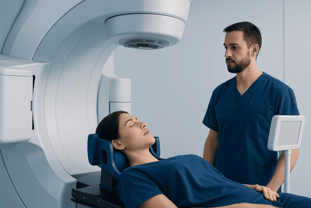
SRS begins with detailed imaging, most commonly magnetic resonance imaging and computed tomography. These images help doctors define the exact size, shape, and position of the lesion. The treatment team also identifies nearby sensitive areas, such as the optic nerves, brainstem, spinal cord, hearing pathways, or major blood vessels, so that the radiation plan can protect them as much as possible.
The term stereotactic refers to a three-dimensional coordinate system that allows the treatment team to locate the target with millimeter-level accuracy. During planning, specialized software calculates how radiation beams should enter the body and overlap at the target. Each beam may carry a relatively small amount of radiation, but where the beams converge, the dose becomes strong enough to treat the abnormal tissue.
Some systems use a rigid head frame for brain treatments, while others use a custom mask or image-guided tracking. For spine radiosurgery, body positioning devices and real-time imaging may be used to reduce movement and maintain accuracy. The patient usually lies on a treatment table while the machine moves around them or directs radiation from multiple angles.
Conditions That May Be Treated

SRS is used for selected brain and spine conditions when the lesion is well defined and can be safely targeted. It is commonly considered for small brain metastases, which are cancer deposits that have spread to the brain from another part of the body. It may also be used after surgical removal of a brain metastasis to treat the surgical cavity and reduce the chance of local recurrence.
Benign or non-cancerous tumors may also be suitable for SRS, depending on their size, growth pattern, and location. Examples include vestibular schwannomas, meningiomas, pituitary adenomas, and some skull base tumors. In these cases, the goal is often to stop or slow growth rather than make the lesion disappear quickly.
SRS may be used for certain vascular and functional conditions. For example, it can be an option for some arteriovenous malformations in the brain when surgery or embolization is not appropriate. In carefully selected patients, focused radiation techniques may also be used for trigeminal neuralgia, a severe facial pain condition, when other treatments have not provided enough relief.
Spine stereotactic radiosurgery or stereotactic body radiation therapy may be considered for selected spinal tumors or metastases. Because the spinal cord is very sensitive to radiation, planning must be especially meticulous. The suitability of spine SRS depends on spinal stability, the degree of nerve compression, previous radiation, and the overall cancer treatment plan.
Planning and the Treatment Day
Before SRS, the care team reviews the diagnosis, imaging, symptoms, general health, medicines, and previous treatments. Some patients need additional imaging, blood tests, or specialist consultations. If a head frame is used, it is placed on the day of treatment with local anesthetic to reduce discomfort. If a mask-based system is used, a custom mask is prepared to help keep the head still.
The planning process is one of the most important parts of SRS. Doctors outline the treatment target and nearby normal structures on imaging scans. Medical physicists and dosimetrists then help create a plan that delivers the prescribed dose to the target while respecting safety limits for healthy tissue. The plan is checked carefully before treatment begins.
During treatment, the patient remains awake in most cases. The treatment itself is painless; patients do not feel the radiation. Depending on the system and plan, the session may take less than an hour or several hours, including preparation and imaging checks. If treatment is divided into a few sessions, it may be called fractionated stereotactic radiotherapy or hypofractionated stereotactic treatment rather than single-session radiosurgery.
Benefits and Limitations
The main benefit of stereotactic radiosurgery is precision. It allows doctors to deliver a high radiation dose to a small target while reducing exposure to nearby healthy brain or spinal tissue. This can be especially valuable when a lesion is deep, near important structures, or difficult to reach with open surgery. Many patients are treated as outpatients and return home the same day.
Another advantage is that SRS may be completed in one session or a short course, which can be convenient compared with several weeks of conventional radiation therapy. Recovery time is often brief, and there is no surgical incision. For cancer care, SRS may help control specific brain or spine lesions while other treatments address disease elsewhere in the body.
However, SRS is not suitable for every lesion. Very large tumors, widespread disease, significant swelling, severe pressure on the brain or spinal cord, or unstable spinal bones may require other approaches. Open surgery may be needed when tissue diagnosis is required, when a mass must be removed quickly to relieve pressure, or when the lesion is causing serious mechanical compression.
It is also important to understand that the response may take time. Some tumors shrink gradually, while others simply stop growing. Vascular malformations may take months or years to close after treatment. Follow-up scans are essential because imaging changes after SRS can be complex and need expert interpretation.
Possible Side Effects and Safety
Many people tolerate SRS well, but side effects can occur. Short-term effects may include fatigue, headache, nausea, scalp tenderness, or temporary swelling around the treated area. When a frame is used, there may be mild soreness or bruising at the pin sites. These effects are usually manageable, and the care team may recommend medicines if needed.
Delayed effects are less common but important to monitor. Radiation-related swelling, tissue irritation, or radiation necrosis can occur weeks, months, or sometimes longer after treatment. Symptoms depend on the treated area and may include headaches, seizures, balance problems, changes in vision, weakness, numbness, or worsening pain in spine cases. These symptoms should be reported promptly.
The risk of side effects depends on several factors, including the dose, target size, target location, prior radiation, previous surgery, and individual medical history. Safety planning includes setting dose limits for sensitive structures and using image guidance during treatment. Patients should discuss expected benefits, alternatives, and risks with their doctor before giving consent.
Recovery, Follow-up, and Self-care
After SRS, most patients are observed for a short period and then go home with instructions. Some can resume normal activities quickly, while others may need rest for a day or two. Patients should follow the care team’s advice about driving, work, exercise, and medications, especially if they have had seizures, neurological symptoms, or sedation.
Follow-up imaging is a central part of care. The timing varies by diagnosis, but MRI is commonly used to assess response after brain SRS. Early scans may show swelling or treatment-related changes that do not necessarily mean the lesion is growing. For this reason, follow-up should be reviewed by clinicians experienced in radiosurgery imaging patterns.
Helpful self-care includes taking prescribed medicines as directed, staying hydrated, getting adequate sleep, and reporting new or worsening symptoms. Patients should keep a written list of symptoms, medications, and questions for follow-up visits. If cancer is the underlying diagnosis, ongoing coordination between the radiation oncology team and the medical oncology team is especially important.
When to See a Doctor and Choosing a Care Team
A person should seek medical advice if they develop new neurological symptoms such as persistent headaches, seizures, weakness, numbness, vision changes, balance problems, hearing changes, facial pain, or back pain with nerve symptoms. Patients already diagnosed with a brain or spine lesion should ask whether SRS is appropriate, what alternatives exist, and what follow-up schedule is recommended.
Choosing a care team for SRS involves more than selecting a machine. Important factors include the team’s experience, quality of imaging, multidisciplinary review, physics support, treatment planning standards, and access to follow-up care. Patients may benefit from asking how the target will be defined, how nearby sensitive structures will be protected, and what side effects are most relevant to their case.
Acibadem International’s multidisciplinary specialists and JCI-accredited hospitals diagnose and treat brain and spine lesions, including selected cases appropriate for stereotactic radiosurgery, for international patients. As with any advanced treatment, the best decision is made after a careful review of imaging, diagnosis, overall health, and personal treatment goals.
Frequently asked questions
Is stereotactic radiosurgery the same as brain surgery?
No. Stereotactic radiosurgery does not involve an incision, opening the skull, or removing tissue. It is called radiosurgery because it delivers a highly focused treatment to a precise target, similar to the precision expected from surgery.
Does SRS hurt during treatment?
The radiation itself is not felt and is not painful. Some patients may feel pressure from a head frame if one is used, or mild discomfort from lying still. The care team can help manage anxiety, positioning discomfort, or headache if they occur.
How soon does stereotactic radiosurgery work?
The effect is usually gradual. Some tumors shrink over months, while others stop growing without disappearing completely. Vascular malformations and some benign tumors may require longer follow-up before the full treatment effect is clear.
Can SRS be repeated?
Repeat SRS may be possible in selected situations, but it depends on the treated area, previous radiation dose, time since treatment, and nearby sensitive structures. Doctors carefully weigh the potential benefit against the risk of radiation-related side effects.
Who is not a good candidate for SRS?
SRS may not be appropriate for very large lesions, severe swelling, urgent pressure on the brain or spinal cord, or lesions that require tissue diagnosis. Some patients may be better treated with surgery, conventional radiation, medicines, or a combined approach.
What follow-up is needed after SRS?
Follow-up usually includes clinical visits and repeat imaging, often MRI for brain lesions. The schedule depends on the condition treated and the patient’s symptoms. Patients should report new neurological symptoms promptly rather than waiting for the next planned scan.
References
- American Society for Radiation Oncology
- National Cancer Institute
- International Stereotactic Radiosurgery Society
- European Association of Neuro-Oncology
- American Association of Neurological Surgeons
This article is for general information only and is not a substitute for professional medical advice. Please consult a qualified doctor about your individual situation.
Joint and spine care in Turkey — expert assessment & treatment
JCI-accredited · board-certified surgeons · reply within 24h
Add us as a Preferred Source to see more of our trusted health content across Google Search, AI Overviews and Discover.

More from the Health Library



Related Specialists

Prof. Dr. Özge Gümüşay
Medical Oncology
Assoc. Prof. Dr. Mehmet Sağır
Aesthetic Plastic & Reconstructive Surgery
Assoc. Prof. Dr. Dilek Altun
Anesthesiology
Dr. Dilara Akman
Infectious Diseases & Clinical Microbiology