PET/CT Scan: What It Shows and How to Prepare
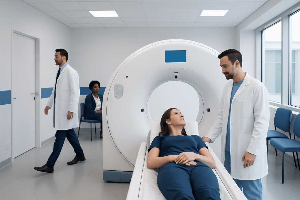
A PET/CT scan combines PET functional imaging with CT anatomical imaging in one appointment. It can help detect disease activity, stage certain cancers, assess treatment response, and guide further care.
Key Takeaways
- A PET/CT scan combines PET functional imaging with CT anatomical imaging in one appointment.
- It can help detect disease activity, stage certain cancers, assess treatment response, and guide further care.
- Preparation often includes fasting, hydration, medication guidance, and special planning for diabetes, pregnancy, or breastfeeding.
- The scan is usually painless, but patients need to lie still while images are taken.
- Results are interpreted by specialists and shared with the referring doctor, who explains what they mean in the context of the patient’s health.
A PET/CT scan combines two imaging methods to show both body structure and metabolic activity. It is commonly used in cancer care and selected heart, brain, infection, and inflammation assessments, with preparation steps that help make the results as accurate as possible.
Overview
A PET/CT scan, also called positron emission tomography/computed tomography, is an advanced imaging test that combines two types of scans in one machine. The PET part shows how active cells and tissues are by detecting a small amount of radioactive tracer. The CT part provides detailed pictures of the body’s internal structures, such as organs, bones, and lymph nodes.
By combining these images, doctors can see both where something is located and how it is behaving. This is especially useful because some conditions cause changes in cell activity before they create a clear change in size or shape. PET/CT therefore adds functional information to the structural detail of CT.
The most commonly used tracer is a form of glucose, often called FDG. Many active cells use glucose for energy, and some diseases may use more glucose than nearby healthy tissue. Other tracers may be chosen for specific medical questions, depending on the organ or condition being investigated.
What a PET/CT Scan Can Show
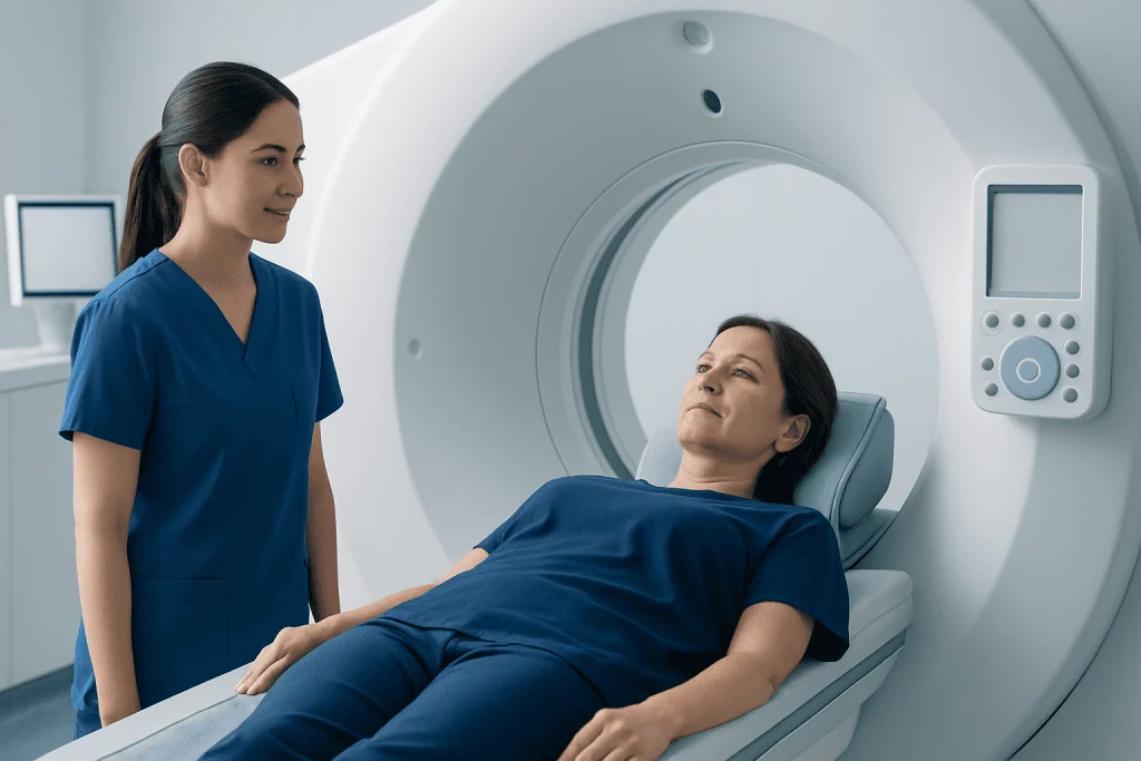
PET/CT is most often used in oncology. It can help doctors identify suspicious areas, determine whether a cancer has spread, plan treatment, choose the best biopsy site, and assess how well treatment is working. It may also help distinguish scar tissue or treated areas from active disease in selected cases.
In addition to cancer care, PET/CT may be used for certain heart and brain conditions. In cardiology, it can assess blood flow and tissue viability in selected patients. In neurology, PET imaging can support evaluation of some types of dementia, seizure disorders, and other complex brain conditions when requested by a specialist.
PET/CT can also help evaluate infection or inflammation when standard tests do not provide enough information. For example, it may be considered in complex fever investigations, certain vascular inflammations, or suspected infection involving implanted medical devices. The exact usefulness depends on the clinical question, the tracer used, and the patient’s overall situation.
How PET/CT Works and Safety

During a PET/CT scan, a small amount of radioactive tracer is usually injected into a vein. The tracer travels through the bloodstream and collects in tissues according to their activity. After a waiting period, the scanner detects signals from the tracer and creates PET images, while the CT scan maps the anatomy.
The amount of radiation used is carefully controlled and kept as low as reasonably possible while still producing useful images. For most patients, the medical benefit of a properly indicated PET/CT scan outweighs the small radiation risk. However, the test should always be requested for a clear medical reason and interpreted by qualified specialists.
Some PET/CT exams include a diagnostic CT with iodine-based contrast dye, while others use a low-dose CT mainly for image correction and localization. If contrast is planned, patients should tell the imaging team about kidney disease, previous contrast reactions, asthma, thyroid conditions, and current medications. This helps the team choose the safest and most appropriate protocol.
How to Prepare for a PET/CT Scan
Preparation can vary depending on the type of PET/CT scan and the tracer used, so patients should follow the instructions from their imaging center. For many FDG PET/CT scans, patients are asked not to eat for several hours before the appointment. Water is usually encouraged unless the care team gives different instructions.
Because FDG is related to glucose use, blood sugar control is important. People with diabetes should receive individualized instructions about meal timing, insulin, tablets, and appointment scheduling. They should not make major changes to diabetes treatment without medical guidance, because both high and low blood sugar can affect safety and image quality.
Patients are usually advised to avoid strenuous exercise before the scan, because active muscles can take up more tracer and make interpretation harder. Comfortable clothing without metal parts is helpful, and jewelry, belts, watches, or removable metal items may need to be taken off. Patients should bring previous imaging reports and a list of medications if requested.
Before the scan, patients should tell the team if they are pregnant, might be pregnant, or are breastfeeding. PET/CT may be postponed, modified, or replaced with another test depending on the situation. Breastfeeding patients may be given specific instructions about expressing and discarding milk for a period after certain tracers, when appropriate.
What Happens During the Appointment
At the imaging center, staff confirm the patient’s identity, medical history, and the reason for the scan. Blood glucose may be checked for FDG PET/CT. The tracer is then injected through a small intravenous line, which may feel like a routine blood test or injection.
After the injection, patients usually rest quietly while the tracer circulates. Talking, walking, chewing gum, or moving around may be limited because muscle activity can affect the images. The waiting period varies, but patients are typically asked to relax in a calm room before scanning begins.
During the scan, the patient lies on a narrow table that moves slowly through the PET/CT scanner. The test is not painful, but lying still is important to avoid blurred images. The scanner is open at both ends, and staff can usually see and speak with the patient throughout the examination.
The imaging time depends on the area being studied and the scan protocol. After the scan, most patients can return to normal activities unless they received sedating medication or were given special instructions. Drinking fluids may help the body clear the tracer through urine.
Understanding PET/CT Results
PET/CT results are not interpreted by the level of brightness alone. Areas of increased tracer uptake can occur for several reasons, including cancer, infection, inflammation, recent surgery, healing tissue, or normal activity in certain organs. This is why the patient’s history, symptoms, laboratory results, previous imaging, and treatment timeline all matter.
A nuclear medicine physician or radiologist reviews the PET and CT images together and prepares a report for the referring doctor. The report may describe the location, size, and activity of findings, and may compare them with earlier scans. In cancer care, PET/CT findings may influence staging, treatment planning, or the decision to do additional tests.
Sometimes PET/CT cannot provide a final diagnosis by itself. A suspicious area may still require targeted ultrasound, MRI, endoscopy, blood tests, or biopsy. Patients should discuss the report with the doctor who requested the scan, because that doctor can explain what the findings mean for the next step in care.
When to See a Doctor and Practical Follow-up
A PET/CT scan is usually arranged by a specialist or referring doctor after reviewing the patient’s symptoms, diagnosis, and previous test results. Patients should contact their doctor if they have new or worsening symptoms, unexplained weight loss, persistent fever, a new lump, or ongoing pain that requires evaluation. The need for PET/CT depends on the clinical context and should be individualized.
Patients should also speak with their care team before the scan if they have diabetes, kidney disease, a history of contrast allergy, claustrophobia, pregnancy, breastfeeding, or difficulty lying flat. These issues do not always prevent the test, but they may require extra planning. Clear communication helps the imaging team adapt the appointment safely.
For international patients, Acibadem International’s multidisciplinary specialists and JCI-accredited hospitals can support the diagnosis and treatment of conditions that may require PET/CT imaging. As with any advanced test, the most appropriate plan is the one guided by a qualified physician who understands the patient’s full medical history.
Frequently asked questions
Is a PET/CT scan painful?
The scan itself is not painful. Most patients feel only a brief needle sensation when the tracer is injected, then need to lie still during imaging. Some people may feel mild discomfort from staying in one position, but staff can help with positioning whenever possible.
Why do patients often need to fast before a PET/CT scan?
For many FDG PET/CT scans, fasting helps lower the effect of recent food intake on glucose metabolism. This can improve image quality and make the scan easier to interpret. Preparation instructions can differ by tracer and medical condition, so patients should follow the specific guidance they receive.
Can people with diabetes have a PET/CT scan?
Yes, many people with diabetes can have PET/CT safely with proper planning. Blood sugar levels, medication timing, and appointment scheduling may need adjustment. Patients should not stop or change diabetes medicines unless their doctor or imaging team gives clear instructions.
How long does a PET/CT appointment take?
The full appointment often takes longer than the scan itself because there is a resting period after the tracer injection. The exact time depends on the tracer, body area, and whether contrast CT is included. The imaging center can provide an estimated schedule before the appointment.
Does a PET/CT scan always mean cancer is suspected?
No. PET/CT is commonly used in cancer care, but it also has roles in selected heart, brain, infection, and inflammation evaluations. The reason for the scan should be explained by the referring doctor based on the patient’s symptoms and previous test results.
Are there side effects from the radioactive tracer?
Side effects from commonly used PET tracers are uncommon, and the tracer leaves the body naturally over time, mainly through urine. Patients may be advised to drink fluids after the scan if appropriate. Pregnant or breastfeeding patients need special guidance because radiation precautions may differ.
When will PET/CT results be available?
Results are usually reviewed by a nuclear medicine physician or radiologist and sent to the referring doctor. Timing varies by hospital and case complexity. Patients should arrange a follow-up appointment to discuss what the report means and whether any further tests or treatment are needed.
References
- American College of Radiology
- Radiological Society of North America
- Society of Nuclear Medicine and Molecular Imaging
- National Cancer Institute
- International Atomic Energy Agency
This article is for general information only and is not a substitute for professional medical advice. Please consult a qualified doctor about your individual situation.
Oncology care in Turkey — second opinion and treatment plan
JCI-accredited · board-certified surgeons · reply within 24h
Add us as a Preferred Source to see more of our trusted health content across Google Search, AI Overviews and Discover.