

PET-CT Scan in Cancer: Purpose, Preparation, and Results

PET-CT combines positron emission tomography and computed tomography to show both how tissues function and where changes are located. In cancer care, PET-CT is often used for staging, treatment planning, checking response to therapy, and looking for recurrence.
Key Takeaways
- PET-CT combines positron emission tomography and computed tomography to show both how tissues function and where changes are located.
- In cancer care, PET-CT is often used for staging, treatment planning, checking response to therapy, and looking for recurrence.
- Preparation commonly includes fasting, blood sugar checks, avoiding strenuous exercise, and informing the team about diabetes, pregnancy, breastfeeding, or allergies.
- PET-CT results are interpreted together with medical history, physical examination, lab tests, biopsy findings, and other imaging.
- A positive PET-CT finding does not always mean cancer, and a negative scan does not always exclude cancer; follow-up may be needed.
A PET-CT scan combines metabolic and anatomical imaging to help doctors detect cancer activity, assess spread, plan treatment, and monitor response. The test is generally painless, and careful preparation helps make the images as accurate as possible.
Overview
A PET-CT scan is an imaging test that combines two technologies in one examination: positron emission tomography (PET) and computed tomography (CT). PET shows areas of increased or decreased metabolic activity in the body, while CT provides detailed anatomical pictures of organs, bones, and tissues. When the images are combined, doctors can see not only where an abnormality is located, but also how active it appears to be.
In cancer care, the most commonly used PET tracer is a small amount of radioactive glucose, called fluorodeoxyglucose or FDG. Many cancer cells use glucose faster than normal cells, so they may appear as areas of higher uptake on the scan. However, infection, inflammation, recent surgery, and some normal organs can also show increased uptake, which is why expert interpretation is essential.
PET-CT is not a screening test for all cancers. It is usually requested when there is a specific clinical question, such as whether a known cancer has spread, whether treatment is working, or whether a suspicious finding needs further evaluation. The scan is one part of a wider diagnostic process and is often considered alongside biopsy results, blood tests, endoscopy, MRI, ultrasound, or conventional CT scans.
Why PET-CT Is Used in Cancer Care
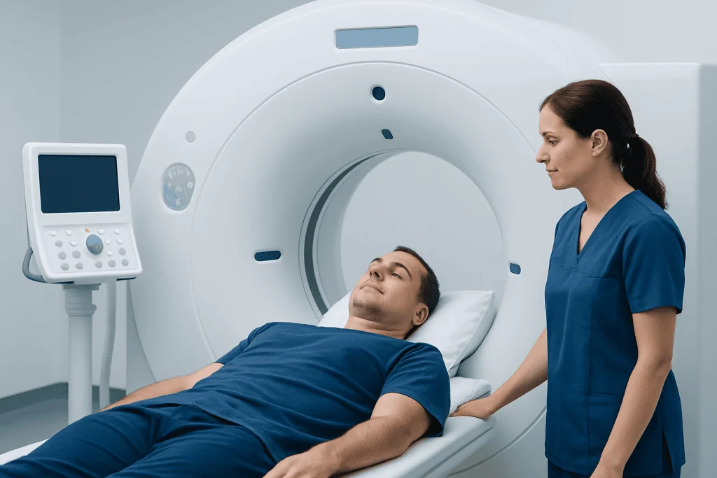
PET-CT can be useful at several points during the cancer journey. It may help doctors determine the stage of cancer, meaning whether disease is limited to one area or has involved lymph nodes or distant organs. Accurate staging is important because it guides decisions about surgery, radiotherapy, chemotherapy, immunotherapy, targeted therapy, or combinations of treatments.
The scan may also help define the best target for a biopsy, especially when there are several abnormal areas and the doctor needs tissue from the most informative site. In radiotherapy planning, PET-CT can help specialists identify active tumor areas and protect nearby healthy tissues as much as possible. During or after treatment, it can provide information about whether cancer activity has decreased, remained stable, or progressed.
Common reasons a doctor may request PET-CT include:
- Staging certain cancers after diagnosis
- Evaluating lymph nodes or possible distant spread
- Assessing response to chemotherapy, radiotherapy, immunotherapy, or targeted treatment
- Investigating suspected recurrence after previous treatment
- Clarifying findings seen on other imaging tests
- Helping guide biopsy or radiation treatment planning
Not every cancer type is best evaluated with PET-CT, and some tumors have low FDG uptake. For example, certain slow-growing tumors, small lesions, or cancers in organs with naturally high background activity may be harder to assess. The oncology and imaging teams choose the most appropriate test based on cancer type, location, stage, and the question that needs answering.
How to Prepare for a PET-CT Scan

Preparation is important because muscle activity, recent meals, and blood sugar levels can affect how the tracer is distributed in the body. Most patients are asked to fast for several hours before the scan, although plain water is usually encouraged. Patients should follow the exact instructions provided by the imaging center, as protocols may vary depending on the type of PET tracer and the reason for the examination.
People with diabetes should inform the appointment team in advance. Blood glucose levels may need to be checked before the injection, and medication timing may need individual planning. Patients should not change insulin or diabetes medicine schedules without medical advice, but they should make sure the imaging team knows about their treatment plan.
General preparation advice may include:
- Avoid strenuous exercise for about 24 hours before the scan, unless instructed otherwise
- Fast for the recommended time before the appointment
- Drink water as advised to support hydration
- Wear comfortable clothing without metal parts if possible
- Bring previous imaging reports, pathology results, and a list of medications
- Tell the team about pregnancy, possible pregnancy, breastfeeding, kidney problems, allergies, or anxiety in enclosed spaces
Some PET-CT examinations include CT contrast dye, which can improve anatomical detail. If contrast is planned, the team may ask about kidney function, previous contrast reactions, asthma, thyroid disease, or certain medications. Patients should always share their full medical history so the scan can be performed safely and appropriately.
What Happens During the Scan
On arrival, the patient may be asked to complete safety questions and may have a blood sugar check. A small intravenous line is placed, and the PET tracer is injected. The amount of radioactivity is carefully controlled and is generally low; it is designed for diagnostic imaging and decreases naturally over time.
After the injection, there is usually a waiting period so the tracer can circulate and be taken up by tissues. During this time, patients are often asked to rest quietly, keep warm, and avoid talking, chewing, walking around, or using a mobile phone excessively, because muscle activity can affect image quality. The waiting period can feel long, but it is an important part of the test.
For the scan itself, the patient lies on a narrow table that moves slowly through the PET-CT scanner. The scanner is shaped like a wide ring rather than a closed tunnel, and the examination is painless. It is important to remain as still as possible while images are being taken. Some patients may receive CT contrast through the IV, which can cause a brief warm sensation or metallic taste.
The total appointment may take a few hours, including preparation, tracer uptake time, scanning, and post-scan checks. The actual image acquisition is usually shorter than the full visit. After the scan, most patients can return to usual activities unless they have been given sedation or specific instructions by the medical team.
Understanding PET-CT Results
PET-CT images are reviewed by specialists trained in nuclear medicine and radiology. They assess areas of tracer uptake, compare them with CT anatomy, and consider whether the pattern is typical of cancer, inflammation, infection, healing tissue, or normal physiological activity. A report is then sent to the referring doctor, who explains the findings in the context of the patient’s overall care plan.
Patients may see terms such as FDG uptake, metabolic activity, hypermetabolic lesion, SUV, or standardized uptake value. SUV is a measurement that helps describe how much tracer is concentrated in an area, but it is not a cancer diagnosis by itself. The meaning of an SUV depends on the cancer type, lesion size, timing of the scan, blood glucose level, recent treatments, and many other factors.
A positive PET-CT finding does not always mean cancer. Infections, inflammatory diseases, healing after surgery, radiation changes, and recent injections can cause increased uptake. Similarly, a negative scan does not always rule out cancer, especially if lesions are very small, slow-growing, or not strongly FDG-avid. This is why follow-up imaging, biopsy, or clinical monitoring may be recommended.
When PET-CT is used after treatment, timing matters. Scanning too soon after radiotherapy, surgery, or certain drug treatments can make interpretation more difficult because inflammation may still be present. The oncology team chooses the timing based on the cancer type, treatment received, and the decision that needs to be made next.
Safety, Radiation, and Possible Side Effects
PET-CT uses ionizing radiation from both the PET tracer and the CT component. The amount is kept as low as reasonably achievable while still producing useful diagnostic images. For many patients with cancer, the benefit of accurate diagnosis, staging, or treatment assessment outweighs the small radiation-related risk, but the decision is individualized.
The PET tracer itself usually does not cause noticeable side effects and leaves the body mainly through urine. Drinking water after the scan, if allowed, helps the body clear the tracer. Patients may be advised to limit close contact with pregnant people and young children for a short period, depending on local instructions and the tracer used.
If CT contrast is used, side effects are uncommon but may include warmth, flushing, nausea, or itching. Rarely, allergic-type reactions can occur, and kidney function may need to be considered in patients with kidney disease. The imaging team is trained to screen for risk factors and manage reactions if they occur.
Pregnant patients or those who may be pregnant should inform the medical team before the examination. Breastfeeding patients should also ask for specific instructions, as temporary interruption or precautions may be recommended for some tracers. Clear communication helps the team choose the safest and most appropriate imaging plan.
When to Speak With a Doctor
Patients should speak with their doctor if they are unsure why PET-CT has been recommended, how the result might affect treatment, or whether another imaging test may be more appropriate. It is reasonable to ask what question the scan is intended to answer and what the possible next steps could be depending on the result.
Before the appointment, patients should contact the imaging center or referring doctor if they have diabetes, are pregnant or breastfeeding, have kidney disease, have had a previous contrast reaction, or feel they may not be able to lie still. The team can often adjust preparation instructions or provide additional support.
After the scan, patients should ask when and how they will receive the results. If the report includes unclear terms or unexpected findings, the referring specialist is the best person to explain what they mean for the individual diagnosis and treatment plan. For international patients, Acibadem International’s multidisciplinary specialists and JCI-accredited hospitals provide evaluation and treatment planning for cancer conditions, including advanced diagnostic imaging such as PET-CT, when clinically appropriate.
Frequently asked questions
Is a PET-CT scan painful?
The scan itself is not painful. Patients may feel a brief needle prick when the intravenous line is placed, and some may notice a warm sensation if CT contrast is used. The most important requirement is lying still during the imaging.
Does a PET-CT scan show all cancers?
No. PET-CT is very useful for many cancers, but some tumors are too small, too slow-growing, or not metabolically active enough to show clearly with FDG. Doctors choose imaging tests based on the cancer type, location, and clinical question.
What does it mean if a PET-CT scan lights up?
An area that “lights up” means it has taken up more tracer than surrounding tissue. This can happen with cancer, but also with infection, inflammation, healing tissue, and some normal organs. The finding must be interpreted by specialists and discussed with the treating doctor.
How long does it take to get PET-CT results?
Timing varies by hospital and clinical situation. The images need to be processed, reviewed by specialists, and reported to the referring doctor. Patients should ask the imaging center or their doctor when to expect results and how they will be explained.
Can a person eat before a PET-CT scan?
Many FDG PET-CT scans require fasting for several hours before the appointment, while plain water is usually allowed. Eating too close to the scan can affect tracer distribution and image quality. Patients should follow the specific preparation instructions provided by their imaging center.
Is PET-CT safe for people with diabetes?
PET-CT can often be performed in people with diabetes, but preparation needs planning because blood glucose levels can affect the scan. Patients should inform the team when booking and should not change diabetes medication without medical advice. The imaging center may provide individualized instructions for meals, insulin, and tablets.
Will PET-CT results decide the treatment plan by themselves?
Usually, PET-CT is one part of the decision-making process. Treatment planning also considers biopsy results, cancer type, stage, symptoms, blood tests, other imaging, general health, and patient preferences. The oncology team uses all available information to recommend the most appropriate approach.
References
- National Cancer Institute
- American College of Radiology
- Society of Nuclear Medicine and Molecular Imaging
- European Association of Nuclear Medicine
- International Atomic Energy Agency
This article is for general information only and is not a substitute for professional medical advice. Please consult a qualified doctor about your individual situation.
Oncology care in Turkey — second opinion and treatment plan
JCI-accredited · board-certified surgeons · reply within 24h
Add us as a Preferred Source to see more of our trusted health content across Google Search, AI Overviews and Discover.

Medical Oncology Department
Medical treatment of cancer with chemotherapy, immunotherapy and targeted therapies under a multidisciplinary tumor board.
60 specialists in this unitMore from the Health Library


Related Specialists

Prof. Dr. Aytekin Akyüz
Neurology
Assoc. Prof. Dr. Elif Ganime Aygün
Gynecology & Obstetrics
Assoc. Prof. Dr. Ata Can
Orthopedic Surgery & Traumatology
Dr. Tülin Turan Hizli
Pediatrics