

How Orthopedic Conditions Are Diagnosed: X-Ray, MRI, and Exam
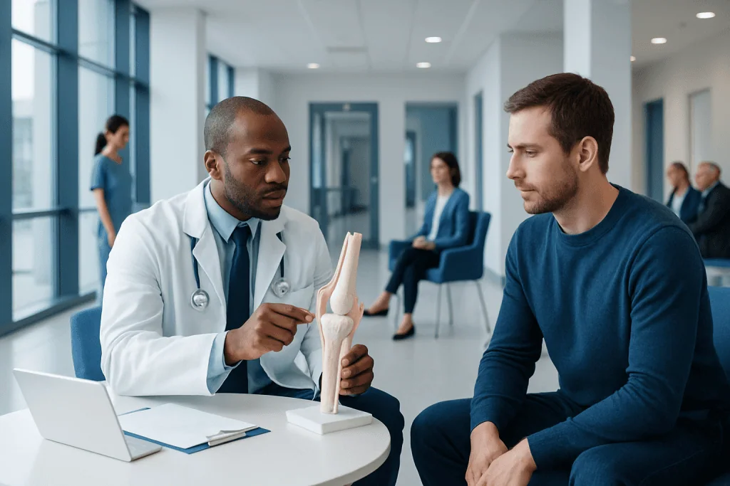
Orthopedic diagnosis starts with symptoms, medical history, and a careful physical exam. X-rays are especially useful for evaluating bones, alignment, fractures, and arthritis changes.
Key Takeaways
- Orthopedic diagnosis starts with symptoms, medical history, and a careful physical exam.
- X-rays are especially useful for evaluating bones, alignment, fractures, and arthritis changes.
- MRI shows soft tissues such as cartilage, ligaments, tendons, muscles, discs, and nerves in greater detail.
- Doctors may also use CT, ultrasound, or lab tests depending on the suspected condition.
- Not every person with joint or back pain needs advanced imaging right away.
- An accurate diagnosis helps guide treatment, recovery planning, and decisions about surgery or rehabilitation.
Orthopedic diagnosis usually combines a medical history, physical examination, and imaging tests such as X-ray or MRI. Each part provides different information, helping doctors identify the cause of bone, joint, muscle, ligament, or spine symptoms and plan appropriate treatment.
Overview: How orthopedic conditions are diagnosed
Orthopedic conditions affect the bones, joints, muscles, tendons, ligaments, and spine. Because many of these structures work together, similar symptoms can come from different causes. For example, knee pain may be related to arthritis, a meniscus tear, a ligament injury, inflammation, or pain referred from another area. This is why diagnosis is usually based on more than one test.
In most cases, an orthopedic diagnosis begins with a discussion of symptoms and a physical examination. The doctor may then recommend imaging such as an X-ray or MRI, depending on the area involved and the suspected problem. Sometimes additional tests, such as ultrasound, CT, nerve studies, or blood work, are also helpful.
The goal is not simply to name a condition, but to understand what structure is affected, how severe the problem is, and how it is influencing movement and daily life. A clear diagnosis helps determine whether treatment should focus on rest, medication, rehabilitation, injections, or procedures such as orthopedic surgery.
Medical history and symptom review
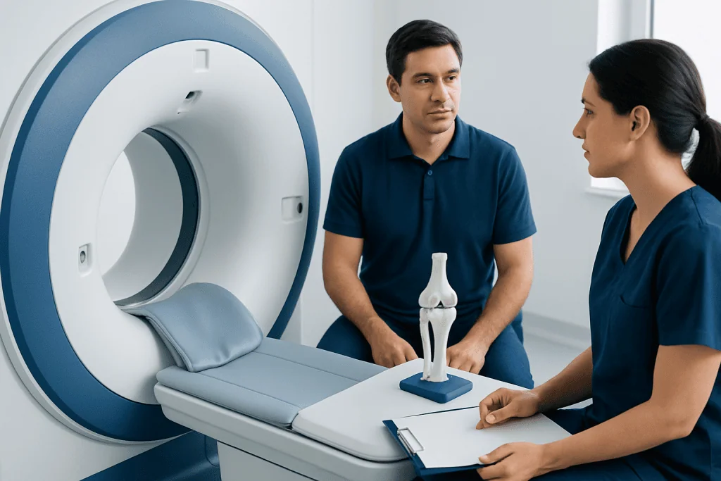
A detailed medical history is an important first step in orthopedic diagnosis. The doctor will usually ask when the pain or limitation began, whether it started suddenly or gradually, and what activities make it better or worse. Information about swelling, stiffness, weakness, numbness, instability, clicking, locking, or difficulty walking can offer important clues.
Past injuries, sports participation, work demands, and previous treatments also matter. A person who developed shoulder pain after lifting may have a different problem from someone whose pain came on gradually over months. Age, general health, medications, and any history of inflammatory disease can also influence the likely diagnosis.
The pattern of symptoms often helps guide the next step. Night pain, pain with weight-bearing, loss of range of motion, or symptoms that travel down an arm or leg can point to different causes. In some cases, the history alone may strongly suggest a condition such as a sprain, tendon irritation, or herniated disc, even before imaging is performed.
What happens during the orthopedic physical exam

The physical exam helps the doctor assess how the affected area looks, moves, and functions. This usually includes inspection for swelling, bruising, deformity, muscle wasting, or asymmetry. The doctor may gently press on certain structures to identify tenderness and determine whether pain is coming from a joint, tendon, bone, or surrounding soft tissue.
Range of motion is another key part of the exam. The doctor may ask the person to move the joint actively and may also move it passively to compare comfort and flexibility. Strength testing, balance assessment, walking analysis, and special maneuvers can help evaluate instability, impingement, tendon injury, nerve irritation, or cartilage problems.
Neurological checks may be included when symptoms involve the neck, back, arms, or legs. Reflexes, sensation, and muscle power can show whether nerves are being affected. For spine-related symptoms, the exam helps distinguish between local pain and problems that involve nerve roots or the spinal cord. These findings guide whether simple imaging, advanced imaging, or further specialist evaluation is needed.
When X-rays are used
X-rays are one of the most common first-line imaging tests in orthopedics. They are especially useful for visualizing bones and joint alignment. Doctors often request X-rays when they suspect fractures, dislocations, arthritis, bone deformities, or changes in the spine such as alignment problems or degenerative wear.
An X-ray can show whether a bone is broken, whether a joint space has narrowed, or whether bone spurs are present. It can also help compare one side of the body with the other when needed. In many situations, an X-ray is the most practical starting point because it is quick and provides important structural information.
However, X-rays do not show soft tissues well. Ligaments, tendons, cartilage, discs, and many muscle injuries are not clearly visible on standard X-rays. A normal X-ray does not always mean that nothing is wrong. If symptoms suggest a soft tissue injury, stress injury, or a problem such as meniscus tear, the doctor may recommend another imaging method.
- Best for: fractures, dislocations, bone alignment, arthritis changes
- May be used first for: knee pain, hip pain, shoulder injuries, back pain after trauma
- Limitations: less detail for ligaments, tendons, cartilage, discs, and early stress injuries
When MRI is needed
MRI, or magnetic resonance imaging, provides detailed images of soft tissues as well as bones. It is often used when the doctor needs more information than an X-ray can provide. MRI is especially helpful for evaluating ligaments, tendons, cartilage, menisci, muscles, spinal discs, and nerves.
For example, MRI may be recommended if a person has persistent knee pain with suspected cartilage or ligament damage, shoulder symptoms suggesting a rotator cuff tear, or back pain with leg numbness that may be related to a disc problem. It can also help assess the extent of inflammation, bone marrow changes, and some occult fractures that do not appear clearly on initial X-rays.
MRI is not always the first test, and it is not necessary for every orthopedic complaint. In some cases, symptoms improve with time, activity modification, and rehabilitation, making advanced imaging unnecessary. When surgery is being considered or when the diagnosis remains uncertain, MRI can be very valuable in treatment planning, including for spine surgery or knee replacement discussions in selected patients.
Other tests doctors may use
Although X-ray and MRI are widely known, they are not the only tools used in orthopedic diagnosis. CT scans can provide detailed cross-sectional images of bones and are sometimes preferred for complex fractures, joint surfaces, or preoperative planning. Ultrasound may be used to examine tendons, fluid collections, bursae, or some muscle injuries, and it can also guide certain injections.
Laboratory tests may be requested if infection, gout, autoimmune disease, or other inflammatory causes are being considered. Blood tests cannot diagnose most routine mechanical orthopedic problems, but they can be important when swelling, fever, unexplained pain, or multiple joint symptoms are present. In some cases, joint fluid may be analyzed to check for crystals or infection.
Nerve conduction studies and electromyography may help when numbness, tingling, weakness, or suspected nerve compression is part of the picture. Bone density testing may also be relevant in people with fracture risk or suspected osteoporosis. The choice of tests depends on the symptoms, exam findings, and how likely each condition is.
How doctors decide which test is best
Choosing the right test depends on the location of symptoms, the suspected diagnosis, symptom severity, and how long the problem has been present. A person with a possible fracture after a fall may need an X-ray first, while someone with a sports injury and joint instability may later need MRI if ligament damage is suspected. Back pain without warning signs may often be managed initially without immediate advanced imaging.
Doctors also consider whether test results would change treatment. If a result is unlikely to alter the plan, imaging may not be necessary right away. This thoughtful approach helps avoid unnecessary testing while still identifying conditions that need prompt treatment, such as fractures, infections, severe nerve compression, or progressive joint damage.
Imaging findings are always interpreted together with the history and exam. Some abnormalities may appear on scans even in people without symptoms, especially age-related wear in the spine or joints. For that reason, the most useful diagnosis is one that matches both the imaging result and the person’s actual symptoms and function.
After diagnosis: treatment planning and when to seek medical care
Once the diagnosis is clear, the doctor can recommend an appropriate treatment plan. Depending on the condition, this may include rest, activity modification, physiotherapy, bracing, medication, injections, or surgery. Many orthopedic problems improve with conservative care, while others need closer follow-up to prevent ongoing pain or loss of function. Structured rehabilitation, including physical therapy and rehabilitation, is often an important part of recovery.
Medical care is especially important if pain follows a significant injury, if a limb cannot bear weight, if there is visible deformity, or if symptoms are severe or getting worse. Prompt assessment is also recommended for fever with joint pain, rapidly increasing swelling, numbness, weakness, loss of bladder or bowel control, or pain that persists despite self-care. Early diagnosis can help prevent complications and support a smoother recovery.
For people seeking international care, Acibadem International’s multidisciplinary specialists and JCI-accredited hospitals evaluate orthopedic symptoms using clinical examination, imaging, and individualized treatment planning. A careful diagnosis is the foundation for choosing the most suitable next step for each patient.
Frequently asked questions
What is usually the first step in orthopedic diagnosis?
The first step is usually a medical history and physical examination. The doctor asks about symptoms, prior injuries, daily activities, and performs movement and strength tests before deciding whether imaging is needed.
Why would a doctor order an X-ray instead of an MRI first?
X-rays are often the best initial test for suspected fractures, joint alignment problems, and arthritis changes. They are quick and useful for evaluating bones, while MRI is generally reserved for more detailed assessment of soft tissues or unclear cases.
Can an MRI show more than an X-ray?
Yes. MRI provides much more detail about ligaments, tendons, cartilage, muscles, discs, and nerves. It can also reveal some injuries or inflammation that may not appear on a standard X-ray.
Do all orthopedic problems need imaging?
No. Some conditions can be diagnosed mainly from the history and physical exam, especially when symptoms are mild or improving. Imaging is most helpful when the diagnosis is uncertain, symptoms are severe, or results would change treatment.
What if the X-ray is normal but pain continues?
A normal X-ray does not rule out all orthopedic conditions. If symptoms continue, the doctor may consider MRI, ultrasound, CT, lab tests, or a reassessment to look for soft tissue injuries, inflammation, or less obvious bone problems.
How do doctors diagnose sports injuries?
Doctors usually combine the injury history, physical exam, and targeted imaging when needed. X-rays may rule out fractures, while MRI or ultrasound may be used to assess ligaments, tendons, cartilage, or muscle damage.
When should someone seek urgent orthopedic assessment?
Urgent assessment is important after major trauma, if a limb looks deformed, or if a person cannot move or bear weight on the area. Immediate care is also needed for numbness, weakness, fever with joint pain, or loss of bladder or bowel control with back symptoms.
References
- American Academy of Orthopaedic Surgeons
- American College of Radiology
- National Institute of Arthritis and Musculoskeletal and Skin Diseases
- Radiological Society of North America
- OrthoInfo
This article is for general information only and is not a substitute for professional medical advice. Please consult a qualified doctor about your individual situation.
Joint and spine care in Turkey — expert assessment & treatment
JCI-accredited · board-certified surgeons · reply within 24h
Add us as a Preferred Source to see more of our trusted health content across Google Search, AI Overviews and Discover.

Orthopedics & Spine
Care for bones, joints and the musculoskeletal system, including joint replacement, sports injuries and trauma.
125 specialists in this unitMore from the Health Library

Related Specialists

Dr. Muhammed Taha Taş
Emergency Service
Dr. Mehmet Akif Akman
Emergency Service
Prof. Dr. Agop Çıtak
Pediatrics
Assoc. Prof. Dr. Selçuk Görmez
Cardiology