Intravascular Ultrasound: How Doctors See Inside the Arteries
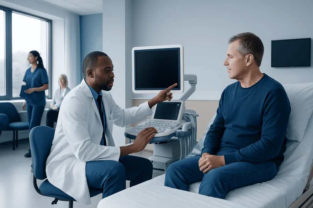
Intravascular ultrasound uses a tiny ultrasound probe on a catheter to create images from inside an artery. It is commonly used during coronary and other vascular procedures to better understand plaque and vessel anatomy.
Key Takeaways
- Intravascular ultrasound uses a tiny ultrasound probe on a catheter to create images from inside an artery.
- It is commonly used during coronary and other vascular procedures to better understand plaque and vessel anatomy.
- IVUS can help doctors choose the right stent size and check whether a stent is fully expanded and well positioned.
- The test is minimally invasive and is usually performed during angiography or catheter-based treatment.
- IVUS adds information that standard X-ray angiography may not show, especially about the artery wall and plaque buildup.
Intravascular ultrasound, often called IVUS, is a specialized imaging technique that helps doctors look at the inside of an artery from within. It can provide detailed information about plaque, vessel size, and blood vessel structure, helping guide diagnosis and treatment during vascular procedures.
Overview: What Intravascular Ultrasound Is
Intravascular ultrasound is an imaging method that allows doctors to see the inside of blood vessels using sound waves. A very small ultrasound probe is attached to the tip of a thin catheter, which is gently guided through an artery. As the probe moves through the vessel, it creates cross-sectional images of the artery wall and the space where blood flows.
This technology is different from a standard external ultrasound, which creates images from outside the body through the skin. Because the ultrasound probe is placed inside the artery, intravascular ultrasound can produce much more detailed views of the vessel’s inner structure. It can show how narrow an artery is, how much plaque is present, and how the plaque is distributed around the vessel wall.
Doctors often use IVUS during catheter-based heart and vascular procedures, especially when they need more information than an angiogram alone can provide. Angiography shows the outline of blood flow using contrast dye and X-ray imaging, while IVUS shows the actual artery wall and plaque. This added detail can help make treatment more precise and tailored to the individual patient.
How IVUS Works and What It Shows
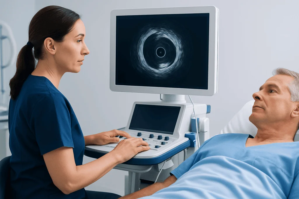
During IVUS, the catheter carrying the miniature ultrasound transducer is inserted into a blood vessel, usually through the wrist or groin, and advanced to the area being examined. The transducer emits sound waves that bounce off the vessel wall and nearby structures. A computer then converts these returning echoes into detailed images.
These images can help doctors evaluate several important features of the artery. IVUS can show the diameter of the vessel, the thickness of the artery wall, the amount and location of plaque, and whether calcium is present. It can also help identify whether narrowing is short and focal or more widespread along the vessel.
One of the most valuable uses of IVUS is during procedures such as angioplasty and stent placement. It helps the care team measure the artery more accurately and determine whether the stent is the right size and has opened fully. In many cases, this information supports safer, more effective treatment planning alongside coronary angiography and related catheter-based care.
When Doctors Use Intravascular Ultrasound

IVUS is most commonly used in cardiology, especially in people with suspected or known coronary artery disease. It may be recommended when an angiogram shows a narrowing that is difficult to interpret, when the exact extent of plaque is unclear, or when doctors want more confidence about the best treatment approach. It is also used to assess the results of an intervention after a stent has been placed.
In the heart, IVUS can be especially helpful for complex lesions, long areas of narrowing, left main coronary artery disease, or cases in which the blood vessel anatomy is not straightforward. It may also help evaluate why symptoms continue after a previous procedure, such as if a stent has not fully expanded or if new plaque has developed nearby.
Beyond the coronary arteries, IVUS may also be used in other blood vessels, including some peripheral arteries. In patients being evaluated for conditions such as coronary artery disease, it can provide detail about the vessel wall that standard imaging may miss. Depending on the situation, IVUS may complement tests and treatments such as angioplasty and stent placement.
What to Expect During the Procedure
IVUS is usually performed in a catheterization laboratory as part of an angiogram or an interventional procedure. Before the test, the medical team explains the process, reviews medications, and checks for allergies, especially to contrast dye if angiography is being done at the same time. Patients are generally awake but receive medication to help them relax.
After the skin is cleaned and numbed, the doctor inserts a thin tube into a blood vessel, often in the wrist or groin. Through this access point, the IVUS catheter is guided to the artery of interest. The patient may feel pressure at the insertion site, but the imaging itself usually does not cause pain. The ultrasound probe collects images as it is slowly moved through the vessel.
The procedure length varies depending on why IVUS is being used and whether treatment is performed during the same session. Afterward, the catheter is removed and the insertion site is monitored for bleeding. Many people return home the same day if no additional concerns arise, though recovery instructions depend on the overall procedure and the person’s health status.
Benefits, Limits, and Safety
The main benefit of intravascular ultrasound is that it provides a detailed view from inside the artery. This can improve understanding of how severe a blockage really is and can help guide important treatment decisions. In stent procedures, IVUS can support more accurate sizing and placement, which may reduce the chance of incomplete expansion or poor fit.
Another advantage is that IVUS helps doctors look beyond the flow channel seen on angiography and evaluate the vessel wall itself. Some plaque can be hidden when only the flow of contrast dye is visible on X-ray. By showing the architecture of the artery, IVUS can reveal information that may influence whether a patient needs medication alone, a stent, or another form of care.
Like any invasive catheter-based procedure, IVUS has some risks, although serious complications are uncommon when performed by experienced teams. Possible risks include bleeding, bruising, damage to the blood vessel, irregular heart rhythm, or a reaction related to the broader catheterization procedure. IVUS also has limits: it does not replace every heart test, and in some cases doctors may choose other tools, such as pressure-based assessment or different imaging technologies, depending on the clinical question.
How IVUS Compares With Other Imaging Tests
IVUS is one of several tools doctors use to examine blood vessels. Standard coronary angiography is often the first test during catheter-based evaluation because it shows how blood flows through the arteries and where significant narrowing may be located. IVUS adds more structural detail by showing the artery wall, plaque burden, and vessel dimensions from within.
Another related technology is optical coherence tomography, or OCT. OCT can provide very high-resolution images of the inner lining of the vessel, while IVUS generally penetrates deeper into the artery wall and may be especially useful in larger vessels or in the presence of certain types of plaque. The choice between these tools depends on the clinical situation, the vessel being studied, and the doctor’s goals during the procedure.
Noninvasive tests such as CT coronary angiography, stress testing, or echocardiography also play important roles, but they answer different questions. IVUS is used when highly detailed inside-the-artery information is needed during an invasive procedure. For some people with advanced narrowing, this information may help guide further treatment, including coronary artery bypass surgery when appropriate.
Aftercare, Follow-Up, and When to Seek Medical Advice
After IVUS, follow-up care depends mainly on the larger procedure that was performed. If IVUS was used during a diagnostic angiogram, recovery may be relatively quick, with advice to rest, drink fluids, and watch the access site. If a stent or another intervention was performed, the care team may recommend additional medications, activity restrictions for a short time, and closer follow-up.
Patients should contact a doctor promptly if they notice increasing pain, swelling, bleeding, numbness, fever, chest discomfort, shortness of breath, or other concerning symptoms after going home. These symptoms do not always signal a serious problem, but they should be assessed without delay. Keeping follow-up appointments is important so that the medical team can review recovery and long-term heart health.
Longer-term care usually focuses on the condition that led to the procedure, such as atherosclerosis or other vascular disease. This may include managing blood pressure, cholesterol, diabetes, smoking cessation, regular physical activity, and a heart-healthy eating pattern. For international patients who need advanced evaluation, Acibadem International’s multidisciplinary specialists and JCI-accredited hospitals diagnose and treat cardiovascular conditions using modern imaging and interventional techniques.
Frequently asked questions
Is intravascular ultrasound the same as a regular ultrasound?
No. A regular ultrasound is performed from outside the body through the skin, while intravascular ultrasound places a tiny ultrasound probe inside an artery on a catheter. This allows doctors to see much more detail about the artery wall and plaque.
Why would a doctor use IVUS during a heart procedure?
A doctor may use IVUS when more detail is needed than an angiogram alone can provide. It can help measure the artery, assess plaque, and confirm that a stent is properly sized and fully expanded.
Is IVUS painful?
Most people do not feel pain from the imaging itself. They may feel brief pressure or discomfort at the catheter insertion site, but medication is usually given to make the procedure more comfortable.
How long does an IVUS procedure take?
The time varies depending on whether IVUS is being used only for imaging or as part of a larger treatment procedure. In many cases, it is completed during the same session as angiography or stent placement.
Are there risks with intravascular ultrasound?
IVUS is generally considered safe when performed by experienced specialists, but it is still an invasive procedure. Possible risks include bleeding, bruising, blood vessel injury, or complications related to catheterization, though serious events are uncommon.
Can IVUS detect plaque that an angiogram misses?
Yes, in some situations it can. Angiography mainly shows the flow channel inside the artery, while IVUS also shows the artery wall and plaque burden, which can reveal disease not fully appreciated on X-ray images alone.
References
- American Heart Association
- National Heart, Lung, and Blood Institute
- European Society of Cardiology
- Society for Cardiovascular Angiography and Interventions
- Radiological Society of North America
This article is for general information only and is not a substitute for professional medical advice. Please consult a qualified doctor about your individual situation.
Ultrasound in Turkey — costs, top hospitals & a free quote
JCI-accredited · board-certified surgeons · reply within 24h
Add us as a Preferred Source to see more of our trusted health content across Google Search, AI Overviews and Discover.

Check-up & Preventive Medicine
Comprehensive health screenings designed for early detection and prevention.
3 specialists in this unit