

Asbestosis vs Mesothelioma: How Doctors Tell Scarring From Cancer After Asbestos Exposure
Key Takeaways
- Asbestosis is non-cancerous scarring inside the lung; mesothelioma is a cancer of the pleural lining, and a pathologist distinguishes them without difficulty once tissue is available.
- Asbestosis is usually diagnosed from exposure history, high-resolution CT and restrictive lung function tests, whereas mesothelioma almost always requires a biopsy by thoracoscopy.
- The NHS puts the typical delay before asbestosis at 20 to 30 years, and the Mayo Clinic gives 20 to 60 years for mesothelioma, so recent good health after old exposure proves nothing.
- Asbestosis does not transform into mesothelioma, but the heavy exposure that causes scarring independently raises the lifetime risk of both mesothelioma and lung cancer.
- Smoking does not appear to raise mesothelioma risk, yet combined with asbestos it greatly multiplies lung cancer risk, making cessation the highest-impact step an exposed person can take.
- Pleural plaques are a footprint of past exposure, not early cancer and not asbestosis, and on their own they usually need reassurance and symptom awareness rather than repeated scanning.
Asbestosis and mesothelioma are different diseases with the same cause. Asbestosis is non-cancerous scarring of lung tissue that stiffens the lungs and slowly reduces breathing capacity. Mesothelioma is a cancer of the pleura, the lining around the lungs. Doctors separate them using exposure history, CT imaging, lung function tests and, when a growth or fluid is suspected, a biopsy. Both appear decades after exposure, and a person can have either, neither or both.
A retired pipefitter sits in an outpatient clinic with a folded CT report in his jacket pocket. He has read it four times. Two phrases keep catching his eye: pleural thickening and lower-zone reticulation. Somewhere in the forty years since he lagged boilers in a shipyard, his lungs changed, and the report does not say which of the two words he fears most applies to him.
That is the moment the asbestosis vs mesothelioma question stops being an internet search and becomes personal. The two conditions share a cause, a long silence and an early symptom of breathlessness on stairs, which is why they blur together in conversation. Inside the chest they could hardly be more different: one is scar tissue, the other is a malignant tumor.
This explainer walks through how clinicians pull those two apart, what each diagnosis actually means for daily life, and where the popular understanding goes wrong.
Asbestosis vs mesothelioma: the one-sentence difference
Strip away the medical vocabulary and the distinction fits on an index card. Asbestosis is scarring. Mesothelioma is cancer. Everything else, from the tests ordered to the specialists involved, follows from that split.
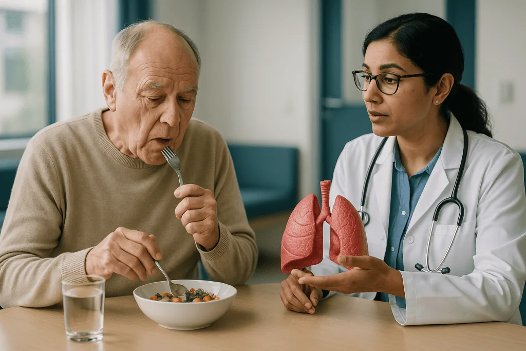
Asbestosis belongs to a family of conditions called pneumoconioses, which means lung disease caused by inhaling mineral dust. The scarring, known as fibrosis, develops within the lung tissue itself, mostly in the lower parts of both lungs. Scarred lung is stiff lung. It cannot expand fully and it transfers oxygen into the blood less efficiently, so breathlessness creeps in during exertion long before it appears at rest. The Mayo Clinic describes asbestosis as a chronic condition that is not cancer, yet one that raises the risk of developing cancer later.
Mesothelioma is a malignancy, a cancer, arising from the mesothelium. The mesothelium is the thin double layer of cells that lines the chest cavity, wraps the lungs and also lines the abdomen. According to the NHS, the form that affects the lining of the lungs, called pleural mesothelioma, is by far the most common, with the abdominal form, peritoneal mesothelioma, seen less often. Unlike scarring, a tumor grows, spreads along the lining and can press on the lung, the chest wall and nearby structures.
Why does the confusion persist? Because both diseases hide for decades, both announce themselves with shortness of breath and both appear in the same group of people: tradespeople, shipyard workers, miners and, less often, their family members. The overlap is in the story, not in the biology. A pathologist looking at tissue under a microscope would never mistake one for the other. The challenge for a clinic team is getting to that point safely and without unnecessary procedures.
What asbestos actually does inside the lung
Asbestos is not one substance but a group of six naturally occurring silicate minerals that split into fibers thinner than a human hair. Their value in construction, the same heat resistance and durability that made them useful in brake linings, insulation and roofing, is exactly what makes them dangerous once inhaled. The WHO notes that all forms of asbestos are carcinogenic to humans and that roughly 125 million people worldwide are still exposed at work.
Picture a fiber drifting down the airways. Larger particles are trapped by mucus and coughed out. The thinnest, longest fibers slip past those defenses into the alveoli, the microscopic air sacs where gas exchange happens. Immune cells called macrophages arrive to engulf the intruder, as they would a bacterium. A fiber, though, is indigestible and often longer than the cell attempting to swallow it. The macrophage releases inflammatory chemicals, dies and is replaced by another, and the cycle repeats for years.
Two outcomes follow from that failed clean-up, and they explain the asbestosis vs mesothelioma divide.
The first is fibrosis. Chronic inflammation around the alveoli triggers the deposition of collagen, the same protein that forms a skin scar. Laid down over decades, it thickens the walls between air sacs and the blood vessels beside them. The result is asbestosis: lungs that look honeycombed on a CT scan and behave like a leather bellows that has dried out.
The second is malignant change. Some fibers migrate outward through the lung to the pleura, or are carried there through lymphatic channels. In the pleura they cause persistent irritation, oxidative stress and direct physical damage to dividing cells and their DNA. The Mayo Clinic explains that cancer begins when a series of genetic changes turns normal cells into ones that grow uncontrollably, and in mesothelioma that process unfolds in mesothelial cells. Scarring is the lung trying to wall off a wound. Cancer is a cell that has lost the instructions to stop.
Asbestos exposure symptoms: why scarring and cancer feel alike at first
The early experience of both diseases is frustratingly similar, which is why no one should attempt to sort them at home. Breathlessness is the shared headline. In asbestosis it usually arrives gradually, noticed first on hills or when carrying groceries, and it worsens slowly over years as more lung becomes stiff. In pleural mesothelioma it can appear more abruptly, often because fluid has collected between the two layers of the pleura, a pleural effusion, and is compressing the lung from outside.
Listen to the chest and the clues start to diverge. The NHS describes fine crackling sounds at the lung bases in asbestosis, produced as stiffened air sacs snap open on each breath. A large effusion, by contrast, produces a dull, quiet area where breath sounds should be. Chest pain also behaves differently: asbestosis is not typically painful, while mesothelioma frequently causes a persistent ache in the chest wall or under the ribs because the tumor involves the lining that carries pain fibers.
Other features tilt the balance. A persistent cough, wheeze and a feeling of tightness are described by both the Mayo Clinic and the NHS in asbestosis. Unexplained weight loss, night sweats, fever and a swollen abdomen point more toward a malignant process, and abdominal swelling in particular raises the possibility of peritoneal mesothelioma. Fingertip changes called clubbing, where the nails curve and the ends of the fingers broaden, are seen in long-standing lung scarring.
None of these signs is a diagnosis. A clinician weighs them against the exposure history, the time since exposure and the pattern on imaging. What matters most for a reader is this: new or worsening breathlessness in anyone with a known asbestos history deserves an appointment, whichever disease turns out to be responsible. The tests, not the symptoms, settle the question.
How doctors tell asbestosis from mesothelioma: the tests, in order
The pathway almost always begins with a conversation rather than a machine. Where did you work, for how long, and how dusty was it? Did you cut, drill or strip asbestos products? Did anyone in your household bring dust home on work clothes? The Mayo Clinic lists a detailed occupational history as the first step in evaluating asbestosis, and the same is true for mesothelioma. Exposure decades ago still counts.
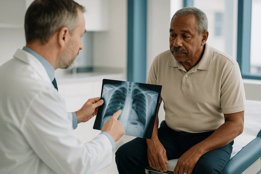
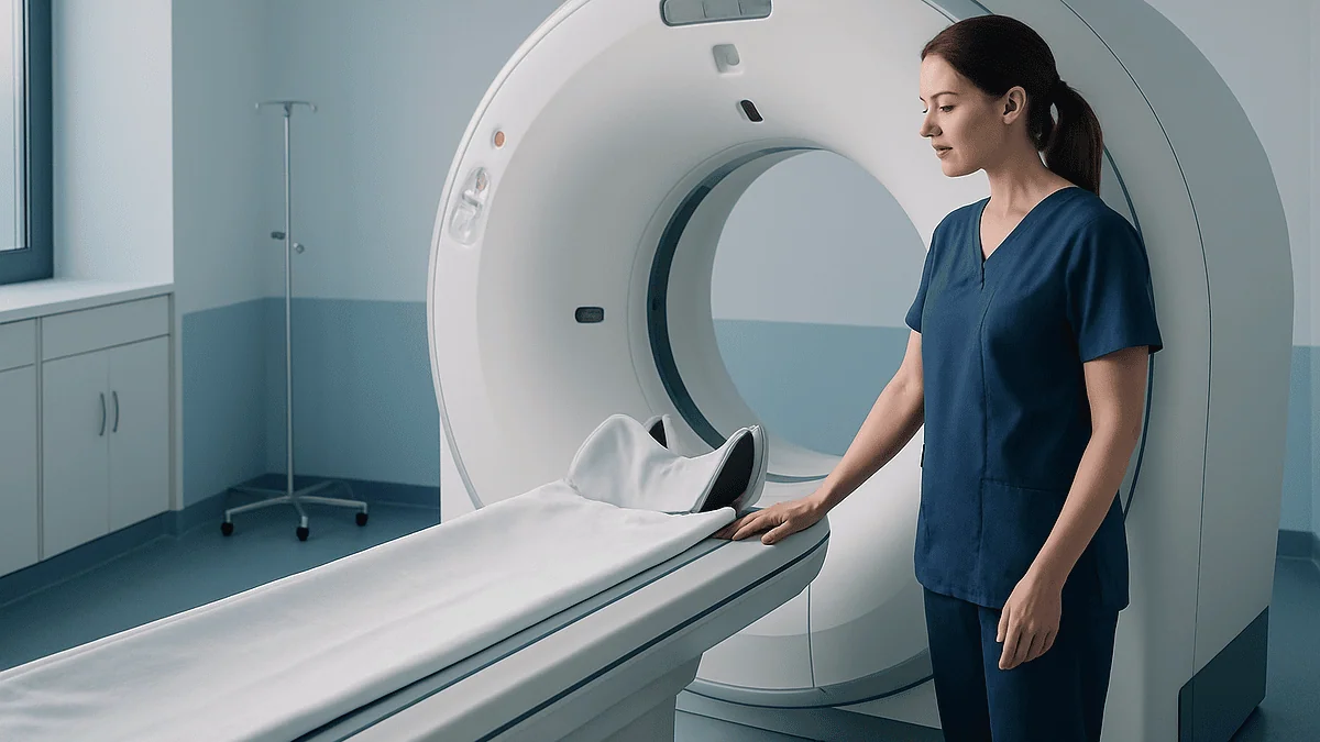
A physical examination follows, listening for those basal crackles and checking for reduced chest expansion, clubbing or dullness from fluid. Then come the tests, and the order matters.
- Chest X-ray. Cheap and quick, it can reveal shadowing at the lung bases, thickened pleura or fluid. According to MedlinePlus, X-ray changes in asbestosis may be subtle or absent early on, so a normal film does not close the case.
- High-resolution CT scan. This is the workhorse. The NHS describes CT as providing far more detail than X-ray, showing the reticular, honeycomb pattern of fibrosis in asbestosis and, separately, nodular or irregular pleural thickening that raises concern for a tumor.
- Lung function tests. Breathing into a spirometer measures how much air the lungs hold and how quickly it moves. Asbestosis produces a restrictive pattern, meaning reduced total volume with relatively preserved airflow, and a reduced ability to transfer gas across the alveolar wall.
- Pleural fluid analysis. If an effusion is present, a needle drains a sample under local anesthetic. Cancer cells in the fluid support mesothelioma, though the NHS notes fluid alone is often inconclusive.
- Biopsy. The decisive step for suspected mesothelioma. A small camera inserted between the ribs, a thoracoscopy, lets the surgeon see the pleura and take tissue. The pathologist then classifies the cells as epithelioid, sarcomatoid or biphasic, which guides treatment planning.
Asbestosis is usually confirmed without a biopsy when the history, CT pattern and lung function line up. Mesothelioma is almost never confirmed without one. That single asymmetry, tissue or no tissue, is the practical heart of telling the two apart.
Asbestosis vs mesothelioma at a glance
Side by side, the differences are easier to hold onto than in paragraphs. The table below condenses what the NHS, Mayo Clinic and MedlinePlus describe for each condition.
| Feature | Asbestosis | Mesothelioma |
|---|---|---|
| What it is | Non-cancerous scarring (fibrosis) of lung tissue | Cancer of the pleura or peritoneum |
| Where it sits | Inside the lung, mainly the lower lobes | The lining around the lung or abdomen |
| Typical delay after exposure | Around 20 to 30 years (NHS) | 20 to 60 years (Mayo Clinic) |
| Exposure pattern | Usually heavy, prolonged exposure | Can follow lower or shorter exposure |
| Key early symptom | Slowly progressive breathlessness, dry cough | Breathlessness with fluid, chest pain, weight loss |
| Examination clue | Fine crackles at lung bases, clubbing | Dullness from effusion, tender chest wall |
| CT appearance | Reticular lines, honeycombing, traction changes | Nodular or circumferential pleural thickening, effusion |
| Lung function | Restrictive pattern, reduced gas transfer | Variable, often reduced by fluid or tumor bulk |
| Confirmed by | History plus CT plus lung function; biopsy rarely needed | Tissue biopsy, usually by thoracoscopy |
| Course | Chronic, slowly progressive; treatment supports breathing | Malignant; treatment aims to control growth and symptoms |
| Raises risk of the other? | Marks heavy exposure, so mesothelioma risk is higher | Does not cause asbestosis |
A few cautions about reading a table like this. The latency ranges overlap heavily, so time since exposure alone cannot decide anything. Exposure intensity is a tendency, not a rule: heavy exposure raises the risk of both, and the WHO stresses that no safe threshold has been established for the cancer-causing effects of asbestos. And the CT descriptions are what a radiologist looks for, not something a patient can read off a report. When a report mentions pleural thickening, that phrase covers everything from harmless calcified plaques to a tumor, and only the full clinical picture, sometimes with tissue, tells which.
Why both diseases take decades to appear
The most disorienting feature of asbestos disease is its patience. A person can leave a dusty job in their twenties, raise a family, retire, and only then develop symptoms. The NHS states that asbestosis typically becomes apparent 20 to 30 years after exposure, and the Mayo Clinic gives a range of 20 to 60 years for mesothelioma. Those numbers are averages drawn from large groups; individuals vary widely around them.
The biology explains the wait. Asbestos fibers are biopersistent, a term meaning the body cannot dissolve or clear them. Once lodged, they remain for life, and the inflammatory process they trigger is low-grade and continuous rather than acute. Scar tissue accumulates a fraction at a time. Lungs have enormous spare capacity, so a considerable amount of fibrosis can build before breathlessness is noticed on ordinary activity. By the time symptoms prompt a visit, the changes on CT are often well established.
Cancer needs even longer. Mesothelial cells must acquire several independent genetic errors before they escape normal growth controls, and each error is a rare, random event driven by decades of irritation. The long latency is one reason the disease is often diagnosed at a later stage than many other cancers.
Two practical consequences follow. First, the absence of symptoms in the years after exposure is no reassurance at all, and a former worker who feels fine should still tell every new doctor about the exposure so it stays in the record. Second, because the delay is so long, the health effects of asbestos persist in a population for generations after the material is banned or restricted. The WHO attributes more than 200,000 deaths each year to occupational asbestos exposure, and many of those deaths trace back to workplaces that closed long ago.
Time since exposure, then, is a clue that raises suspicion. It is never the answer on its own.
Who is investigated further, and who is asked to watch and wait
Not every asbestos-exposed person with an abnormal scan is heading for a biopsy. Clinicians sort findings into three broad groups, and the sorting decides what happens next.
The first group has pleural plaques. These are smooth, often calcified patches of thickening on the pleura that are a marker of past exposure rather than a disease in their own right. The NHS explains that plaques do not usually cause symptoms and are not cancerous. People in this group are commonly reassured, advised to stop smoking if they smoke, and asked to report any new breathlessness or pain. Routine repeat scanning is not standard practice for plaques alone.
The second group has features of asbestosis: basal fibrosis on CT, restrictive lung function and a consistent history. Investigation focuses on confirming the pattern and measuring how much breathing capacity has been lost. A biopsy is rarely needed and carries risk in scarred lungs, so it is reserved for cases where the picture does not fit. Follow-up usually involves periodic lung function testing so any decline can be tracked, along with attention to the heart, because long-standing lung scarring can strain the right side of it.
The third group has findings that cannot be explained by scarring or plaques: irregular or nodular pleural thickening, a new effusion, chest pain or weight loss. These people are usually referred promptly to a respiratory or thoracic team, because the question now is whether a tumor is present, and that is answered with tissue.
Who is asked to wait? Someone with stable plaques and no symptoms; someone whose CT change is minimal and whose lung function is normal, where a repeat scan in a defined interval is safer than an invasive test. Waiting in these cases is an active clinical decision, reviewed against clear criteria, not neglect. The treating team sets the interval and the triggers for bringing the appointment forward.
Is mesothelioma worse than asbestosis? And can asbestosis turn into mesothelioma?
These two questions arrive together in almost every consultation, and both deserve straight answers.
Is mesothelioma worse? In terms of prognosis, yes. Mesothelioma is an aggressive cancer, and the NHS is candid that it is usually diagnosed at a stage where removing it entirely is not possible, with treatment directed at controlling the disease and its symptoms for as long as possible. Asbestosis, by contrast, is a chronic condition. The NHS notes that although it cannot be reversed, many people live for many years with it, and the main threats to health come from its complications rather than the scarring itself: respiratory infections, strain on the heart and, critically, a raised risk of lung cancer and mesothelioma.
That comparison should not be read as asbestosis being trivial. Advanced fibrosis can become severely disabling, with breathlessness at rest and a need for supplemental oxygen. It is a serious diagnosis with a different tempo.
Can asbestosis turn into mesothelioma? Not in the way a mole can turn into melanoma. Scar tissue does not transform into a tumor. What the two share is the fiber burden that caused them. A person with asbestosis has, by definition, had heavy exposure, and heavy exposure independently raises the lifetime risk of mesothelioma and of ordinary lung cancer. The Mayo Clinic lists both as complications of asbestosis for exactly this reason. So the honest phrasing is that asbestosis and mesothelioma can coexist, and having one signals that vigilance for the other is warranted, without one causing the other.
Smoking changes the arithmetic sharply for lung cancer but not for mesothelioma. The NHS advises that asbestos exposure combined with smoking greatly increases the risk of lung cancer compared with either alone, which is why stopping smoking is the single most powerful step an exposed person can take. Mesothelioma risk, unusually among chest cancers, appears largely independent of tobacco.
How asbestosis is managed once scarring is confirmed
There is no medicine that dissolves established lung scar, and clinicians will say so plainly. The NHS and Mayo Clinic both describe the aim of asbestosis care as slowing decline, easing symptoms and preventing the complications that do the most harm. That framing disappoints some people at first hearing, yet the components are practical and make a measurable difference to daily life.
Stopping smoking sits at the top. Tobacco accelerates the loss of lung function in fibrotic lungs and multiplies the already elevated lung cancer risk. Clinicians can refer to support services and, where appropriate, discuss medicines that reduce cravings; those choices belong to the prescribing clinician and the patient together.
Pulmonary rehabilitation is the next pillar. This is a structured program of supervised exercise, breathing techniques and education, typically run over several weeks, that trains the muscles and the mind to work efficiently with a reduced oxygen supply. It does not add lung tissue, but it reliably reduces breathlessness for a given level of activity.
Oxygen therapy is considered when blood oxygen levels fall below a threshold at rest or during exertion, assessed by the respiratory team with a walking test or overnight monitoring. The Mayo Clinic notes that some people use oxygen only during activity, others continuously. The decision rests on measured levels, not on how breathless a person feels.
Vaccination against influenza and pneumococcal infection is routinely recommended, because a chest infection that a healthy person shakes off in a week can tip scarred lungs into failure. Prompt treatment of infections follows the same logic.
Some people with severe, progressive fibrosis are assessed for lung transplantation. That is a specialist conversation with strict criteria and its own risks, and only the transplant team can say whether it applies.
Finally, avoiding any further asbestos exposure is non-negotiable. Older homes, garages and workshops can still contain intact asbestos products, and disturbing them adds to a fiber burden that is already doing harm.
How mesothelioma is treated, and what each option is trying to do
Treatment for mesothelioma is planned by a multidisciplinary team, meaning a group that includes a respiratory physician, thoracic surgeon, oncologist, radiologist, pathologist and specialist nurse. They weigh the cell type from the biopsy, how far the tumor has spread, the person’s general fitness and their own priorities. No single plan fits everyone, and the NHS is clear that in most cases the goal is to control the cancer and relieve symptoms rather than to remove it completely.
Each modality has a distinct job.
Surgery may aim to remove as much visible tumor as possible from the pleura, sometimes together with the lining of the diaphragm and heart, and in selected cases the lung itself. It is major surgery suitable for a minority of people whose disease is confined and whose fitness allows it. Smaller procedures, such as thoracoscopy with talc pleurodesis, are used to stop fluid re-accumulating: talc powder is introduced to make the two pleural layers stick together so there is no space for fluid to collect. An indwelling pleural catheter, a thin tube left in place so fluid can be drained at home, is an alternative.
Chemotherapy uses drugs that damage rapidly dividing cells, given in cycles over several months, to shrink or slow the tumor. Immunotherapy, specifically the class known as immune checkpoint inhibitors, works differently: it releases a brake on the body’s own T cells so they can recognize and attack cancer cells. Both are systemic treatments and both have side effects that the oncologist monitors closely.
Radiotherapy directs high-energy beams at a specific area, most often to ease pain from a tumor pressing on the chest wall, or to treat the sites where instruments have entered the chest.
Palliative and supportive care runs alongside all of this from diagnosis, not only at the end. It addresses pain, breathlessness, appetite and the emotional weight of the diagnosis, for the patient and the family. Choosing between or combining these options is the treating team’s call, made with the person in front of them.
What the following weeks usually look like after a suspicious scan
The period between an abnormal scan and a settled diagnosis is often the hardest stretch, precisely because nothing is yet certain. Knowing the usual shape of those weeks can take some of the edge off.
In the first days, the report is reviewed by the referring doctor and, if a tumor is a possibility, an urgent referral to a respiratory or thoracic team is made. Many health systems operate a fast-track pathway for suspected cancer, with a specialist appointment within a short, defined window. A specialist nurse is frequently the first point of contact and remains so throughout.
At that first specialist visit, expect the exposure history to be taken again in detail, an examination, blood tests and often a repeat or more detailed CT. If fluid is present, it may be drained the same day or soon after, which usually brings immediate relief from breathlessness and provides a sample for the laboratory.
If the fluid or imaging leaves doubt, a thoracoscopy is arranged. It is performed under general anesthetic or heavy sedation, usually involves a short hospital stay, and leaves one or two small incisions between the ribs. Soreness at the wound sites is common for several days. Pathology results typically follow within one to two weeks, because the tissue must be processed, stained and examined with specialized markers to distinguish mesothelioma from other cancers that can reach the pleura.
Results are given at a face-to-face appointment, ideally with a companion present. If mesothelioma is confirmed, the case is discussed at the multidisciplinary meeting and a treatment plan is proposed, usually within a further week or two. If the diagnosis is asbestosis or benign pleural disease, the plan shifts to lung function monitoring and supportive measures, and the pace slows.
Timelines vary between health systems and individuals, and these are typical patterns rather than guarantees. What people find most helpful is asking, at each step, what the next step is and when to expect it.
What people often get wrong about asbestosis and mesothelioma
Misunderstandings about asbestos disease cluster around a handful of ideas, most of them understandable and most of them wrong.
The first is that they are the same thing with different names. They are not. One is scarring within the lung, the other is cancer of its lining. A person can have either alone, both together, or neither despite heavy exposure.
The second is that only heavy industrial exposure matters. Asbestosis does generally follow prolonged, dense exposure, but mesothelioma has been recorded after far lower doses, including in people who washed a spouse’s work overalls or lived near a processing plant. The WHO’s position that no safe exposure threshold has been identified for asbestos-related cancer reflects this.
The third is that asbestos is a problem of the past. Bans and restrictions vary between countries, and the material remains in place in vast numbers of buildings constructed before those rules. Intact, undisturbed asbestos releases few fibers; drilling, sanding or demolition releases many. The risk today is concentrated in renovation and maintenance work.
The fourth is that a clear chest X-ray rules everything out. MedlinePlus notes that early asbestosis may not show on X-ray, and early pleural tumors are easily missed. CT is far more sensitive, and even CT cannot confirm cancer without tissue.
The fifth is that smoking causes mesothelioma. Smoking does not appear to raise mesothelioma risk, but it dramatically raises lung cancer risk in asbestos-exposed people, and the NHS singles out this combination as especially dangerous. Stopping still matters enormously.
The sixth is that pleural plaques are early cancer. They are a footprint of exposure, not a tumor, and do not become one.
The last is that a diagnosis of asbestosis means life is nearly over. It means the lungs are damaged and need protecting, and it changes what the future looks like. It does not carry the outlook of a malignancy, and many people live with it for a long time.
Questions to ask your care team
Consultations about asbestos disease are dense with information, and people often leave having forgotten the question that mattered most. Writing questions down beforehand, and bringing someone to take notes, helps. The list below is a starting point; your own situation will add others.
- Based on my scan and tests, are you seeing scarring, pleural plaques, fluid, a possible tumor, or a combination? Which of these is driving my symptoms?
- Do I need a biopsy, and if so, what kind? What would change in my care if the result is one thing rather than another?
- If the diagnosis is asbestosis, how much of my lung function has been affected, and how often will you measure it?
- Am I a candidate for pulmonary rehabilitation? How do I get referred?
- Do my oxygen levels need checking at rest, on exertion or overnight?
- Which vaccinations do you recommend for me, and when?
- If mesothelioma is confirmed, what cell type is it and what stage? What is the treatment aiming to achieve in my case: control, symptom relief, or an attempt at removal?
- What are the likely side effects of the treatments you are proposing, and what are the alternatives, including doing less?
- Who is my main point of contact between appointments, and how do I reach them?
- Should my family members who shared my home or workplace be told to mention this exposure to their own doctors?
- Is there a specialist nurse, counselor or support service I can be connected with?
- Is my condition something I should report for occupational health or compensation purposes, and who can help with that paperwork?
None of these questions has a universal answer. Their value is in prompting a conversation in which the treating team explains its reasoning, so that decisions about tests and treatment are made together and understood.
When to call your doctor
Anyone with a history of asbestos exposure, whether or not a diagnosis has been made, should book an appointment for new or worsening breathlessness, a cough that lasts more than a few weeks, persistent chest or shoulder pain, unexplained weight loss, night sweats or new abdominal swelling. These symptoms have many causes, most of them unrelated to asbestos, but in an exposed person they warrant a proper look rather than a wait-and-see approach at home.
Some situations should not wait for a routine appointment. Seek urgent or emergency care the same day if you notice:
- Sudden or severe shortness of breath, especially at rest or when lying flat.
- Chest pain that is sharp, worsening or accompanied by breathlessness, which can signal a large effusion, a collapsed lung or a heart problem.
- Coughing up blood.
- Blue or gray lips or fingertips, confusion or extreme drowsiness, which can indicate dangerously low oxygen.
- A high fever with worsening cough or breathlessness in someone with known asbestosis, since infections can deteriorate quickly in scarred lungs.
- Rapid swelling of the abdomen or legs, or a sudden drop in the amount of urine.
- For people on treatment for mesothelioma, a fever during chemotherapy or immunotherapy, new severe diarrhea, a rash that spreads quickly, or any symptom your oncology team has told you to report immediately.
People with an indwelling pleural catheter should contact their team if the drainage site becomes red, painful or leaks, if the fluid changes color or smells, or if drainage suddenly stops while breathlessness returns.
Hospital teams would far rather be called about a symptom that turns out to be minor than learn afterward that someone waited. Keep the contact number for your respiratory or oncology nurse where you can find it. Every decision about tests, treatment and follow-up sits with your treating team, and the way to keep them able to make good decisions is to tell them promptly when something changes.
Frequently asked questions
Is mesothelioma worse than asbestosis?
In terms of outlook, yes. Mesothelioma is an aggressive cancer that the NHS describes as usually being found at a stage where complete removal is not possible, so treatment focuses on control and symptom relief. Asbestosis is chronic, irreversible scarring; it can become disabling, but the NHS notes many people live with it for many years. The two conditions differ in tempo and in what treatment can realistically achieve.
Is mesothelioma 100% fatal?
No single figure applies to everyone, and clinicians avoid absolute statements. Mesothelioma is a serious cancer that is rarely removable in full, and most people eventually die from it or its complications. How long someone lives varies widely with cell type, stage at diagnosis, general fitness and response to treatment; some people live for years after diagnosis. Your oncology team can discuss what the evidence suggests for your specific situation.
How did Steve McQueen get mesothelioma?
Steve McQueen was diagnosed with pleural mesothelioma and died of complications in 1980. He is widely reported to have been exposed to asbestos in several ways: stripping insulation from pipes during his time in the Marines, working around asbestos-containing brake linings and racing gear, and possibly through film-set materials. His case is often cited because it illustrates the decades-long delay between exposure and disease.
What is the life expectancy of asbestosis?
There is no fixed asbestosis life expectancy, because progression varies enormously between people. The NHS states that although the scarring cannot be reversed, many people live for many years with the condition. The main risks to life come from complications such as chest infections, strain on the heart, lung cancer and mesothelioma, so stopping smoking, vaccination and regular monitoring matter more than any single number.
Can asbestosis turn into mesothelioma?
Scar tissue does not transform into a tumor, so asbestosis does not turn into mesothelioma in the way a mole can become melanoma. What links them is the fiber burden. Having asbestosis means exposure was heavy, and heavy exposure independently raises the risk of mesothelioma and lung cancer. The Mayo Clinic lists both as complications of asbestosis for this reason. The two conditions can coexist in the same person.
Can you have asbestosis and mesothelioma at the same time?
Yes. Both arise from inhaled asbestos fibers, and a person with heavy exposure can develop scarring within the lung and, separately, a tumor of the pleural lining. When this happens, clinicians usually identify the scarring on CT and the tumor by biopsy, and the mesothelioma generally dominates treatment planning. Existing fibrosis may influence which treatments are safe, particularly major surgery, so the team considers both together.
What asbestos exposure symptoms should prompt a doctor's visit?
In anyone with past asbestos exposure, new or worsening breathlessness, a cough lasting several weeks, persistent chest or shoulder pain, unexplained weight loss, night sweats or abdominal swelling warrant an appointment. These symptoms are not a diagnosis and most have other causes, but the combination of exposure history and any of them justifies examination, imaging and lung function testing rather than watchful waiting at home.
Do pleural plaques mean I have asbestosis?
No. Pleural plaques are smooth, often calcified areas of thickening on the lung lining that mark past asbestos exposure. The NHS explains they are not cancer, do not usually cause symptoms and are not the same as asbestosis, which is scarring within the lung tissue itself. Plaques do confirm that exposure occurred, so they are a reason to mention your history to every doctor and to report new chest symptoms promptly.
Can a chest X-ray tell asbestosis from mesothelioma?
Usually not on its own. An X-ray can show fluid, pleural thickening or basal shadowing, but MedlinePlus notes early asbestosis may not be visible and early pleural tumors are easily missed. High-resolution CT is much more sensitive and can show the honeycomb pattern of fibrosis or the nodular thickening that suggests a tumor. Confirming mesothelioma still requires tissue from a biopsy, which an X-ray cannot replace.
I was exposed to asbestos once, briefly. Should I be screened?
Routine screening is not generally recommended after a single brief exposure, because the risk from a one-off event is low and repeated scans carry their own small risks. The WHO notes no safe threshold has been established, so the exposure is worth recording in your medical notes. Tell your doctor, do not smoke, and report any new breathlessness, cough or chest pain. Your clinician can judge whether imaging is warranted.
References
- NHS: Asbestosis
- NHS: Mesothelioma
- MedlinePlus Medical Encyclopedia: Asbestosis
- WHO: Asbestos – elimination of asbestos-related diseases
This article is for general information only and is not a substitute for professional medical advice. Please consult a qualified doctor about your individual situation.

More from the Blog

When Is a Bronchiectasis Flare-Up an Emergency? How Exacerbations Are Treated
A bronchiectasis flare-up, also called an exacerbation, is usually treated with a longer-than-usual antibiotic course, guidelines typically advising 14 days, plus more intensive airway…
Airway Clearance for Bronchiectasis: What a Physiotherapy Session Actually Involves
Airway clearance for bronchiectasis is a set of breathing techniques, positions and sometimes handheld devices that help loosen and cough up mucus from permanently…

Life After a Pneumothorax: Diving, Altitude, Exercise and the Activities to Clear First
Diving after a pneumothorax is usually advised against, because trapped gas expands as a diver ascends and a second collapse underwater can be life-threatening;…

How Pulmonary Function Tests Guide Pulmonary Disease Treatment: Reading Your Spirometry Report
Pulmonary function test results are read in three steps: the FEV1/FVC ratio shows whether airways are narrowed (obstruction), the FVC percent-of-predicted shows whether the…

When Pneumonia Does Not Improve With Treatment: What Doctors Check Next
When pneumonia does not improve after a few days of antibiotics, doctors usually re-examine the patient, repeat blood tests and imaging, and look for…

What to Expect From Bronchitis Treatment: Symptom Relief, Recovery Pace and Review Visits
Bronchitis treatment for the common acute form is mostly about comfort and time: rest, fluids, and relief of cough and aches while the airway…