

Before Breast Cancer Treatment Begins: Imaging, Biopsy Review and Staging Tests Explained
Key Takeaways
- Imaging can only raise suspicion of breast cancer; a needle biopsy is the only test that confirms it, and no routine blood test can do either.
- Grade (1 to 3) describes how abnormal cells look under the microscope, while stage (0 to IV) describes how far the cancer has spread, so a grade 3 cancer can still be stage I.
- Every breast cancer is tested for estrogen receptor, progesterone receptor and HER2, and those three results determine which treatment options are biologically relevant.
- For small, node-negative cancers without symptoms, guideline bodies advise against routine CT, bone or PET scans because incidental false alarms outnumber true findings.
- Sentinel node biopsy typically removes one to three armpit nodes and spares the rest when they are clear, which lowers the risk of lymphedema.
- Germline genetic tests examine the genes you were born with, whereas multigene tumor tests examine the cancer's own gene activity; they answer entirely different questions.
Before breast cancer treatment begins, most people have a diagnostic mammogram and ultrasound, sometimes a breast MRI, and a needle biopsy whose tissue is tested for type, grade, hormone receptors and HER2. Blood tests check general health rather than the cancer. Scans of the rest of the body are added only when stage or symptoms warrant them. This workup usually spans several weeks and is what allows the care team to plan treatment.
The mammogram appointment took twenty minutes. The phone call afterward took two. And then, somehow, three weeks opened up on the calendar with nothing in them but words: biopsy, receptor status, staging, multidisciplinary meeting. One reader described sitting in her car in the hospital parking garage rereading the appointment letter, trying to work out whether all those tests before breast cancer treatment meant things were worse than she had been told, or simply that nobody had explained the sequence.
Usually it is the second. The weeks between an abnormal image and the first day of treatment are not dead time. They are when radiologists, pathologists, surgeons and oncologists assemble the specific facts about one specific cancer, so that what follows fits it rather than fitting an average.
This guide walks through that sequence in the order it typically unfolds, explains what each result changes, and is honest about where the evidence draws firm lines and where it leaves judgment to the team.
What actually happens during the tests before breast cancer treatment
The word most people remember from the first appointment is not a test name. It is ‘wait.’ Wait for the radiologist to compare views, wait for the lab, wait for a meeting where several specialists look at everything at once. That waiting has a structure, and understanding it changes how the weeks feel.
The tests before breast cancer treatment answer four questions in a fixed order. Is there a cancer? Imaging raises the suspicion; only a biopsy, the removal of a small piece of tissue for examination under a microscope, can confirm it. What kind is it? The pathologist, a doctor who studies tissue, describes the cell type, how abnormal the cells look and which proteins sit on or inside them. How far has it reached? That is staging, which combines the size of the tumor, whether lymph nodes are involved and whether there is any sign of spread elsewhere. Is the body ready for treatment? Blood tests and a general health review answer that last part.
Each answer changes the next step. A cancer that turns out to be hormone-driven, small and confined to the breast is planned differently from one that is larger or has reached a node. That is why the team does not schedule surgery or drug treatment on the day of the mammogram, and why a multidisciplinary meeting, where surgeons, oncologists, radiologists and pathologists review the case together, is standard practice in breast care, as the NHS describes in its guidance on tests and next steps.
The honest framing is this: the tests are the treatment plan being written. Skipping them would be faster and worse.
Which imaging tests come first, and why the order matters
Imaging almost always starts with a diagnostic mammogram, a low-dose X-ray of the breast taken from several angles with more focused views than a routine screening mammogram. Radiologists, the doctors who interpret scans, study the shape of a mass, the pattern of any calcium deposits and changes in the surrounding tissue. A mammogram cannot say ‘cancer’; it can only say ‘this needs a closer look.’
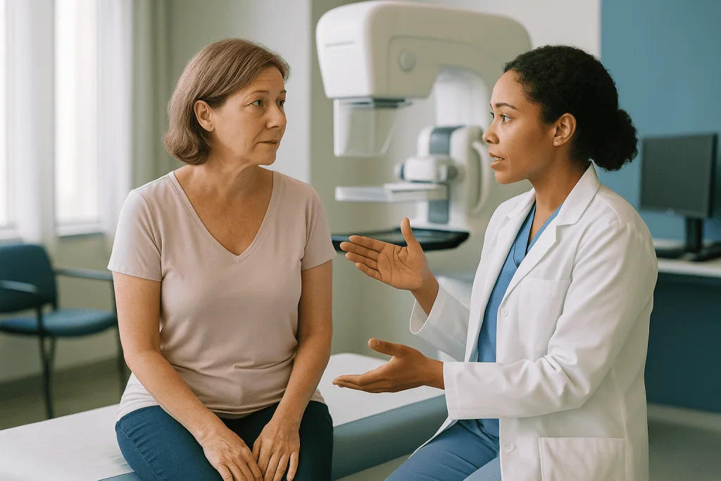
Ultrasound usually comes next, often in the same visit. It uses sound waves rather than radiation and is particularly good at telling a fluid-filled cyst from a solid lump, and at guiding a needle precisely to the right spot. The radiologist will also sweep the probe across the armpit, because the lymph nodes there, small bean-shaped filters in the immune system, are the first place breast cancer tends to travel. A node that looks thickened or rounded may be sampled with a needle at the same sitting.
Breast MRI is the test people most often ask for and most often do not need. It uses magnets and a contrast dye injected into a vein and is very sensitive, which cuts both ways: it finds small additional areas that matter, and it also flags harmless changes that lead to extra biopsies. Mayo Clinic notes that MRI is reserved for particular situations, such as dense breast tissue, uncertainty about the true extent of a tumor, some lobular cancers or a high inherited risk, rather than used for everyone.
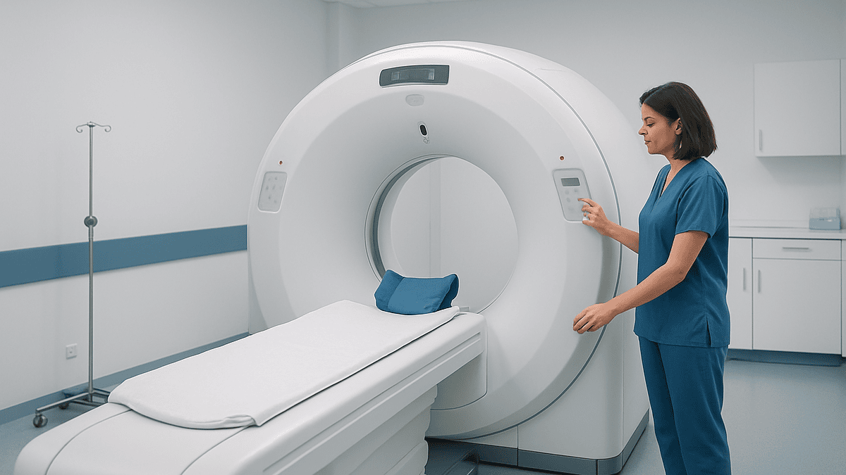
Chest X-rays, CT scans and bone scans belong to a different category. They look for disease outside the breast and are decided by stage, not by default. That distinction gets its own section later, and it is where a great deal of unnecessary worry lives.
Breast biopsy results explained: reading the pathology report
A core needle biopsy is the workhorse. After numbing the skin with local anesthetic, the radiologist or surgeon passes a hollow needle into the abnormal area, guided by ultrasound, mammogram (a stereotactic biopsy) or MRI, and removes several slivers of tissue about the width of a matchstick. A tiny metal marker is usually left behind so the exact spot can be found later, even if treatment shrinks the tumor. MedlinePlus describes the procedure as brief, with bruising and soreness the most common aftereffects.
The report that follows carries a handful of terms worth knowing. ‘Invasive’ means the cells have broken through the milk duct or lobule wall into surrounding tissue; ‘in situ’ (ductal carcinoma in situ, or DCIS) means they have not. ‘Ductal’ and ‘lobular’ describe where the cancer began. Grade, scored 1 to 3, describes how different the cells look from normal breast cells and how actively they appear to be dividing; grade 1 is closest to normal, grade 3 furthest from it. Grade is not stage. It describes behavior under the microscope, not how far the cancer has spread.
Results commonly take several days to a week or longer, according to MedlinePlus, and the receptor tests described next can add time on top. If the first sample is too small, or the microscope findings do not match what imaging showed, a repeat biopsy is requested. That is not a sign something went wrong; it is the team refusing to plan around an incomplete picture.
Ask for a copy of the report. Reading the words in your own hands, with the team’s explanation beside them, tends to be steadier than remembering them from a phone call.
What receptor and HER2 testing tells the team about your cancer's type
Two breast cancers can look identical on a mammogram and behave like different diseases. The difference lives in three proteins, and testing for them is now as routine as the biopsy itself.

Estrogen receptor (ER) and progesterone receptor (PR) are proteins inside the cancer cell that respond to the body’s hormones. When a tumor is ‘positive’ for them, hormone signals are helping it grow, and treatments that block or lower those hormones become part of the conversation. Most breast cancers are hormone receptor–positive, according to the National Cancer Institute.
HER2 is a protein on the cell surface that, when present in excess, tells the cell to divide. Roughly one in five breast cancers make too much of it, per NCI. Laboratories measure HER2 in two steps: a staining test that scores the amount of protein and, when that result is borderline, a second test that counts copies of the HER2 gene itself. MedlinePlus explains that the result is reported as positive, negative or, increasingly, ‘low,’ a category oncologists are still learning to use.
A cancer negative for all three, ER, PR and HER2, is called triple-negative. It is planned differently, because hormone-blocking and HER2-targeted approaches do not apply.
Some reports also include Ki-67, a marker of how many cells are actively dividing. Its usefulness is debated, and guideline bodies treat it as supporting information rather than a decision-maker on its own.
None of this tells you what treatment you will have. It tells your team which options are biologically sensible, which is the question they need answered before anyone talks about sequence. The decision remains theirs to make with you, once every result is in.
Blood tests for breast cancer: what they can and cannot show
People often arrive hoping a blood test will settle the diagnosis. It will not. No routine blood test detects breast cancer, and none confirms or rules it out; that is the biopsy’s job.
What blood tests do before treatment is check the body that will receive it. A complete blood count measures red cells, white cells and platelets, which matters because some drug treatments lower these counts. Kidney and liver function tests establish a baseline, since the liver processes many cancer medicines and some scans use contrast dye that the kidneys must clear. A pregnancy test is offered where relevant, because it changes both imaging and treatment choices. Mayo Clinic lists these general-health checks as preparation rather than diagnostic tools.
Tumor markers deserve a specific mention because they generate so much confusion. These are substances, such as CA 15-3 or CEA, that some cancers release into the blood. They can be raised by benign conditions and normal in people with cancer, so major oncology guidelines do not recommend them for diagnosing or staging early breast cancer. If your team orders one, the usual purpose is to record a starting value for later comparison in specific situations, not to answer whether the cancer is present or how far it has gone.
Blood tests for inherited gene changes, such as BRCA1 or BRCA2, are a separate matter covered in their own section; they test your genes, not the tumor’s.
The practical upshot: a normal blood panel is reassuring about your fitness for treatment and says almost nothing about the cancer. An abnormal one usually prompts a repeat or a closer look at one organ, and the team will explain what they are checking. Either way, the result sits inside a bigger picture, not on top of it.
Breast cancer staging tests: who has body scans and who is asked to wait
Staging is the step that most shapes treatment and most often gets over-imaged. The system used worldwide is TNM: T for tumor size and reach, N for lymph nodes involved, M for metastasis, meaning spread to distant organs. Those letters are combined, along with grade and receptor status, into stages 0 through IV, as the National Cancer Institute’s patient guidance describes.
Much of staging is already done by the time you finish the tests above. Mammogram, ultrasound and, where used, MRI estimate T. Armpit ultrasound and node biopsy estimate N. That leaves M, and here guidelines draw a deliberate line.
For a small cancer that appears confined to the breast, with no involved nodes and no symptoms elsewhere, guideline bodies do not recommend routine CT scans, bone scans or PET scans. The chance of finding distant disease in that setting is low, and the chance of finding an unrelated shadow that leads to more scans and more anxiety is comparatively high. Being told ‘we are not scanning your whole body’ is, in this context, a statement about your stage, not a shortcut.
Body imaging is typically ordered when the cancer is larger, when several nodes are involved, when drug treatment is planned before surgery, or when a symptom such as persistent bone pain or breathlessness raises a specific question. The team then chooses between a CT scan (a detailed X-ray series of chest, abdomen and pelvis), a bone scan (a tracer injected into a vein that highlights areas of bone turnover) or a PET-CT (a sugar-based tracer that lights up metabolically active tissue), depending on what they need to see.
Stage can be revised after surgery, when the pathologist measures the removed tumor and examines the nodes directly. ‘Clinical stage’ before surgery and ‘pathologic stage’ after it are both legitimate, and the second refines the first.
How lymph nodes are checked, from ultrasound to sentinel node biopsy
Whether cancer has reached the armpit nodes is one of the most consequential facts in the whole workup, and it is checked in layers.
The first layer is touch and ultrasound at the initial visit. If a node looks abnormal, a needle biopsy under ultrasound guidance samples it. A positive result answers the question early and influences whether treatment begins with drugs or with surgery.
If the nodes look normal, the answer usually waits for surgery, where the sentinel node biopsy is performed. The sentinel node is the first node, or first few nodes, that lymph fluid from the tumor drains into. Johns Hopkins describes the technique: a blue dye, a small amount of radioactive tracer, or both are injected near the tumor before or during the operation; the surgeon then follows the dye or a handheld detector to the node that takes it up and removes it for examination. Typically one to three nodes are taken. If they are clear, the remaining nodes are left alone, which lowers the risk of lymphedema, the arm swelling that can follow more extensive node removal.
The dye can turn urine green or blue for a day or so and leave a faint stain at the injection site for weeks; both are expected. Rare allergic reactions to blue dye are the main risk the anesthesia team watches for.
If a sentinel node contains cancer, the team decides what comes next: sometimes further node removal, sometimes radiation to the armpit, sometimes neither, depending on how much disease was found and what other treatment is planned. Large trials have shifted practice toward doing less surgery in the armpit in selected cases, and your surgeon can explain how your findings fit those criteria. It is a judgment for the team, not a fixed rule.
When genetic testing and multigene tumor tests enter the picture
Two very different tests share the word ‘genetic,’ and conflating them causes real distress.
Germline genetic testing looks at the genes you were born with, using a blood or saliva sample. It asks whether an inherited change in a gene such as BRCA1, BRCA2 or PALB2 raised your lifetime risk. NCI notes that most breast cancers are not linked to inherited mutations; testing is offered based on age at diagnosis, family history, ancestry, triple-negative status or a personal history of other cancers. The result matters before treatment because it can influence surgical choices and, in some cases, drug options, and because it carries implications for relatives. A genetic counselor, a specialist trained to explain inherited risk, usually guides this conversation, and results can take a few weeks.
Tumor genomic testing looks at the cancer itself. Multigene expression tests, sometimes called recurrence-score assays, measure the activity of a panel of genes in the biopsy or surgical specimen to estimate how likely a hormone receptor–positive, HER2-negative cancer is to return and, in turn, how much a person might gain from chemotherapy in addition to hormone treatment. The National Cancer Institute describes these tests as helping to identify people who can reasonably avoid chemotherapy and those for whom it is more clearly worthwhile. They are not used for every subtype, and they add turnaround time; the team orders them when the answer would actually change the plan.
Neither test delivers a verdict. Both feed information into a discussion in which your values, age, other health conditions and preferences sit alongside the numbers. If a test is not offered, ask why; the reason is usually that your subtype or stage puts you outside the group it helps, not that something was overlooked.
Tests before breast cancer treatment at a glance
The table below puts the main tests side by side. Waiting times are deliberately omitted, because they vary by service, by the extra tests requested and by how many results the team wants in hand before meeting you. Your care team can give the ranges that apply where you are treated.
| Test | What it answers | What happens to you | Who usually has it |
|---|---|---|---|
| Diagnostic mammogram | Size, shape and calcifications of the abnormal area | Compression X-ray views from several angles | Nearly everyone |
| Breast and armpit ultrasound | Solid versus cyst; node appearance; needle guidance | Gel and a handheld probe, no radiation | Nearly everyone |
| Core needle biopsy | Cancer or not; type; grade; ER, PR, HER2 | Local anesthetic, hollow needle, marker clip left in place | Everyone with a suspicious finding |
| Breast MRI | True extent; additional areas; dense tissue | Lying face down in the scanner with a contrast injection | Selected cases only |
| Blood tests | Fitness for treatment; organ baselines | Standard venous sample | Nearly everyone before treatment |
| CT, bone scan or PET-CT | Spread beyond breast and nodes | Tracer or contrast injection, then the scanner table | Higher stage, involved nodes or symptoms |
| Sentinel node biopsy | Cancer in the first draining nodes | Dye or tracer injection; nodes removed at surgery | Most people with invasive cancer and normal-looking nodes |
| Germline genetic test | Inherited risk genes | Blood or saliva sample after counseling | Those meeting family or subtype criteria |
| Multigene tumor test | Recurrence risk; likely benefit from chemotherapy | No extra procedure; uses existing tissue | Selected hormone-positive, HER2-negative cancers |
Two patterns stand out. The tests that everyone has are the ones that answer ‘what is it,’ and they are quick. The tests that take longest are the ones only some people need, and needing them is a statement about biology, not about how thorough your team is being. A short list can be the correct list, and a long one is not a bad omen so much as a set of specific questions the team has decided are worth answering before anything begins.
What tests are done before breast cancer surgery?
Once the plan points toward surgery, a second, shorter round of checks begins. These are about safety under anesthesia and a smooth recovery, and they are what people usually mean when they search for tests before breast cancer surgery.
A pre-assessment visit, often with a nurse and an anesthesiologist, covers your medical history, allergies, previous anesthetics and every medicine and supplement you take. Blood-thinning medicines, some herbal supplements and certain diabetes drugs may need to be adjusted around the operation; the team will tell you exactly what to do and when, and any change should come only from them. An electrocardiogram, a tracing of the heart’s electrical rhythm, is commonly requested for older adults or anyone with heart or lung conditions. Repeat blood counts may be drawn if the earlier ones are several weeks old.
Two procedures are specific to breast surgery. If the tumor cannot be felt, a localization step marks it so the surgeon can find it: a fine wire placed under imaging guidance on the morning of surgery, or a small seed or reflector placed days beforehand. For sentinel node mapping, the tracer injection described earlier is timed before the operation. If reconstruction is planned, a plastic surgeon may add measurements and photographs for the operative record.
You will be told when to stop eating and drinking, usually the night before, and given instructions on washing and what to bring. Ask whether you should expect a drain, a thin tube that removes fluid from the wound for a few days, and how it will be managed at home. Knowing that in advance turns a startling detail into an expected one, which is a small mercy on a day with few of them.
What the weeks between diagnosis and treatment usually look like
The interval between the first abnormal image and the first day of treatment is measured in weeks, not days, and the NHS describes a series of appointments rather than a single verdict. A rough shape, with the caveat that every service and every case differs:
The first week or two usually holds the diagnostic mammogram, ultrasound and biopsy, often compressed into one or two visits. The following week brings pathology and receptor results and a results appointment. The multidisciplinary meeting typically follows within days of those results, and the treatment recommendation comes to you after it. If MRI, node biopsy, genetic testing or body scans are added, each can stretch the timeline by days to a few weeks. Pre-operative assessment slots in before a surgical date.
What to do with that time is a fair question, and the evidence-based answers are undramatic. Keep taking prescribed medicines unless the team tells you otherwise. Eat normally; no diet changes the pathology. Move; regular walking helps sleep and mood and is safe. If you smoke, the anesthesia team will want to know, and stopping even in the weeks before surgery improves wound healing, as NHS pre-operative guidance notes. Write questions down as they occur to you rather than trying to summon them in the room.
Things to avoid are fewer than the internet suggests. Do not start new supplements without telling the team, since some interfere with anesthesia or bleeding. Do not begin any remedy you read about, however natural it sounds, before the plan is agreed. And do not treat a few weeks of testing as delay; guidelines build this window in because planning on partial information is the greater risk.
What people often get wrong about testing and staging
The wait means the cancer is racing ahead. Most breast cancers grow over months to years before they are detectable, and the weeks spent completing tests are built into every major guideline. Your team would move faster if the biology demanded it.
A blood test could have caught this earlier. No routine blood test detects breast cancer. Imaging finds it; tissue confirms it.
Grade 3 means stage 3. Grade describes how the cells look; stage describes how far they have spread. A grade 3 cancer can be stage I, and a grade 1 cancer can be stage III.
Everyone should have a PET scan to be safe. For early-stage cancer without symptoms, body scans are more likely to find something irrelevant that leads to further tests than to change the plan, which is why guidelines advise against them in that setting.
A survival percentage for my stage and age will tell me my outcome. Published survival statistics describe groups of people diagnosed years ago, before several current treatments existed, and they blend every subtype, grade and health status together. They cannot be read as a personal forecast. Your team can discuss your specific outlook once staging and receptor results are complete, and that conversation will be more accurate than any table found online.
A second biopsy or an MRI means they missed something. It usually means the first result and the imaging did not fully agree, or the sample was too small for all the required tests. Resolving that before treatment is the system working.
You are never the same afterward. Many people describe lasting changes, physical and emotional, and many describe returning to a life that feels fully their own. Both are true, and neither is predictable from a scan. Support services exist for exactly this, and asking for them early is a reasonable step.
Questions to ask your care team
Bring these to the results appointment or the first meeting after the multidisciplinary review. Not every question has a settled answer yet, and ‘we will not know until after surgery’ is itself useful information.
- What type and grade of breast cancer is this, and what are the ER, PR and HER2 results?
- What is the clinical stage now, and which findings could change it after surgery?
- Did the ultrasound show anything in the lymph nodes, and was a node sampled?
- Are any further tests recommended before a plan is finalized, and what would each one change?
- Why is or is not a breast MRI being suggested in my case?
- Do I meet criteria for genetic counseling and germline testing, and would the result affect the surgical options?
- Would a multigene tumor test be useful for my subtype, and how long would it take?
- Is treatment likely to start with surgery or with drug treatment first, and what determines that order?
- Which of my current medicines or supplements need attention before surgery, and who will give me those instructions?
- Who is my point of contact between appointments, and how quickly should I expect a reply?
- Can I have copies of my imaging and pathology reports?
- What support, such as a specialist nurse, counseling or physical therapy for the arm, is available from now onward?
A practical note from years of listening to these conversations: take someone with you, or ask whether you may record the discussion, because the answers to two or three of these questions tend to crowd out the rest. Ask the team to write down anything with a number in it, from stage to timelines to appointment dates, so the record is theirs and not your memory’s. And if an answer arrives as ‘it depends,’ ask what it depends on; that follow-up usually reveals which result the team is still waiting for, and why.
When to call your doctor
Most of the testing period passes without medical emergencies, and the team expects some soreness, bruising and worry. A few situations warrant a same-day call to your breast care contact or, out of hours, urgent care.
After a biopsy or node sampling, contact the team if the site keeps bleeding through a dressing despite firm pressure, if it becomes increasingly red, hot, swollen or leaks pus, or if you develop a fever. These can signal a hematoma (a collection of blood under the skin) or infection, both described by MedlinePlus as uncommon but recognizable complications. A hard, rapidly enlarging lump at the site or an arm that suddenly swells also needs review.
After contrast dye for MRI or CT, call if you develop hives, swelling of the face or lips, wheezing or difficulty breathing. A severe reaction is a medical emergency and warrants calling emergency services rather than waiting for a clinic to open.
Between appointments, any new symptom that is persistent and unexplained deserves a mention to the team rather than a wait until the next scheduled visit: bone pain that wakes you at night, breathlessness at rest, a new severe headache, persistent nausea, or yellowing of the eyes or skin. These are not common findings in early breast cancer, and most turn out to have ordinary causes, but they are the precise things that would change whether body scans are needed, so the team wants to hear about them.
Finally, if the wait itself is becoming unmanageable, with sleep gone, panic, or thoughts of harming yourself, say so. Breast care nurses and psycho-oncology services treat that as a clinical concern in its own right, and crisis lines are available around the clock. It is part of the care, not a distraction from it.
Frequently asked questions
What tests are done before breast cancer surgery?
A pre-assessment visit reviews your history, medicines and allergies, often with repeat blood counts and, for older adults or those with heart or lung conditions, an electrocardiogram. If the tumor cannot be felt, a wire, seed or reflector is placed to mark it, and sentinel node tracer is injected before the operation. Fasting and washing instructions are given at the same visit.
How long do breast biopsy results take?
MedlinePlus describes results as typically taking several days to a week or longer. Receptor tests for ER, PR and HER2 are run on the same tissue and can add time, as can a borderline HER2 result that needs a second gene-based test. Your team can give the range that applies in your service.
Can blood tests for breast cancer show whether I have it?
No. There is no routine blood test that detects, confirms or rules out breast cancer; that requires a biopsy. Blood tests before treatment check your general fitness, including blood counts and kidney and liver function. Tumor markers can be raised by benign conditions and normal in cancer, so guidelines do not use them for diagnosis or early-stage staging.
Do I need a CT or PET scan as part of breast cancer staging tests?
Usually not for a small cancer confined to the breast with normal-looking nodes and no symptoms. Guideline bodies advise against routine body scans in that setting because incidental findings lead to more tests without changing treatment. Scans are typically reserved for larger tumors, involved nodes, planned pre-surgery drug treatment or a specific symptom.
What is the survival rate for stage 1 breast cancer by age group?
A single figure cannot answer this honestly. Published statistics describe large groups diagnosed years ago and blend every subtype, grade and health status together, so they are not a personal forecast. Once your staging and receptor results are complete, your treating team can discuss what the evidence suggests for your specific situation, which is far more informative than a table.
What are the do's and don'ts before breast cancer treatment begins?
Keep taking prescribed medicines unless your team says otherwise, eat normally, stay active with walking, and tell the team about every supplement you use. Avoid starting new supplements or remedies before the plan is agreed, since some affect bleeding or anesthesia. If you smoke, stopping before surgery supports wound healing. Write down questions as they occur.
Are you ever the same after breast cancer?
Many people report lasting physical or emotional changes, and many report returning to a life that feels fully their own; both experiences are common and neither can be predicted from a scan. Fatigue, body-image concerns and anxiety about recurrence are well recognized, and breast care nurses, counseling and physical therapy exist precisely for them. Asking early is reasonable.
Why was I asked to have a second biopsy or an MRI?
Usually because the first sample was too small for all the required tests, or because the microscope findings did not fully match what imaging showed. An MRI may be added when the true extent of the tumor is uncertain, breast tissue is dense or the cancer is lobular. Resolving those questions before treatment is the process working as intended.
Does waiting a few weeks for test results let the cancer spread?
Most breast cancers grow over months to years before they become detectable, and the weeks needed to complete imaging, biopsy review and staging are built into every major guideline. Teams move faster when a cancer’s features demand it. Planning treatment on incomplete information carries a greater risk than the short, structured wait itself.
What does hormone receptor–positive mean on my pathology report?
It means the cancer cells carry estrogen and/or progesterone receptors, proteins that respond to the body’s hormones, so hormone signals are helping the tumor grow. According to the National Cancer Institute, most breast cancers fall into this group. The finding makes hormone-blocking approaches biologically relevant; whether and how they are used is decided by your treating team.
References
This article is for general information only and is not a substitute for professional medical advice. Please consult a qualified doctor about your individual situation.

More from the Blog

How Neck Cancer Treatment Is Planned When Lymph Nodes Are Involved
When neck cancer involves lymph nodes, a multidisciplinary team plans treatment after biopsy, imaging and staging establish how many nodes are affected, their size,…
How Ureteroscopy With Biopsy Confirms a Ureteral Tumor and Sets the Treatment Plan
Ureteroscopy with biopsy passes a thin telescope through the bladder into the ureter so a urologist can see a suspected tumor directly, collect urine…

Life in the Weeks After SBRT: Fatigue Patterns, Activity and When Follow-Up Scans Happen
Life after SBRT treatment is usually quieter than people expect, but not effect-free. Tiredness often builds over the first one to two weeks, peaks,…

What Remission Means in Leukemia and How Blood Counts and Marrow Checks Track It
In leukemia, remission means tests can no longer find the disease at a set level of sensitivity; it is a description of what the…

How Treatment Differs Between Acute and Chronic Lymphocytic Leukemia
Acute lymphocytic leukemia (ALL) is treated urgently with intensive, phased chemotherapy and targeted drugs that usually run for about two to three years, often…

Who May Be Considered for a Bone Marrow Transplant: Disease Stage, Age and Organ Fitness
A bone marrow transplant is usually considered for people whose blood cancer, marrow failure or inherited blood disorder is unlikely to be controlled by…