

Before Parotidectomy Surgery: Imaging, Fine Needle Biopsy and the Anesthesia Evaluation

Key Takeaways
- The facial nerve runs directly through the parotid gland and divides into five branches, which is why every pre-operative scan and biopsy is partly about predicting what that nerve will face during surgery.
- Most parotid tumors are benign, according to MedlinePlus, yet benign and malignant lumps are usually equally painless, so the absence of pain does not settle the diagnosis.
- Fine needle aspiration replaced open surgical biopsy in the parotid because the thin needle carries a far lower risk of scattering tumor cells or injuring the facial nerve.
- MRI is generally preferred over CT for showing the deep lobe of the parotid and the tumor's relationship to the facial nerve, while CT is better for bone and for people who cannot have an MRI.
- Cytology gives a category rather than a final name; the definitive diagnosis, the histology, comes only when the whole removed specimen is examined after surgery.
- The NHS explains that patients are usually asked to stop eating about six hours and clear fluids about two hours before a general anesthetic, though your own written instructions always take priority.
Before parotidectomy surgery, most people have three things: imaging (usually ultrasound first, then MRI or CT) to map the lump and its relation to the facial nerve, a fine needle aspiration biopsy to sample cells and guide the extent of surgery, and a pre-anesthesia evaluation covering medical history, medicines, fasting and airway safety. The surgical team weighs these together before recommending an operation.
It usually starts with a hand resting against the jaw. Shaving, putting on an earring, leaning into a phone call, and there it is: a firm, painless swelling just in front of the ear that was not there a year ago. The family doctor examines it, says the word parotid, and refers onward. From that moment, the question shifts from what is this to what happens before parotidectomy surgery, and why does it seem to take so many appointments to get there.
The honest answer is that the parotid is a crowded neighborhood. The gland wraps around the nerve that moves half your face, sits beside the jaw joint and the ear canal, and contains a mix of tumor types that look alike from the outside. Surgeons want to know three things before they operate: where the lump is, what it is made of, and whether the body is ready for an anesthetic. Each pre-operative step answers one of those.
This guide walks through that sequence in plain language, with the evidence behind each step and the questions worth bringing to your care team.
What actually happens before parotidectomy surgery?
Think of the weeks before a parotidectomy as a three-part conversation between you and your team. Imaging answers the where. A needle biopsy answers the what. The anesthesia evaluation answers whether now is the right moment for your body.
Order matters a little, but not rigidly. Most pathways begin with an examination and an ultrasound scan of the neck, because it is quick, painless and shows whether the lump is solid or fluid-filled. MedlinePlus lists ultrasound, CT and MRI among the tests used to work up a salivary gland mass, alongside biopsy. When the ultrasound raises questions about depth, size or spread, an MRI or CT follows to show the deep lobe of the gland and the tissues around it.
Somewhere in the same stretch, a fine needle aspiration biopsy is usually arranged. Fine needle aspiration means drawing a few cells out through a thin needle so a pathologist, a doctor who examines cells and tissue under a microscope, can classify them. This one test often determines how much of the gland the surgeon plans to remove.
Once the team leans toward surgery, the anesthesia evaluation happens. A nurse or anesthesiologist reviews your heart, lungs, breathing during sleep, medicines, allergies and previous anesthetics, and orders blood tests or a heart tracing only when your history calls for them.
None of these steps is a formality. The parotid contains the facial nerve, and the difference between a superficial benign tumor and a deep or malignant one changes the operation, the counseling and sometimes the choice to operate at all. Slow-looking preparation is how surgeons avoid surprises once the skin is open.
What is a parotidectomy and why is it done?
The parotid is the largest of the salivary glands, a soft pad of tissue in front of and below each ear that produces saliva when you chew. Running straight through it is the facial nerve, which controls the muscles of expression on that side: raising the eyebrow, closing the eye, smiling, pursing the lips. Surgeons describe the part of the gland lying outside the nerve as the superficial lobe and the part beneath it as the deep lobe.
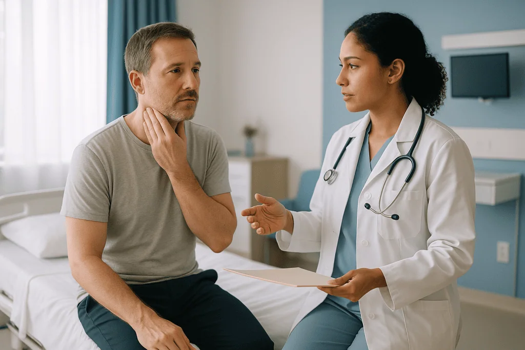
A parotidectomy is the removal of part or all of that gland. A superficial parotidectomy takes the outer lobe while preserving the nerve. A total parotidectomy removes both lobes, again with the nerve carefully dissected free whenever the disease allows. Johns Hopkins Medicine describes the operation as one performed under general anesthesia through an incision that typically runs in front of the ear and curves into the neck.
Why remove a lump that does not hurt? MedlinePlus notes that most salivary gland tumors are benign, meaning they are not cancer, and that the parotid is the most common site. Benign tumors are still removed for good reasons: they keep growing, they can become harder to remove safely as they enlarge, and the most common type, pleomorphic adenoma, carries a small long-term risk of transforming into a malignant tumor if left for many years.
Other indications include confirmed or suspected salivary gland cancer, chronic infection or stones that repeatedly inflame the gland, and occasionally cysts or lymph nodes inside the gland that need diagnosis. In each case the pre-operative work-up is what lets the team explain which version of the operation they are proposing and why.
Who is usually offered parotidectomy, and who is asked to wait?
Most people referred with a solid parotid lump will, in time, be offered surgery. The imaging and biopsy are what move a person from the possibly group into the yes, and here is why group.
Surgery is usually recommended when a mass is solid on ultrasound and the needle sample shows a neoplasm, a word that simply means an abnormal growth of cells, whether benign or malignant. It is also recommended when the biopsy is suspicious or inconclusive but the imaging is worrying, because the only way to settle the diagnosis is to remove the tissue and examine it whole. Mayo Clinic notes that surgery is the main treatment for most salivary gland tumors, with radiation therapy sometimes added for cancers.
Waiting, or watchful monitoring, is discussed in a smaller set of situations. A lump that appeared during an acute infection may be swelling rather than tumor, and the team may re-scan after the inflammation settles. Some benign tumors in older adults or in people with significant heart or lung disease are followed with repeat ultrasound rather than removed, because the risks of an anesthetic may outweigh the risks of a slow-growing lump. A newly diagnosed medical problem found at the anesthesia evaluation, such as uncontrolled blood pressure or a heart rhythm change, can also pause the schedule until it is treated.
Pregnancy is another common reason to defer a non-urgent parotid operation. Delay in these cases is not neglect; it is the team choosing the safer sequence.
What decides the timing is a combination of what the lump looks like, what the cells show, and how the rest of you is doing. The recommendation always rests with the surgeon and anesthesiologist who have examined you.
Ultrasound of the parotid gland: what does the first scan show?
Ultrasound uses sound waves, not radiation, and the parotid sits close enough to the skin that a standard probe sees most of it clearly. The scan takes minutes. Gel goes on the cheek and neck, the probe slides over the lump, and the radiologist or sonographer builds a picture in real time.
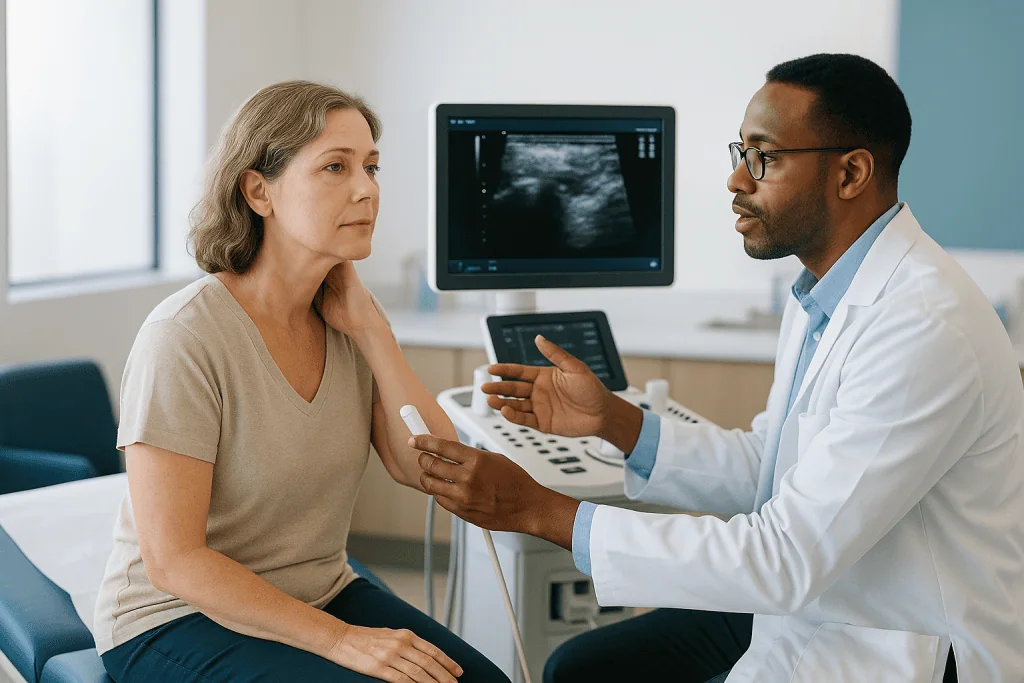
Three findings matter most. First, whether the lump is solid or cystic. A fluid-filled cyst has different implications from a solid growth. Second, its edges. Smooth, well-defined borders are more often seen with benign tumors; irregular or infiltrating margins raise concern. Third, the lymph nodes of the neck. Enlarged or oddly shaped nodes nearby prompt closer investigation, because salivary cancers can spread to them.
Ultrasound also helps the team plan the biopsy. Watching the needle on screen while it enters the lump improves the chance of sampling the right spot and keeps the needle away from vessels.
The scan has limits, and knowing them explains why more imaging often follows. The deep lobe of the parotid lies behind the jawbone, where sound waves cannot reach well. A lump that seems to extend deeper than the probe can see, a very large mass, or any suspicion of cancer are typical reasons a surgeon then requests an MRI or CT. Ultrasound also cannot distinguish tumor types reliably; a Warthin tumor, a pleomorphic adenoma and a low-grade cancer may all appear as similar solid nodules.
Patients sometimes ask whether they can skip straight to the big scan. In practice the ultrasound is rarely wasted: it is fast, it guides the needle, and for a small superficial benign-looking lump it may be all the imaging the surgeon needs.
MRI for parotid tumor vs CT: which imaging is used and why?
When ultrasound leaves questions, the choice is between MRI and CT. Both create cross-sectional pictures of the head and neck; they differ in what they show best and in what they ask of the patient.
MRI, magnetic resonance imaging, uses a strong magnet and radio waves rather than X-rays. It excels at showing soft tissue, so it distinguishes tumor from normal gland, shows the deep lobe clearly, and can suggest whether a mass is pushing on or growing along the facial nerve. Many surgeons prefer it for parotid tumors for that reason. The trade-off is time in a narrow tube, noise, and the need to screen for metal implants or devices.
CT, computed tomography, uses X-rays and is faster. It shows bone well, which matters if a tumor seems close to the jaw or skull base, and it is often the practical choice for people who cannot lie still for long or who have incompatible implants. Contrast dye is commonly given through a vein for either scan; the anesthesia and radiology teams check kidney function and allergy history first.
| Question the surgeon has | Ultrasound | MRI | CT |
|---|---|---|---|
| Solid or cystic? | Very good | Good | Good |
| Deep lobe involvement? | Limited | Very good | Good |
| Relationship to facial nerve? | Limited | Best available | Fair |
| Bone involvement? | Poor | Fair | Very good |
| Radiation exposure? | None | None | Yes |
| Guides the needle biopsy? | Yes, in real time | Rarely | Occasionally |
Neither scan gives a definitive diagnosis on its own. Mayo Clinic lists imaging as one part of the work-up, alongside physical examination and biopsy. The pictures tell the surgeon how to plan the incision and how much dissection to expect; the cells tell them what they are dealing with.
What is a fine needle aspiration parotid gland biopsy like?
The needle used for a fine needle aspiration is thinner than the one used to draw blood. That surprises many people, who imagine something closer to a core of tissue. Instead, a doctor, often a radiologist or pathologist, passes the fine needle into the lump, usually while watching on ultrasound, and moves it gently back and forth to draw cells into the hub. The sample is smeared on slides or placed in fluid and sent to the laboratory.
Local anesthetic may or may not be used; the needle itself is so slim that some centers skip it. The whole procedure typically takes a few minutes, with two or three passes to gather enough cells. Afterward a small dressing goes over the spot. MedlinePlus describes a salivary gland biopsy as a way of removing cells or tissue for examination, and notes that bruising, mild soreness and a small risk of bleeding or infection are the main things to expect.
Why not simply cut a piece out? Because of two problems specific to the parotid. Open incisional biopsy risks scattering tumor cells along the wound track, which is a particular concern with pleomorphic adenoma, a tumor known to recur if its capsule is breached. It also risks injuring the facial nerve before the surgeon has planned the operation. The fine needle sidesteps both.
A parotid gland biopsy by needle is not compulsory in every case, and some surgeons will proceed on imaging alone for a small, classic-looking superficial lump. Most, however, want the cell result. It shapes the conversation about how much gland to remove, whether lymph nodes need to be addressed, and how to counsel you about the facial nerve.
What do fine needle biopsy results mean before parotid surgery?
Cytology, the study of individual cells, gives categories rather than certainties. Understanding the categories takes some of the anxiety out of waiting for the report.
Many laboratories now use a structured reporting system for salivary gland cytology that groups results into tiers: non-diagnostic (too few cells to say), non-neoplastic (inflammation or normal gland), atypia of undetermined significance, benign neoplasm, salivary gland neoplasm of uncertain malignant potential, suspicious for malignancy, and malignant. The middle tiers are the ones that frustrate patients. They do not mean the pathologist was careless; they reflect a genuine feature of salivary tumors, which include several benign and low-grade malignant types that look alike under the microscope.
What does each result do to the plan? A clear benign neoplasm result usually supports a superficial parotidectomy with nerve preservation. A malignant or suspicious result prompts more imaging of the neck, discussion at a multidisciplinary tumor board, and planning for a wider operation that may include lymph nodes. An indeterminate result generally still leads to surgery, because removing the tumor whole is the only way to reach a final diagnosis.
Sometimes the team requests a core needle biopsy instead of, or after, a fine needle sample. A core biopsy uses a slightly larger needle to remove a thin sliver of tissue rather than loose cells, giving the pathologist architecture as well as cells. It is chosen when fine needle results have been non-diagnostic or when lymphoma, a cancer of the immune system that can appear in the parotid, is suspected.
The final answer, called the histology, comes only after surgery, when the whole specimen is examined. Pre-operative cytology is a guide for planning, and your surgeon will frame it that way.
What does the pre-anesthesia evaluation before surgery involve?
Parotidectomy is done under general anesthesia, which MedlinePlus describes as medicines that keep you asleep and free of pain during surgery, with breathing usually supported through a tube in the airway. The pre-anesthesia evaluation exists to make that as safe as possible for you specifically.
It usually begins with questions. The nurse or anesthesiologist asks about heart disease, high blood pressure, diabetes, lung disease including asthma, sleep apnea, reflux, previous operations and how you responded to anesthesia, bleeding tendencies, and family history of problems with anesthetics. They ask what you take, including over-the-counter products and supplements, and about smoking, alcohol and recreational drug use. Honesty here is protective, not judged.
Then comes a focused examination. The airway assessment matters especially for head and neck surgery: how wide you can open your mouth, the state of your teeth, neck movement, and whether anything about the lump itself might make placing the breathing tube harder. A large deep-lobe tumor can push into the throat, so the team looks for that.
Tests are selective rather than routine. Blood counts, kidney function and blood sugar are checked when history or medicines warrant them. An electrocardiogram, a tracing of the heart’s electrical activity, is commonly ordered for older adults or anyone with cardiac risk factors. Chest X-rays are reserved for people with lung symptoms.
The visit ends with practical instructions: fasting times, which medicines to continue on the morning of surgery, and what to bring. If something unexpected turns up, such as a new murmur or poorly controlled blood sugar, surgery may be postponed while it is addressed. That pause is the evaluation doing its job.
Why the facial nerve dominates the conversation before parotidectomy surgery
Ask anyone who has had a parotidectomy what their surgeon talked about most, and the answer is almost always the facial nerve. This is the seventh cranial nerve, which exits the skull just behind the ear, enters the parotid and fans out into five main branches across the face. Every parotidectomy involves finding and protecting it.
Johns Hopkins Medicine explains that the surgeon identifies the nerve early in the operation and traces its branches so the gland can be lifted away from them. Temporary weakness of some facial movements after surgery is common, because handling and stretching a nerve during dissection can stun it for a time. Permanent weakness is uncommon in surgery for benign tumors and is more likely when a cancer has grown into the nerve and part of it must be sacrificed.
Facial nerve monitoring during parotidectomy is widely used. Small electrodes placed in the facial muscles connect to a machine that sounds when a branch is touched or stimulated. Monitoring is an aid rather than a guarantee; the surgeon’s anatomical knowledge remains the main protection.
Why does this belong in a discussion of pre-operative preparation? Because the imaging and biopsy exist partly to predict what the nerve will face. An MRI that shows a tumor hugging the nerve, or a cytology report suggesting malignancy, changes how the surgeon counsels you and may lead to a conversation about nerve grafting or reconstruction.
Before surgery, the team will usually document your baseline facial movement, sometimes with photographs, so any change afterward can be measured. If you already have any asymmetry, mention it; it matters for the record.
Medicines and supplements before parotid surgery: what your team will ask about
Bring an accurate list. Not a memory of the list, the list itself, including anything bought without a prescription. The pre-anesthesia team reviews every item for three concerns: bleeding, interaction with anesthetic drugs, and the effect of fasting on how a medicine behaves.
Bleeding is the first concern. Anticoagulants, medicines that slow clotting, and antiplatelet drugs, which stop platelets from clumping, are common in people with heart disease or previous clots. The team weighs the bleeding risk of continuing against the clotting risk of pausing, often in consultation with the prescribing cardiologist or physician. Whether, when and how to adjust any of these is a decision for those clinicians, never for the patient alone.
Some supplements also affect clotting or interact with anesthesia. Fish oil, vitamin E, ginkgo, garlic extracts and St John’s wort are among those commonly asked about. The NIH Office of Dietary Supplements recommends telling your healthcare providers about all supplements you take, particularly before surgery, precisely because of these interactions.
Diabetes medicines need attention because you will not be eating on the morning of surgery. Insulin and several oral classes are typically adjusted around the fasting window by the prescribing team. Blood pressure medicines are often continued, but a few classes are sometimes held on the day; the anesthesiologist will specify.
Herbal remedies, weight-loss medicines and hormonal therapies all belong on the list too. A newer class of injectable diabetes and weight-management medicines that slow stomach emptying is of particular interest to anesthesiologists because it can affect fasting safety; mention it if it applies to you.
Nothing here should be changed on your own initiative. The rule is simple: disclose everything, then follow the written instructions your surgical and prescribing teams give you.
Parotidectomy preparation: fasting, smoking and the final days before surgery
The last week before a parotidectomy is mostly ordinary life with a few deliberate adjustments. Getting them right lowers your risk in ways that no scan or blood test can.
Smoking is the largest modifiable factor. The NHS advises stopping smoking before an operation because it raises the risk of chest infection, slows wound healing and can affect how anesthetic drugs work. The benefit begins within days of stopping and grows with time, so there is no point at which it is too late to help. Nicotine replacement and support programs are things to discuss with your team rather than to arrange alone.
Alcohol matters too; heavy drinking affects bleeding, liver handling of anesthetics and post-operative confusion. Tell the team honestly how much you drink.
Fasting instructions will be specific to you and take priority over anything written here. As a general guide, the NHS explains that patients are usually asked not to eat for about six hours before general anesthesia and to stop clear fluids about two hours before. The reason is aspiration: stomach contents can enter the lungs while the protective reflexes are switched off. Sipping water until the stated time is usually encouraged rather than forbidden, because a dry, thirsty patient is not safer.
Practical steps round it out. Shower the night before or on the morning as instructed. Leave jewelry, piercings and nail polish at home; the anesthesiologist watches fingertip color and monitors clip to fingers. Arrange someone to take you home and stay the first night. Pack loose, front-fastening tops so you will not have to pull anything over a fresh incision and drain. Bring your medicine list and any inhalers or devices you use.
Finally, keep an eye on your own health. A cold, fever or new cough in the days before surgery should be reported; the team may prefer to reschedule rather than anesthetize someone with an active chest infection.
What do the days and weeks after parotidectomy usually look like?
Knowing the shape of recovery makes the preparation feel purposeful. Johns Hopkins Medicine describes parotidectomy as an operation after which many people stay in hospital overnight, with a small drain placed under the skin to prevent fluid collecting. The drain typically comes out within a day or two, once the output falls, and the incision is closed with fine stitches or glue hidden in the skin crease in front of the ear.
The first week brings swelling, bruising and a tight feeling along the jaw. Numbness of the earlobe is very common because a sensory nerve, the greater auricular nerve, runs across the surgical field and is often stretched or divided; the earlobe may stay numb for months, and in some people permanently. Any facial weakness that is present usually shows itself immediately; if it is due to nerve stretch rather than injury, movement returns gradually over weeks to months.
Most people return to desk work and light activity within a couple of weeks, though your surgeon will give a range based on your operation. Heavy lifting and strenuous exercise are usually held longer to protect the wound.
Two later effects are worth knowing about. Frey syndrome is sweating or flushing of the cheek while eating, caused by regrowing salivary nerve fibers connecting to sweat glands in the skin; it can appear months later and is usually mild. First bite syndrome is a sharp pain in the jaw at the start of a meal, more often after deep-lobe surgery, which tends to lessen over time.
The final pathology report returns in the weeks after surgery. That appointment is where the pre-operative cytology is confirmed or refined, and where any further treatment, if needed, is discussed.
What people often get wrong about parotid lumps and surgery
A painless lump means it is harmless. Not reliably. Most parotid tumors are benign, as MedlinePlus notes, but benign and malignant lumps are often equally painless. Pain, rapid growth, skin changes or facial weakness raise concern, yet their absence does not settle the question. That is what imaging and biopsy are for.
The needle biopsy will spread cancer. This fear comes from older experience with open surgical biopsies, which do carry a seeding risk in the parotid. The fine needle used today is thin enough that tumor spread along its track is considered very rare, and it is the reason needle sampling replaced incisional biopsy in this gland.
If the biopsy says benign, surgery is unnecessary. A benign cytology result lowers concern but does not remove the reasons for surgery: continued growth, the small long-term risk of malignant change in pleomorphic adenoma, and the fact that cytology is a guide rather than the final diagnosis. Some benign tumors are indeed observed, but that is a shared decision, not an automatic consequence of the report.
Everyone loses facial movement afterward. Temporary weakness of one or more branches is common; lasting weakness after surgery for benign disease is uncommon. The surgeon will give you their honest assessment based on your imaging, not a blanket figure.
Removing a salivary gland leaves you with a dry mouth. There are three pairs of major salivary glands plus hundreds of minor ones. Losing one parotid, or part of one, rarely produces a noticeable change in saliva.
The scan can tell what kind of tumor it is. Imaging shows size, depth and relationships. Only cells and tissue name the tumor, and the definitive name comes after it is removed.
Questions to ask your care team before parotidectomy
A good pre-operative consultation is a conversation, not a briefing. These questions help you steer it toward what matters to you.
- What did my ultrasound and MRI or CT show about the size and depth of the lump, and does it involve the deep lobe?
- What category did my fine needle aspiration fall into, and how confident are you in that result? Would a core biopsy add anything?
- Are you planning a superficial or total parotidectomy, and what would change that plan during the operation?
- How does the tumor sit in relation to the facial nerve on my scans, and what is your realistic assessment of temporary and lasting weakness in my case?
- Will facial nerve monitoring be used, and will my baseline facial movement be documented beforehand?
- Which of my medicines and supplements should I continue, and who will give me written instructions about any that need adjusting?
- What are my fasting times, and can I sip water until the stated hour?
- Would watchful waiting with repeat imaging be a reasonable option for me, and what would you want to see to change that advice?
- How long do you expect me to stay in hospital, and how is the drain managed?
- What sensations should I expect around the ear and cheek in the first months, and which ones would you want to hear about promptly?
- When will the final pathology be available, and who will explain it to me?
- If the final report shows cancer, what are the possible next steps and who would be involved?
Write the answers down or bring someone to listen with you. The details about nerve risk and the plan for indeterminate biopsy results are the ones people most often wish they had asked about more carefully.
When to call your doctor
Some symptoms should not wait for the next scheduled appointment, whether they arise before or after your operation.
Before surgery, contact your team promptly if the lump grows quickly over days to weeks, becomes painful, or the skin over it reddens, breaks down or feels hot. Report any new weakness or drooping on that side of the face, difficulty closing the eye, numbness of the cheek, or trouble swallowing or opening the mouth. These signs can point to a tumor involving the nerve or growing inward and may change how urgently the team wants to proceed. Also let them know about fever, a new cough or chest infection, a fall, or any hospital visit in the run-up to the anesthetic.
After a fine needle biopsy, call if swelling at the site keeps enlarging rather than settling, if bleeding does not stop with firm pressure, or if the area becomes increasingly red, hot and tender over the following days.
After parotidectomy, seek help urgently for rapidly increasing swelling under the wound, which may indicate bleeding beneath the skin; for difficulty breathing or swallowing; for a fever with spreading redness or pus from the incision; or for a new inability to close the eye on the operated side, which risks damage to the cornea and needs same-day advice about eye protection. Sudden, severe facial weakness that was not present when you left hospital, calf pain or swelling, and chest pain or breathlessness should also be treated as emergencies.
If anything about the wound, your face or your general health feels wrong and you cannot reach your surgical team, use your local urgent or emergency service. The team would always prefer an unnecessary call to a missed complication.
Frequently asked questions
How long does it take to get from a parotid lump to surgery?
There is no fixed interval; it depends on how quickly the ultrasound, any MRI or CT, the fine needle biopsy and the anesthesia evaluation can be scheduled and reviewed together. Suspected cancers are generally fast-tracked, while a small benign-appearing lump may follow a more routine timeline. Ask your surgical team what their expected pathway looks like for your situation rather than relying on general figures.
Is a fine needle aspiration parotid biopsy painful?
Most people describe it as a brief sting or pressure rather than significant pain, because the needle is thinner than the one used for blood tests. Some centers use a little local anesthetic and others do not. Mild soreness and a small bruise at the site are common for a day or two. MedlinePlus lists bleeding and infection as uncommon risks of salivary gland biopsy.
Do I always need an MRI for a parotid tumor before surgery?
Not always. For a small, superficial, benign-appearing lump, some surgeons operate on ultrasound and biopsy findings alone. MRI is usually requested when the lump seems large or deep, when the ultrasound cannot see its full extent, when cancer is suspected, or when the surgeon wants a clearer view of how the tumor sits relative to the facial nerve. CT may substitute if MRI is unsuitable for you.
What if my biopsy result is indeterminate?
Indeterminate results are common in salivary cytology because several benign and low-grade malignant tumors look similar under the microscope. An indeterminate result usually still leads to surgery, since removing the tumor whole is the only way to reach a definitive diagnosis. Your team may discuss a repeat aspiration or a core needle biopsy first, and will explain how the uncertainty shapes the planned operation.
Will I have to stop my blood thinner before parotidectomy?
That decision belongs to your surgical team together with the clinician who prescribes the medicine, and it depends on why you take it and your individual clotting risk. Anticoagulants and antiplatelet drugs are reviewed at the pre-anesthesia evaluation, and you will receive written instructions. Do not adjust or pause any prescribed medicine on your own; disclose everything you take and follow the plan you are given.
What does parotidectomy preparation involve on the day before surgery?
Typically it means following your written fasting instructions, showering as directed, removing jewelry and nail polish, confirming who will take you home and stay overnight, and packing your medicine list along with loose front-fastening clothes. The NHS advises stopping smoking before any operation because it raises the risk of chest infection and slows healing. Report any fever, cough or new illness to your team.
Is facial nerve monitoring used in every parotidectomy?
It is widely used but not universal, and practice varies between surgeons. Monitoring involves small electrodes in the facial muscles that alert the surgeon when a nerve branch is stimulated. It is an aid to the surgeon’s anatomical dissection rather than a guarantee against injury. Asking whether monitoring will be used, and how your baseline facial movement will be recorded, is a reasonable pre-operative question.
Why might my surgeon want a core biopsy instead of a fine needle sample?
A core biopsy removes a thin sliver of tissue rather than loose cells, so the pathologist can see how the cells are arranged. It is chosen when fine needle results have been non-diagnostic, or when lymphoma is suspected, because lymphoma is classified by tissue architecture and special stains. It uses a slightly larger needle under ultrasound guidance and is still far less invasive than an open biopsy.
Can I drink water before general anesthesia?
Usually yes, up to a stated cut-off. The NHS explains that patients are generally asked to stop clear fluids about two hours before a general anesthetic and food about six hours before, though your own instructions take priority. Sipping water until the permitted time is often encouraged because dehydration does not make anesthesia safer. Milk, juice with pulp and anything solid count as food.
Does removing part of the parotid cause a dry mouth?
Rarely to a noticeable degree. You have three pairs of major salivary glands plus hundreds of minor glands lining the mouth, so losing one parotid or part of one is usually compensated for by the rest. A more common change after surgery is numbness of the earlobe from the greater auricular nerve, and occasionally cheek sweating while eating, known as Frey syndrome.
References
- MedlinePlus: Salivary gland tumors
- MedlinePlus: General anesthesia
- NHS: Having an operation (surgery): Preparation
This article is for general information only and is not a substitute for professional medical advice. Please consult a qualified doctor about your individual situation.

More from the Blog
Surgical Oncology Preparation: Staging Scans, Blood Tests and the Anesthesia Assessment
Preparing for cancer surgery usually means three things happening in parallel: staging scans such as CT, MRI or PET-CT to map where the cancer…
What Neck Cancer Follow-Up Looks Like: Physical Exams, Imaging and Side-Effect Reviews
Follow-up after neck cancer treatment usually combines a hands-on physical exam of the neck, mouth and throat at each visit, a camera check of…
Follow-Up After Cervical Cancer Treatment: Pelvic Exams, Imaging and Recurrence Checks
Cervical cancer follow up after treatment usually combines a review of symptoms, a pelvic exam and, depending on the treatment given, cytology or HPV…
What a Chemotherapy Cycle Looks Like: Treatment Days, Low-Count Days and Recovery
A chemotherapy cycle is one treatment session (or a short run of sessions) followed by a planned rest period, commonly repeating every one to…
Living With Multiple Myeloma: Bone Protection, Kidney Care and Staying Active Safely
People living with multiple myeloma can protect their bones and kidneys through a combination of bone-strengthening medicines chosen by their hematology team, regular dental…
When Radiotherapy Is Added to Lymphoma Treatment and When Chemotherapy Alone Is Enough
Radiotherapy is usually added after chemotherapy for lymphoma when disease was localized or bulky at diagnosis, or when a post-treatment PET scan still shows…