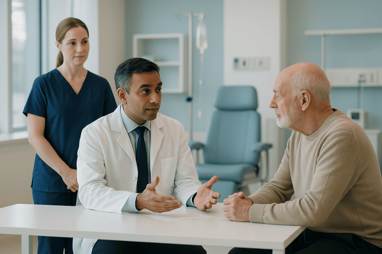
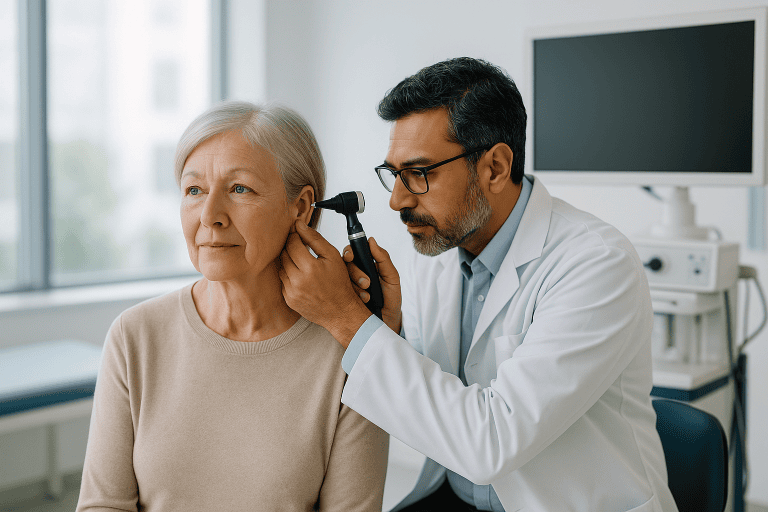
Parotidectomy Surgery
Parotidectomy surgery removes part or all of the parotid salivary gland, most often to treat benign or malignant tumors while protecting facial nerve function.
Quick answer
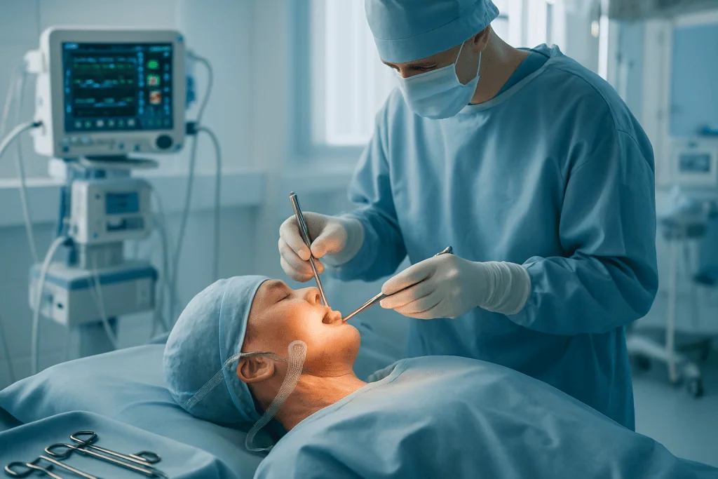
Parotidectomy is an operation to remove part or all of the parotid salivary gland, most often because of a benign or malignant tumour. Performed under general anaesthesia through an incision in front of the ear, it centres on identifying and protecting the facial nerve, which runs through the gland. Extent ranges from superficial removal of the outer lobe to total gland removal, sometimes with neck dissection.
Parotidectomy: What the Operation Is and Why It Is Recommended
Parotidectomy is the surgical removal of part or all of the parotid gland: the largest of the salivary glands, which sits in front of and below each ear and extends towards the angle of the jaw. It is performed most often to remove a tumour of the gland, benign or malignant, and occasionally to treat chronic infection, inflammatory disease or recurrent cysts. Because the facial nerve travels directly through the gland, every parotidectomy is planned around one central priority: removing the disease while protecting the nerve that moves your face.
A recommendation for parotidectomy usually arrives after a period of uncertainty. Perhaps you noticed a painless lump near the jaw, or a swelling in front of the ear that has slowly changed shape. Perhaps an ultrasound picked something up incidentally, or a biopsy report raised the question of whether the growth is benign or malignant. For many patients, the medical question is only part of the decision. There is understandable anxiety about the facial nerve, the prospect of a visible scar, and possible changes in facial movement.
The anatomy explains the anxiety. The facial nerve leaves the skull base just behind the ear, enters the parotid gland as a single main trunk, and divides within the gland into the branches that control the forehead, the eyelids, the cheek, the lips and the lower face. The gland is conventionally described as having a superficial lobe and a deep lobe, with the nerve running in the plane between them. That intimate relationship is the single biggest reason a parotidectomy demands careful planning and an experienced surgical team rather than a quick excision.
Most parotid tumours are benign, but they rarely disappear on their own. Some continue to grow, become harder to remove cleanly, or, in certain tumour types, can undergo malignant change over time. Malignant parotid tumours need timely, coordinated treatment because they can involve nearby nerves, lymph nodes, skin or jaw structures. The goal of care is never simply to remove a mass. It is to treat the disease appropriately while preserving facial movement, facial appearance, comfort and quality of life as far as possible.
At Acibadem, parotidectomy sits within a structured diagnostic and treatment pathway. Ear, nose and throat specialists, head and neck surgeons, radiologists, pathologists, oncologists, reconstructive surgeons, anaesthesiologists and rehabilitation teams work from the same imaging, the same biopsy findings and the same plan, so that the extent of surgery matches the disease, no more and no less.
Parotic meaning: where the name comes from
If you have looked up the parotic meaning, the answer sits in the Greek: para, meaning beside, and otos, meaning ear, so “parotic” and “parotid” both describe something situated beside the ear. That is precisely where the gland lies, wrapped around the back of the jaw just in front of the ear canal and earlobe. The suffix -ectomy means surgical removal, so parotidectomy translates literally as removal of the gland beside the ear. The word is worth understanding because it explains almost everything else about the operation: the placement of the incision around the ear, the closeness of the facial nerve, the numbness patients often notice near the earlobe afterwards, and the care the surgeon must take in a small, crowded anatomical space.
What Is Parotidectomy Surgery?
Parotidectomy surgery is an operation to remove part or all of the parotid salivary gland, most often to treat a tumour of the gland. That includes benign tumours such as pleomorphic adenoma and Warthin tumour, as well as malignant salivary gland cancers. It may also be used in selected cases of chronic infection, inflammatory disease or recurrent parotid cysts, although tumours remain by far the most common reason.
The type of parotidectomy depends on the location, size and behaviour of the lesion, and above all on its relationship to the facial nerve:
- Superficial parotidectomy removes the outer portion of the gland, where many benign tumours develop. It is the most frequently performed version of the operation.
- Partial parotidectomy removes only the segment of gland containing the tumour, together with a cuff of healthy tissue, and may be appropriate for smaller, well-placed lesions.
- Extracapsular dissection removes a carefully selected small benign tumour with a narrow margin, without formally dissecting the full nerve. It is reserved for specific anatomical situations and depends heavily on surgeon judgement.
- Total parotidectomy removes both the superficial and deep portions of the gland, which requires dissection on both sides of the facial nerve branches.
- Extended parotidectomy is used in cancer surgery when involved skin, lymph nodes, soft tissue, nerve segments or surrounding structures must also be removed.
A defining feature of every parotidectomy is identification and preservation of the facial nerve whenever it is oncologically safe. The surgeon locates the main trunk of the nerve, or its individual branches, and separates the gland tissue and tumour from the nerve under magnification, millimetre by millimetre. In most benign disease the nerve can be preserved intact. If a malignant tumour has invaded the nerve, the plan becomes more complex: the team may need to remove an involved segment and reconstruct it, balancing cancer control against function and planning rehabilitation from the outset.
Parotidectomy is performed under general anaesthesia. The incision is usually designed along natural skin creases in front of the ear, curving around the earlobe into the upper neck, which gives the surgeon full access while allowing the scar to mature in a discreet position. Depending on the diagnosis, surgery may be combined with neck dissection, reconstructive procedures or later radiotherapy. One practical reassurance is worth stating plainly: removing one parotid gland does not normally cause a dry mouth, because the remaining salivary glands: the opposite parotid, the submandibular and sublingual glands, and hundreds of minor glands, continue producing saliva.
The operation is usually carried out by ear, nose and throat or head and neck surgeons: the same specialty that performs ear and airway procedures such as mastoid surgery and adenoidectomyoften working alongside reconstructive and oncology colleagues when the case requires it.
Parodectomy and other spellings you may see
Parodectomy is a common misspelling of parotidectomy; both words refer to the same operation on the parotid gland. You may also encounter variants such as “parotectomy” in older texts, or descriptive phrases such as “excision of parotid lesion” in operative notes. The terminology matters far less than the substance. Whatever spelling appears in your paperwork, the questions that actually shape your treatment are the same: which lobe of the gland is involved, how much tissue is planned for removal, and how the facial nerve will be protected.
Who May Need Parotid Surgery?
Parotid surgery is considered when a mass or abnormality in the parotid gland needs removal, for definitive diagnosis, for symptom relief, to prevent further growth, or as cancer treatment. Most parotid tumours first appear as a lump in front of the ear, below the ear or near the angle of the jaw. The lump is often painless and slow-growing, which makes it tempting to ignore. The difficulty is that physical examination alone cannot reliably tell a benign tumour from a malignant one, which is why specialists investigate even a quiet, comfortable lump rather than watching it indefinitely.
Findings that typically lead to evaluation include a visible or palpable swelling near the jaw, fullness in the cheek, pain or tenderness around the gland, repeated swelling during meals, skin changes over the mass, numbness, ear discomfort, difficulty opening the mouth, or enlarged lymph nodes in the neck. Facial weakness, difficulty closing the eye, drooping at the corner of the mouth, reduced forehead movement or an uneven smile, is treated by clinicians as a significant finding, because it can indicate that the nerve is under pressure or, less commonly, directly involved by tumour.
Diagnosis begins with a clinical examination of the head and neck, including formal assessment of facial nerve movement. Imaging then defines the lesion. Ultrasound is useful for superficial masses and for guiding needle biopsy. Magnetic resonance imaging is often the key study, showing the relationship between the tumour, the deep lobe of the gland, the expected course of the facial nerve and the surrounding soft tissues. Computed tomography helps when bone involvement, calcification, deep extension or neck disease is suspected, and in selected cancer cases further imaging is used to evaluate lymph nodes or possible distant spread.
Tissue diagnosis is usually obtained with fine-needle aspiration or core needle biopsy, both performed through the skin, often under ultrasound guidance. These tests help distinguish benign from malignant disease and shape the operative plan. Sometimes the result is indeterminate, the cells cannot be confidently classified, and in that situation parotidectomy serves two purposes at once: it treats the lesion and provides the whole specimen for definitive examination by pathologists.
Parotidectomy may therefore be recommended for a growing benign tumour, a mass with uncertain biopsy findings, a suspected or confirmed salivary gland cancer, a recurrent tumour after previous treatment, or a lesion causing pain, repeated infection, pressure symptoms or cosmetic concern. The decision is individual. A small benign tumour in a medically complex older patient may reasonably be observed, while the same tumour in a younger patient, who would otherwise live with it, and with its slow growth, for decades, is usually removed.
How serious is a parotid tumor?
A parotid tumor can be anything from a slow-growing benign lump that has been present for years to an aggressive cancer, and its seriousness can only be judged from imaging and biopsy, never from how the lump feels or how long it has been there. Benign tumours are more common than malignant ones, but “benign” does not mean “ignorable”: several benign types keep enlarging, and pleomorphic adenoma in particular carries a small long-term possibility of malignant change. Malignant parotid tumours vary enormously among themselves; some low-grade cancers behave almost indolently, while high-grade subtypes can spread to nerves and lymph nodes. Features that raise concern include rapid growth, pain, a mass fixed to skin or deeper tissue, skin ulceration, facial weakness and enlarged neck nodes, although none of these proves cancer on its own. Final answers come from pathology, which is why obtaining tissue and, when needed, removing the tumour completely matters so much.
Conditions Treated: Benign and Malignant Parotid Neoplasms
On imaging requests, coding systems and pathology indexes you may see the entry written as neoplasm parotid, plain shorthand for a new growth, benign or malignant, arising in the parotid gland, and the most frequent reason a parotidectomy is performed. Both categories deserve careful assessment, because early on they can look and feel remarkably similar.
Pleomorphic adenoma is one of the most common benign parotid tumours. It typically grows slowly, sometimes over many years. Surgical removal is generally recommended because continued growth makes later surgery more complex, and because a small risk of malignant transformation accumulates over time. Complete removal with an intact capsule and a margin of healthy gland tissue is important, since rupture or spillage of tumour contents during surgery raises the chance of recurrence, one of the technical reasons experience matters in this operation.
Warthin tumour is another benign tumour, seen more often in older adults and associated with a smoking history. It can occur on both sides, or in more than one site within the same gland. Management is tailored to size, symptoms, diagnostic certainty, general health and patient preference: many patients undergo removal, while selected patients with a secure diagnosis and a stable, untroubling tumour may reasonably be observed.
Malignant salivary gland tumours include mucoepidermoid carcinoma, adenoid cystic carcinoma, acinic cell carcinoma, salivary duct carcinoma, carcinoma ex pleomorphic adenoma and a range of rarer cancers. Treatment usually centres on surgery; some patients also need neck dissection and radiotherapy afterwards. The type and extent of the operation depend on tumour grade, size, location, nerve involvement, lymph node status and whether disease extends beyond the gland. Tumours reaching towards the jaw or the deep spaces of the face may bring colleagues from oral and maxillofacial surgery into the planning.
Parotidectomy is also used for metastatic disease in intraparotid lymph nodesthe gland contains lymph nodes that can receive spread from skin cancers of the scalp, face or ear region. These cases are planned jointly across dermatology, head and neck surgery, pathology, radiation oncology and medical oncology, because the parotid operation is only one part of the overall treatment.
Less commonly, parotidectomy is considered for chronic inflammatory disease, recurrent abscesses, stones with irreversible gland damage, cysts or other benign conditions that keep causing symptoms despite conservative treatment. These cases demand careful selection: operating near the facial nerve carries specific risks, and surgery is only justified when the expected benefit clearly outweighs them.
How the Parotidectomy Operation Is Performed
The parotidectomy operation begins long before the day of surgery, because surgical safety depends on understanding the tumour, the facial nerve, the patient’s general health and the likely aftercare needs. Existing records are typically reviewed in advance, imaging studies, biopsy reports, pathology slides where available, medication lists and previous operative notes if there has been earlier surgery on the same side.
During the preoperative assessment, the surgeon examines the face and neck and formally documents facial nerve function: raising the eyebrows, closing the eyes tightly, smiling, puffing the cheeks, moving the lower lip. Any weakness before surgery matters, because it may point to tumour pressure, inflammation or malignant nerve involvement, and it changes both the surgical conversation and the operative plan. The team also maps the tumour on imaging in relation to the superficial and deep lobes, the jaw, the ear canal, the skin, the major vessels and the lymph nodes of the neck.
Preparation covers the practical details: fasting instructions, anaesthetic assessment, and any additional tests such as blood work, an electrocardiogram or specialist clearance depending on age and health. Patients taking blood thinners or certain supplements need an individual plan around the operation: a decision that belongs entirely to the treating doctors, who balance bleeding risk against cardiovascular safety; no medicine should ever be adjusted without that guidance. When cancer is suspected, the case is usually reviewed by a multidisciplinary tumour board so that surgery, pathology, radiotherapy and systemic treatment decisions are aligned before the first incision.
On the day itself, parotidectomy is performed under general anaesthesia with the patient positioned for safe access to the side of the face and neck. Many teams use intraoperative facial nerve monitoring, fine electrodes that give real-time feedback when instruments work near nerve branches. Monitoring never replaces surgical expertise, but it adds a layer of information during the most delicate stages. A typical operation proceeds in these steps:
- 1. Incision: a curved incision in front of the ear, around the earlobe and into a natural crease of the upper neck.
- 2. Flap elevation: the skin is carefully lifted off the gland to expose the operative field.
- 3. Landmark identification: the surgeon identifies the anatomical landmarks that point to the main trunk of the facial nerve near the ear cartilage and mastoid region.
- 4. Nerve identification: the nerve trunk, or, in selected approaches, an individual branch, is found and confirmed, often with the help of the monitor.
- 5. Dissection: the gland tissue containing the tumour is separated from the nerve branches under magnification, keeping the tumour capsule intact.
- 6. Removal: the superficial lobe, a partial segment or the entire gland is removed according to the plan; in cancer surgery, margins of healthy tissue are taken where feasible.
- 7. Haemostasis and drain: bleeding points are controlled and a small drain is often placed to prevent fluid collecting under the skin.
- 8. Closure: the incision is closed in layers, sometimes with a light pressure dressing.
If cancer involves the neck lymph nodes, or the risk of nodal spread is significant, a neck dissection is performed during the same anaesthetic, removing selected node groups for both treatment and staging. If skin, soft tissue or a nerve segment must be sacrificed because of tumour invasion, reconstruction is planned, coverage, facial contour, nerve grafting or nerve transfer, eyelid procedures, and in some cases staged facial reanimation carried out with colleagues in plastic and reconstructive surgery. Everything removed goes to pathology, where specialists establish the tumour type, examine the margins and any lymph nodes, and identify features that determine whether further treatment is needed.
Hospital stay depends on the extent of surgery and the patient’s condition. Many patients having a straightforward superficial parotidectomy stay one or two nights, mainly for drain management and monitoring; complex cancer surgery, neck dissection or reconstruction extends the stay. Early recovery focuses on wound care, drain output, eating and drinking comfortably, protecting the eye if there is any temporary weakness, and a gradual return to activity while final pathology is awaited.
How long does a parotidectomy surgery take?
A straightforward superficial parotidectomy for a small benign tumour usually takes a few hours, surgeons commonly quote a window of roughly two to four hours of operating time. Total parotidectomy, deep lobe tumours, revision surgery after previous operations, cancer resections, neck dissection and reconstruction each add time, and complex combined procedures can run considerably longer. The duration reflects the nature of the work rather than anything going wrong: the surgeon is dissecting gland tissue off individual nerve branches under magnification, and that cannot be hurried. Add time for anaesthetic preparation beforehand and the recovery unit afterwards, so the day is longer than the operating time alone suggests.
Is a parotidectomy a major surgery?
Yes, parotidectomy is classed as major surgery, because it requires general anaesthesia and meticulous dissection around the facial nerve, and because the consequences of imprecision involve facial movement. At the same time, “major” describes the precision the operation demands, not necessarily a long or difficult hospital experience: for benign disease, many patients are walking and eating the same evening and home within a night or two. For experienced head and neck teams this is a routine, well-standardised procedure with an established sequence of safety checks. The sensible conclusion for a patient is not alarm but selectivity: this is an operation where the experience of the surgical team genuinely matters.
Why Acting Early Matters
Early evaluation of a parotid mass keeps treatment simpler. A smaller tumour is easier to characterise on imaging and easier to remove while preserving normal structures. Delay allows a benign tumour to enlarge, extend into the deep lobe, stretch the surrounding tissue and nerve branches, and turn a contained operation into a more complex dissection with a longer incision. In pleomorphic adenoma specifically, long-standing tumours raise additional concern about malignant change, particularly when growth accelerates or new symptoms appear.
For malignant tumours, delay risks spread within the gland, involvement of the facial nerve, extension into skin or deeper tissue, and spread to lymph nodes, each of which can enlarge the operation, add treatments or narrow the options. Some salivary gland cancers grow slowly and others aggressively, and because behaviour varies so widely by subtype and grade, timely diagnosis is the safeguard. Acting promptly does not mean rushing: it means moving through the right diagnostic steps efficiently, so that decisions are made with complete information rather than under pressure.
Facial weakness, pain, numbness, rapid growth, skin ulceration, a fixed mass or enlarged neck nodes are the findings specialists prioritise. They do not always mean cancer, inflammation and benign disease can mimic several of them, but they change the tempo of the diagnostic work-up. The practical reward of early assessment is choice: the chance to have the least extensive appropriate operation, with less need for reconstruction or combined treatment.
Potential Benefits of Parotidectomy Surgery
The benefits of parotidectomy depend on the diagnosis, but the operation is designed to do three things at once: remove the disease, establish the pathology definitively, and protect long-term facial function wherever possible.
| Benefit | What It Means for You |
|---|---|
| Removal of the parotid tumour | The mass is removed for treatment and full pathological examination, giving a definitive diagnosis and a clear basis for next steps. |
| Facial nerve-focused surgery | The operation is planned around identifying and preserving facial nerve branches when medically safe, protecting movement and expression. |
| Reduced risk from tumour growth | Removing an enlarging benign tumour prevents future distortion, deeper extension and a more difficult operation later. |
| Appropriate cancer control | For malignant tumours, surgery removes the primary cancer and provides staging information that guides radiotherapy or other treatment if needed. |
| Improved comfort and appearance | Relief from swelling, pressure, repeated infection or visible facial asymmetry caused by the lesion. |
| Personalised follow-up planning | Final pathology determines surveillance frequency, imaging needs, rehabilitation and any additional therapy. |
Recovery After Parotidectomy
Recovery varies with the extent of surgery, the tumour type, how the facial nerve responds, and whether neck dissection or reconstruction was performed, but most patients follow a recognisable pattern.
| Time Period | What Patients Can Expect |
|---|---|
| Day 1 | Monitoring after anaesthesia. Mild to moderate discomfort, tightness, numbness near the ear and a drain are common. Facial movement is checked regularly. |
| First week | Swelling and bruising begin to settle. The drain, if used, is removed once output falls. Wound care instructions and activity limits apply. |
| First month | Most daily activities resume gradually. Numbness around the earlobe or cheek may persist. Any temporary facial weakness is monitored and may need eye care or therapy. |
| Longer term | The scar matures over months. Final pathology guides surveillance or additional treatment. Some patients notice sweating or flushing over the cheek during meals, which can be treated if bothersome. |
Two practical points deserve emphasis. First, eye care: if eyelid closure is temporarily weak, protecting the eye with lubrication and simple measures prevents irritation while the nerve recovers. Second, patience with sensation: numbness around the earlobe is expected because small sensory nerves cross the incision line, and feeling typically improves slowly over months rather than weeks.
How long does it take to recover from a parotidectomy?
Most people return to desk-based work and light daily routines within about one to two weeks of a standard parotidectomy, while swelling, numbness and the appearance of the scar continue to settle over subsequent months. The drain usually comes out within the first days. Strenuous exercise and heavy lifting wait until the wound has healed securely and the surgical team agrees. Where temporary facial weakness occurs, its recovery follows the nerve’s own timetable, often weeks, sometimes months, and facial physiotherapy can support the process. Recovery after cancer surgery with neck dissection or reconstruction is naturally longer and is planned as its own pathway.
How long does it take to recover from a partial parotidectomy?
Recovery after a partial or superficial parotidectomy is usually quicker and more comfortable than after total parotidectomy, because less tissue is removed and the dissection around the deeper facial nerve branches is more limited. The hospital stay is short, the drain is typically removed early, and everyday activities resume over days rather than weeks. The same long-term rhythms still apply: numbness near the earlobe fades gradually, the scar softens and pales over months, and follow-up is arranged around the final pathology result. The honest caveat is that “partial” describes the amount of gland removed, not a guarantee of an effortless course, individual healing varies, and the surgical team’s instructions remain the reference point.
What Influences Outcomes and a Good Result?
Outcomes after parotidectomy are shaped by several medical and technical factors, and tumour type is the most important. Benign tumours carry a different recovery and follow-up plan from malignant ones. Among cancers, grade, stage, margins, lymph node involvement, nerve invasion and pathological subtype all influence treatment recommendations and long-term outlook.
Size and location matter too. A small superficial tumour away from the major nerve branches is generally less complex than a deep lobe tumour, a recurrent tumour or one lying directly against the nerve. Prior surgery, previous radiation, infection, scarring and anatomical variation each increase difficulty. Large tumours may have stretched the facial nerve over time, which makes temporary postoperative weakness more likely even when the nerve is fully preserved.
Facial nerve function before surgery is an important predictor. Patients with normal movement before a benign tumour operation usually do well, although temporary weakness can still occur. Weakness that exists before surgery, particularly in suspected cancer, may mean the nerve is already involved, and the team must then balance nerve preservation against complete disease removal, discussing that balance openly beforehand.
The quality of imaging, pathology and planning contributes directly to a safer operation. High-resolution imaging defines the anatomy and anticipates challenges; biopsy interpretation by experienced pathologists calibrates the extent of surgery; and in cancer, multidisciplinary discussion coordinates surgery with radiation oncology, medical oncology, radiology, reconstruction and rehabilitation from the start rather than after the fact.
Patient factors complete the picture. Smoking, diabetes, immune suppression, nutritional status and cardiovascular or lung disease affect healing and anaesthetic risk. So does following the postoperative plan: protecting the incision, pacing activity and attending follow-up. A good result is measured on several axes at once, complete and appropriate removal of disease, preserved or recovering facial movement, a well-healed incision, accurate pathology and a clear follow-up plan; for cancer patients, it also means timely coordination of any additional treatment.
Risks and Possible Side Effects
Every operation has risks, and parotidectomy has specific ones because of the facial nerve and the gland’s position. The most discussed is facial weakness. Temporary weakness, from stretching or irritation of nerve branches during dissection, is the more common form and generally improves over time as the nerve settles. Permanent weakness is less common but possible, particularly with large tumours, revision surgery, deep lobe disease, significant inflammation or cancer directly involving the nerve. This is the risk your surgeon should discuss in most detail, in the context of your specific anatomy.
Numbness around the earlobe, ear and upper neck is expected to some degree, because the small sensory nerves in the area, notably the greater auricular nerve, cross the surgical field. Sensation usually improves, though a patch of altered feeling can persist. Other recognised risks include bleeding, haematoma, infection, a collection of salivary fluid under the skin (sialocele), temporary leakage of saliva through the wound (salivary fistula), delayed healing, a visible scar, a contour depression where gland tissue was removed, and ear discomfort.
Some patients later develop Frey syndrome: sweating, warmth or flushing of the cheek skin during meals. It occurs when regenerating nerve fibres that once served the gland connect instead to sweat glands in the overlying skin. Many cases are mild and need nothing; bothersome cases can be treated. A smaller number of patients describe first-bite syndromea brief, sharp pain in the parotid area at the first bite of a meal, easing with subsequent bites, which also tends to soften over time and can be managed if persistent.
Where eyelid closure is weak after surgery, the eye needs active protection: lubricating drops, ointment, taping at night, or in selected cases temporary eyelid weights and specialist care. If lower facial branches are weak, speech clarity, smile symmetry and eating can be affected for a period; facial physiotherapy and rehabilitation help selected patients regain control and coordination. The essential point is proportion: a benign superficial tumour carries a very different risk profile from an aggressive cancer wrapped around the nerve, and an honest preoperative conversation should set out your individual profile, not a generic list.
How Acibadem Organises Parotidectomy Care
Parotid surgery involves more than the operation itself. It depends on a reliable diagnosis, a clear surgical plan, trustworthy pathology, and continuity of care after discharge. Acibadem’s approach is built around that whole sequence rather than the operating day alone.
Clinically, care is delivered by physicians experienced in salivary gland and head and neck disease, with multidisciplinary tumour boards convened for cancer and complex cases, bringing together head and neck surgeons, radiologists, pathologists, radiation and medical oncologists, reconstructive surgeons and rehabilitation professionals. Diagnostic pathways use high-resolution ultrasound, MRI and CT as indicated, with image-guided biopsy where it changes the plan. In theatre, magnified dissection, facial nerve monitoring, meticulous haemostasis and pre-agreed reconstructive options support work around delicate anatomy; the specific techniques are chosen for the individual case, not applied as a package.
Two patients with a similar lump may need entirely different operations, one a limited superficial parotidectomy for a benign tumour, another a total parotidectomy with neck dissection and postoperative radiotherapy, a third revision surgery for a recurrence with reconstruction planned from the outset. Matching the extent of treatment to the diagnosis, avoiding both undertreatment and unnecessary intervention, is the organising principle. Records are reviewed before decisions are made, and discharge includes written wound care guidance, medication instructions from the treating doctor, the pathology report, and a documented follow-up plan, so that any further care continues from a clear record rather than from scratch.
Preparing for a Consultation or Second Opinion
A consultation about parotidectomy is most productive when the surgical team can review complete information. Useful material includes the actual ultrasound, MRI, CT or PET-CT images, not only the written reports, plus biopsy results, pathology slides or blocks where obtainable, previous operative notes if there has been earlier surgery, a current medication list and a summary of medical conditions. If facial movement has changed over time, photographs or short videos documenting it are genuinely helpful.
Questions worth asking the surgeon include: which type of parotidectomy is recommended and why; how the facial nerve will be identified and whether monitoring will be used; whether a neck dissection is planned; where the incision will run and what the scar is expected to look like; what the likely hospital stay is; and which changes after surgery would need prompt review by the team. If cancer is suspected, add: whether the case will go to a tumour board; whether radiotherapy or systemic treatment may follow; and how the final pathology result could change the plan.
Practical recovery questions matter just as much: whether a drain might still be in place at discharge, when work and exercise can resume, how the wound should be cared for at home, and how follow-up visits will be scheduled around the pathology result. Settling these details before surgery removes most of the uncertainty from the weeks after it.
A Measured Path Forward
Parotidectomy can feel deeply personal because it involves the face, expression, appearance, the way you smile, and, for some patients, the possibility of cancer. Good care starts with a careful diagnosis and a plan that respects both the medical priorities and your own concerns. The evidence of that care is visible in the details: imaging reviewed properly before decisions are made, an operation sized to the disease, a facial nerve treated as the centre of the plan rather than an obstacle to it, and follow-up that continues after the stitches are out. With thorough evaluation, experienced surgical technique and coordinated aftercare, most patients move through parotidectomy understanding exactly what is being done and why, which is, in the end, what makes a difficult decision manageable.
Preparation
- Before surgery, patients usually have imaging tests and a biopsy or fine needle aspiration when needed. Blood tests, anesthesia evaluation, and medication review are completed before admission. Patients are typically asked to stop eating and drinking for several hours before general anesthesia.
Aftercare
- After surgery, the incision and any drain are monitored, and pain is managed with prescribed medication. Patients should keep the wound clean, avoid strenuous activity, and attend follow-up visits to review healing and pathology results. Temporary facial weakness, numbness, or swelling should be reported if worsening.
Frequently Asked Questions
What affects the cost of parotidectomy surgery?
The final cost depends on tumor size and location, whether the operation is superficial or total, facial nerve monitoring needs, pathology testing, hospital stay, anesthesia, surgeon expertise, and whether reconstruction or neck surgery is required.
How can I get a personalised quote from Acibadem?
You can request a free consultation and share your medical reports, imaging, biopsy results if available, and current symptoms. A specialist team reviews the case and prepares a personalised treatment plan and quote.
Does a parotidectomy package usually include tests and hospital stay?
Package contents vary by case. A package may include specialist consultation, preoperative tests, anesthesia, surgery, hospital stay, pathology, and early follow-up, but the exact inclusions should be confirmed in writing before travel.
Can the quote change after arrival?
It can change if new findings appear during examination, imaging, surgery, or pathology review. Examples include unexpected tumor extent, need for additional procedures, or a different postoperative care plan.
Is facial nerve monitoring included in the cost?
Facial nerve monitoring is commonly considered for parotid surgery, especially when tumor position is complex. Whether it is included depends on the surgical plan and hospital package, so it should be clarified during the quote process.
Is this information medical or financial advice?
No. This is general educational information. A specialist consultation is needed to confirm suitability for surgery, and a personalised quote is needed to understand the expected cost for your case.
Medically reviewed by the Acıbadem International Medical Board September 13, 2026
See our medical review board →
Update history
- PublishedJune 8, 2026
- Medical review approvedSeptember 13, 2026
- Last content updateSeptember 12, 2026
Trusted care for international patients
From the Acibadem Blog
When Does a Parotid Gland Tumor Need Parotidectomy Surgery Rather Than Monitoring?
A parotid gland tumor usually needs parotidectomy surgery when biopsy or imaging suggests cancer, when a benign tumor such as a pleomorphic adenoma is…
Parotidectomy Surgery Step by Step: How Surgeons Protect the Facial Nerve
During a parotidectomy, surgeons protect the facial nerve by locating its main trunk with fixed anatomical landmarks near the ear, then tracing each branch…
How Long Is Parotidectomy Recovery? Drain Removal, Incision Care and Resuming Activity
Parotidectomy recovery time is usually measured in weeks rather than days. Most people go home the same day or after one night, the surgical…