

When Does a Parotid Gland Tumor Need Parotidectomy Surgery Rather Than Monitoring?
Key Takeaways
- The parotid is the most common site for salivary gland tumors, and most tumors found there are benign, but a benign biopsy result is a probability, not a guarantee, until the removed tissue is examined.
- Pleomorphic adenoma, the most common benign parotid tumor, is usually removed even when stable because it keeps growing and carries a small long-term risk of malignant change; Warthin tumor, by contrast, is the type most often watched.
- The facial nerve runs directly through the parotid, which is why surgeons identify and protect it before removing any tissue and why new facial weakness is a red flag rather than a minor symptom.
- Fine-needle aspiration is the standard biopsy and has not been shown to spread tumor, whereas an open incisional biopsy is avoided because it scars the nerve plane and can leave tumor behind.
- Temporary facial weakness after parotidectomy is common and usually improves over weeks to months; permanent weakness is uncommon when the nerve is preserved intact.
- Most people are back to routine activities within about one to two weeks of surgery, but nerve recovery, scar maturation and any Frey syndrome follow slower timelines measured in months.
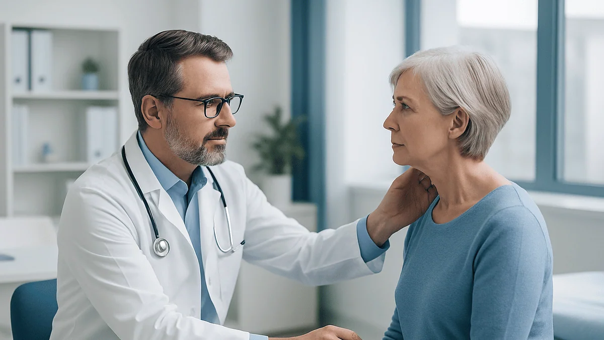
A parotid gland tumor usually needs parotidectomy surgery when biopsy or imaging suggests cancer, when a benign tumor such as a pleomorphic adenoma is growing or causing symptoms, or when the diagnosis stays uncertain. Monitoring is more often considered for small, stable, clearly benign lumps, especially Warthin tumors, or when surgical risk is high. The treating team weighs tumor type, size, nerve position and overall health.
The lump was the size of a grape, just in front of her left ear, and her dentist noticed it before she did. Two appointments later she was sitting in a head and neck clinic with an ultrasound report, a needle biopsy result that read “favors benign,” and one question she could not put down: does this actually have to come out?
It is a fair question, and the honest answer is that it depends on facts that a scan alone cannot always give. Understanding when a parotid tumor needs surgery, and when it is reasonable to watch, means understanding what these lumps are, why the facial nerve makes this gland different from almost any other in the body, and how surgeons and patients weigh a small but real operative risk against a small but real risk of leaving something behind.
This explainer walks through that reasoning the way a good clinic conversation would, with the evidence laid out and the decision left where it belongs, with you and your treating team.
When does a parotid tumor need surgery? The short version
Most parotid lumps are not cancer, yet most of them still end up being removed. That sounds contradictory until you see how the decision is built. Salivary gland tumors are uncommon, the parotid is by far their most frequent site, and the majority found there are benign, according to MedlinePlus. The catch is that “benign” does not mean “harmless forever,” and “probably benign” on a needle biopsy is not the same as “definitely benign” under a pathologist’s microscope after removal.
Three situations push firmly toward parotidectomy, which is the operation to remove part or all of the parotid gland. The first is any suspicion of malignancy on biopsy or imaging. The second is a tumor that is enlarging, painful, pressing on structures, or affecting the facial nerve. The third is diagnostic uncertainty: when the needle sample cannot say what the lump is, removal is often the only way to find out.
Two situations more often support watching. A small, stable lump with a confident benign diagnosis, particularly a Warthin tumor (a benign growth that almost never turns cancerous), may be observed. So may a benign tumor in someone whose other health problems make an operation riskier than the lump itself.
Everything else sits in the middle, which is where most real conversations happen. A pleomorphic adenoma, the most common benign parotid tumor, is usually removed even when it is behaving well, because it tends to keep growing and carries a small long-term risk of becoming cancerous, a pattern described by the Mayo Clinic. Whether that argument outweighs the operative risks for a particular person is exactly the judgment a specialist team is trained to make.
What the parotid gland does, and why the facial nerve changes everything
Picture a soft, wedge-shaped gland tucked between the ear and the angle of the jaw, draining saliva through a small duct that opens inside the cheek near the upper molars. That is the parotid, the largest of the three paired major salivary glands. It makes a thin, enzyme-rich saliva that starts breaking down starches while you chew.
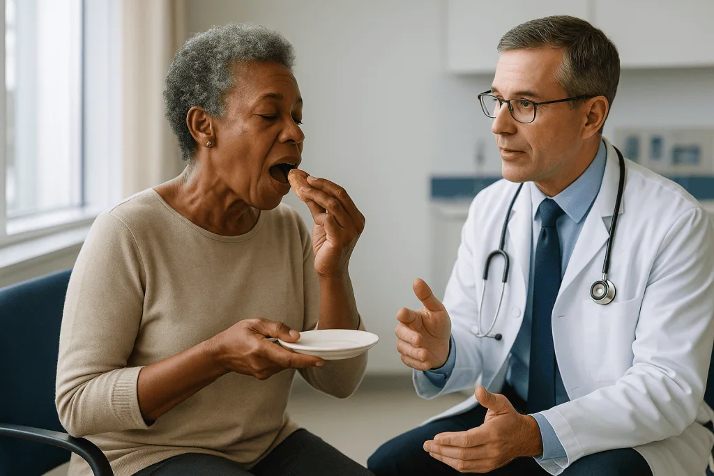
What makes it surgically unusual is a nerve. The facial nerve, which controls every muscle of expression on that side of the face, exits the skull just behind the ear and runs straight through the parotid, fanning out into five main branches like the fingers of a hand. Surgeons describe the gland as having a superficial lobe above the nerve and a deep lobe beneath it, although in reality it is one continuous piece of tissue with the nerve threaded through it.
This anatomy explains almost every feature of parotid care. It explains why surgeons cannot simply scoop out a lump the way they might elsewhere; they have to find the nerve first and work around it. It explains why a needle biopsy is preferred over a cutting biopsy, since an incision into the gland can scar the nerve plane. And it explains why weakness of the face, even subtle weakness, is a warning sign that clinicians take very seriously, because benign tumors push the nerve aside while cancers can grow into it, as the NHS notes.
Around the gland sit the ear canal, the jaw joint, the large neck vessels and a cluster of lymph nodes. Deep-lobe tumors can bulge into the throat rather than outward, which is why a few people first notice a fullness on one side of the soft palate rather than a lump on the cheek.
Benign, malignant, and the awkward in-between: what a parotid lump can be
Not every swelling near the ear is a tumor. Enlarged lymph nodes, blocked ducts, salivary stones, cysts and infections all live in the same postcode, and part of the first visit is ruling them out. Once a true tumor is confirmed, it falls into one of several groups.
Pleomorphic adenoma is the most common. It is benign, slow-growing, and firm, and it has a thin capsule with tiny finger-like projections that can be left behind if the lump is simply shelled out. Those remnants are why recurrence after incomplete removal is a recognized problem, and why surgeons remove a margin of normal gland around it.
Warthin tumor is the second most common benign type. It is strongly linked to smoking, can occur in both glands, and has a very low risk of turning cancerous. Because of that, it is the tumor most often considered for observation when it is small and not troubling the person.
Malignant tumors are less common but varied. Mucoepidermoid carcinoma, adenoid cystic carcinoma, acinic cell carcinoma and carcinoma arising in a long-standing pleomorphic adenoma each behave differently, and their grade (how abnormal the cells look) matters as much as their name. The National Cancer Institute groups them by type, grade and stage when describing treatment options.
Then there is the in-between: lumps whose needle biopsy is “atypical” or “cannot exclude,” and rare tumors whose behavior is hard to predict. These are not failures of diagnosis. Salivary tumors are notoriously varied under the microscope, and a needle sample of a few hundred cells can genuinely be ambiguous. When that happens, removal of the lump becomes both the treatment and the definitive test.
How doctors work out what the lump is before anyone talks about surgery
The work-up follows a fairly standard path, and each step narrows the question of when a parotid tumor needs surgery rather than watching.

The clinic examination comes first. The clinician feels the lump for size, mobility and tenderness, checks the facial muscles branch by branch, looks inside the mouth for a deep-lobe bulge, and feels the neck for lymph nodes. A lump that is fixed to skin or bone, or one accompanied by facial weakness, raises concern well before any test result arrives.
Ultrasound is usually the first imaging test. It is quick, has no radiation, shows whether the lump is solid or fluid-filled, and lets the radiologist guide a needle precisely. Magnetic resonance imaging (MRI) is added when the lump is large, sits in the deep lobe, or shows worrying features, because MRI outlines the tumor’s relationship to the nerve plane and surrounding tissue. Computed tomography (CT) is sometimes used instead, or to check bone.
Fine-needle aspiration, often shortened to FNA, is the key biopsy. A thin needle draws out cells for a cytologist to examine. It is well tolerated, carries minimal risk of spreading tumor, and gives a useful answer in most cases, according to the Mayo Clinic. Its limitation is that it samples cells rather than architecture, so it can miss the pattern that distinguishes some benign tumors from low-grade cancers. Core needle biopsy, which takes a slender cylinder of tissue, is used in some centers when FNA is inconclusive.
Results are then reviewed together, ideally in a multidisciplinary meeting where surgeons, radiologists and pathologists look at the same pictures. That combined view, not any single test, is what produces a recommendation to operate, to observe, or to investigate further.
Who is usually offered parotidectomy, and who is usually asked to wait
Surgeons rarely think in terms of a single rule. They think in terms of a balance sheet, and it helps to see both columns the way they do.
People most often advised to have surgery include those with a cancer diagnosis or a biopsy that cannot rule one out; those with a pleomorphic adenoma, because of its tendency to keep growing and its small long-term malignant risk; those whose tumor is enlarging on serial scans; those with pain, facial weakness, numbness or skin changes; and those whose lump is large enough to be disfiguring or to press on the ear canal, jaw or throat. Younger people with benign tumors are also more often steered toward removal, on the reasoning that decades of continued growth and monitoring lie ahead.
People more often asked to wait, or offered watching as one reasonable option, include those with a small Warthin tumor confirmed on cytology; older adults with a stable benign lump and no symptoms; people whose heart, lung or other conditions make anesthesia and a two- to three-hour operation a meaningful risk; and those who, after a full discussion of the trade-offs, prefer surveillance and accept its uncertainties. The Cleveland Clinic describes surgery as the usual treatment for most parotid tumors while noting that the plan is individualized.
Watching is not the same as doing nothing. It involves scheduled examinations and repeat imaging, with a clear agreement about what change would trigger a return to the surgical conversation. A lump that grows, becomes painful, or changes its appearance on ultrasound moves from one column to the other.
Neither column is a verdict. A person in the “wait” group can choose surgery for peace of mind, and a person in the “operate” group can ask for time. What matters is that the choice is informed and revisited.
Monitoring, superficial parotidectomy, or total parotidectomy: a side-by-side comparison
The options are easier to weigh when they sit next to each other. The table below summarizes how the main approaches differ in purpose, what they involve, and their typical trade-offs, drawing on descriptions from the Cleveland Clinic and the National Cancer Institute.
| Approach | Usually considered for | What it involves | Main trade-offs |
|---|---|---|---|
| Active monitoring | Small, stable, confidently benign lumps (especially Warthin tumor); people at higher surgical risk | Scheduled examinations and repeat ultrasound or MRI; agreed triggers for re-evaluation | Avoids operative risk now; accepts diagnostic uncertainty, possible growth, and a later operation that may be larger |
| Superficial (partial) parotidectomy | Most benign tumors and some low-grade cancers confined to the superficial lobe | Removal of the tumor with a cuff of gland above the facial nerve, which is identified and preserved | Definitive diagnosis and treatment in one step; temporary facial weakness, numbness of the earlobe and Frey syndrome are possible |
| Extracapsular dissection | Selected small, mobile, superficial benign tumors in experienced hands | Removal of the tumor with a narrower margin, without formally exposing the full nerve | Less tissue removed and often less Frey syndrome; requires careful case selection to avoid leaving tumor behind |
| Total parotidectomy | Deep-lobe tumors; higher-grade or larger cancers; some recurrent tumors | Removal of both lobes, working the nerve free from the tissue on either side; sometimes with neck lymph node removal | More complete clearance; longer operation, higher chance of temporary nerve weakness, more noticeable contour change |
| Radical parotidectomy | Cancers that have grown into the facial nerve | Removal of the gland and involved nerve segments, usually with nerve grafting or other reconstruction | Reserved for advanced disease; permanent facial weakness expected, mitigated by reconstruction and rehabilitation |
Two things stand out. The nerve is preserved in every operation except the last, and that last operation is for a situation the earlier ones are trying to prevent. The scale of surgery tracks the nature of the tumor, not the surgeon’s preference, which is why an accurate pre-operative picture matters so much.
How fast do parotid tumors grow, and does speed change the decision?
Speed is one of the most useful clues in the whole assessment, and it is one that patients themselves often supply. A lump that has been the same size for five years is telling a different story from one that appeared over a few months.
Benign parotid tumors are typically slow. A pleomorphic adenoma may take years to become noticeable and can enlarge so gradually that a person only registers the change when they compare old photographs. Warthin tumors behave similarly, and some fluctuate in size, occasionally swelling with inflammation and then settling. The Mayo Clinic describes most salivary gland tumors as slow-growing, painless swellings.
Faster growth does not prove malignancy, but it does shift the odds. High-grade cancers can double in size over weeks to months, and a benign tumor that suddenly accelerates raises the question of malignant change within it. Growth paired with new pain, skin tethering, facial weakness or a firm neck node is the combination that moves a case from routine to urgent.
There is an important caveat about measurement. Ultrasound and MRI measure to the millimeter, but glands, posture and radiologists vary, and a difference of a millimeter or two between scans can be noise rather than growth. Monitoring protocols therefore look for consistent change across more than one interval before drawing conclusions, unless the change is large.
Slow growth is not, on its own, a reason to leave a pleomorphic adenoma indefinitely. The risk with this tumor is less about how fast it grows and more about what it can eventually become, and the technical reality that a larger tumor stretches and thins the nerve, making later surgery harder. That is the paradox clinicians try to explain: sometimes the calmest-looking lump is the one best removed while it is still small.
What actually happens during a parotidectomy, step by step
Most people picture an operation on the cheek. In practice, the incision is designed to hide. It usually starts in a natural crease just in front of the ear, curves around the earlobe, and continues into the hairline behind the ear or into a neck crease, a pattern often called a modified facelift incision. The Cleveland Clinic describes the procedure as taking roughly two to four hours under general anesthesia, depending on extent.
Once the skin flap is lifted, the surgeon’s first goal is not the tumor but the nerve. The main trunk of the facial nerve is found using fixed landmarks at the base of the skull, then followed forward as it branches, with the gland tissue above it gently separated away. A nerve monitor, which is a set of fine electrodes placed in the facial muscles, sounds an alert if a branch is touched or stretched, giving the surgeon real-time feedback.
With the branches identified, the portion of gland containing the tumor is lifted off the nerve and removed in one piece with a margin of normal tissue. If the tumor lies in the deep lobe, the nerve branches are freed and gently retracted so the deeper tissue can be delivered from beneath them. Any suspicious lymph nodes may be removed at the same time.
Before closing, surgeons often place a thin layer of tissue or a small implant over the nerve bed. This is intended to reduce Frey syndrome, a condition in which regrowing nerve fibers meant for saliva connect instead to sweat glands in the skin, causing sweating or flushing of the cheek at mealtimes. A small drain is usually left for a day or so to prevent fluid collecting, and the wound is closed with fine sutures.
The removed tissue goes to a pathologist, whose full report, typically available within one to two weeks, gives the final diagnosis and guides any further care.
How serious is parotid gland surgery? Risks in plain language
Parotidectomy is a major operation performed in a delicate area, but it is also a well-established one with a predictable set of risks that are worth understanding precisely rather than fearfully.
Facial nerve weakness is the one everyone asks about. Temporary weakness of part of the face, most often the corner of the mouth or the lower lip, is common after surgery and results from the nerve being handled, stretched or briefly deprived of blood supply. It usually improves over weeks to months as the nerve recovers. Permanent weakness is uncommon when the nerve is intact at the end of the operation, and is more likely with larger, deeper or recurrent tumors, according to the Cleveland Clinic. Where cancer has invaded the nerve, part of it may need to be removed deliberately, with reconstruction planned in advance.
Numbness of the earlobe and the skin in front of the ear is expected, because a sensory nerve called the great auricular nerve crosses the operative field. Some sensation often returns, but a patch of permanent numbness is frequent and most people adapt to it.
Frey syndrome, described above, may appear months later as sweating or redness of the cheek while eating. It ranges from unnoticeable to bothersome, and there are treatments for those who find it troubling.
Other risks are those of any surgery: bleeding, infection, a collection of saliva or fluid under the skin (which is usually managed with drainage or dressings), a noticeable hollow in front of the ear, and scarring. Salivary leakage through the wound occasionally occurs and almost always settles.
Set against these is the risk of not operating: missing a cancer, allowing a benign tumor to enlarge until surgery becomes more complex, or living with prolonged uncertainty. Weighing one list against the other is the heart of the decision, and it looks different for each person.
Parotid surgery recovery time: what the first days and weeks usually look like
Recovery from parotidectomy tends to be faster than people expect from the length of the operation, but it follows a rhythm worth knowing in advance.
The first night is usually spent in hospital, sometimes two, mainly so the drain can be monitored and removed once output falls. The wound is covered with a light dressing. Pain is generally described as moderate and manageable with the medicines the team prescribes; jaw stiffness and a pulling sensation around the ear are typical. Eating soft foods and chewing gently on the opposite side is often advised for the first few days.
During the first week the earlobe is numb, the skin in front of the ear may feel tight and swollen, and any facial weakness is most apparent. Sutures or skin clips are usually removed at around one week if they are not dissolvable. Most people are told to avoid strenuous exercise, heavy lifting and swimming until the wound has sealed.
By two weeks the majority of people are back to desk work and ordinary daily activity, a range described by the Cleveland Clinic. Swelling continues to settle over several weeks, and the scar fades over months, particularly if it is protected from sun.
Nerve recovery has its own, slower clock. Temporary facial weakness commonly improves over weeks to a few months; the team may suggest gentle facial exercises and eye protection if the eyelid is affected. Frey syndrome, if it develops, tends to appear somewhere between a few months and a year later.
A follow-up appointment is scheduled once the pathology report is available, usually within one to two weeks, to explain the final diagnosis and confirm the plan. That conversation, more than the operation itself, is where the story of the lump finally gets its ending.
When monitoring is the right call: how active surveillance of a parotid tumor works
Choosing to watch a parotid tumor is a positive decision with a structure, not a shrug. Understanding that structure helps people who are on it feel that something is being done, because it is.
Surveillance starts with a confident baseline. That means a needle biopsy result that fits the imaging, an MRI or ultrasound that documents exact dimensions and appearance, and a clinical examination recorded in detail, including facial nerve function. If any of those three does not fit the others, most teams will want to resolve the mismatch rather than watch it.
The follow-up schedule is set by the team and depends on the tumor type and the person’s circumstances. In general, intervals are shorter in the first year or two, when the pattern of behavior is being established, and lengthen once stability is confirmed. Each visit repeats the examination and, at agreed intervals, the imaging.
Triggers for stepping off surveillance are agreed at the outset. They typically include measurable growth confirmed across scans, new pain, any change in facial movement, skin changes, a new neck node, or a change in the imaging character of the lump. A person’s own wish to have the lump removed, whether for comfort, appearance or peace of mind, is also a legitimate trigger at any time.
Surveillance suits some tumors better than others. Warthin tumors, with their very low malignant potential, are the best candidates, as the MedlinePlus overview of salivary tumors implies in describing them as benign. Pleomorphic adenomas are watched less often, and usually only when surgery poses particular risks, because their long-term behavior is less predictable.
The quiet cost of surveillance is psychological. Living with a lump you have been told is probably fine is not the same as living without one, and it is reasonable to weigh that cost alongside the medical ones.
What happens after surgery: the pathology report, follow-up, and when radiotherapy is discussed
The operation answers the first question, what the lump is. The pathology report answers the second, what happens next, and it is worth knowing how to read it in outline.
For a benign tumor with clear margins, follow-up is usually light. Pleomorphic adenomas can recur, sometimes many years later, so a period of periodic review is common, and people are asked to report any new swelling near the scar. Warthin tumors rarely recur but can occur in the other gland, which is one reason a baseline examination of both sides is recorded.
For cancer, the report describes the tumor type, its grade, whether the margins are clear, whether it has spread along nerves or into lymphatic channels, and whether any removed lymph nodes were involved. These details are combined with imaging into a stage. The National Cancer Institute outlines how type, grade and stage together shape the treatment plan.
Radiotherapy after surgery is discussed when features suggest a higher chance of the cancer returning locally: high grade, close or involved margins, nerve invasion, larger size, or positive nodes. It uses focused X-ray beams to treat the area where the tumor lay, typically over several weeks of short daily sessions. Its side effects, which can include dry mouth, skin reaction, taste change and fatigue, are part of the discussion. Chemotherapy has a more limited role in salivary cancers and is generally reserved for advanced or recurrent disease, sometimes within clinical trials.
People often ask about the survival rate for a parotid tumor. There is no single number, because a low-grade, small, node-negative cancer and a high-grade, advanced one are almost different diseases. Outlook is best discussed with the treating team once the full pathology and stage are known, and anyone quoting a percentage without those details is guessing.
What people often get wrong about parotid tumors and parotidectomy
Myth: a needle biopsy can spread a parotid cancer. The fine needles used for salivary gland sampling have not been shown to seed tumor along their track in any meaningful way, and FNA is a standard, recommended first step, as the Mayo Clinic describes. What surgeons do avoid is an open incisional biopsy, because cutting into the gland scars the nerve plane and can leave benign tumor behind.
Myth: if it is benign, it can safely be ignored. That is close to true for a small Warthin tumor and not true for a pleomorphic adenoma, which keeps growing, can eventually undergo malignant change, and becomes harder to remove safely as it enlarges around the nerve.
Myth: the operation means a permanently drooping face. Temporary weakness is common; permanent weakness is uncommon when the nerve is preserved. Careful nerve identification and monitoring exist precisely to keep it that way.
Myth: surgery leaves a large scar across the cheek. The incision runs in front of and behind the ear and into the hairline or a neck crease, which is why most healed scars are hard to spot.
Myth: a normal scan means no surgery is needed. Imaging characterizes a lump; it does not diagnose it. When the biopsy and images disagree, or when the biopsy is inconclusive, removing the lump is often the only way to know what it is.
Myth: parotid tumors are always cancer, or never cancer. Neither. Most are benign, a meaningful minority are malignant, and the mix shifts with age, smoking history, prior radiation exposure and the specific features found on examination.
Myth: taking out one salivary gland leaves the mouth dry. The remaining parotid, the two submandibular glands, the sublingual glands and hundreds of minor glands more than compensate. Persistent dryness after parotidectomy is unusual unless radiotherapy is also given.
Questions to ask your care team before deciding on parotidectomy or monitoring
A good consultation leaves room for questions, and the ones below tend to produce the most useful answers. Write down the replies; the details blur later.
- What does my biopsy show, and how confident is the pathologist? Is a repeat or core biopsy worth considering?
- Which lobe is the tumor in, and how does it sit in relation to the facial nerve on my imaging?
- Based on what you know today, what do you think this lump most likely is, and what else could it be?
- If we monitor, what exactly would the schedule be, and what change would make you recommend surgery?
- If we operate, which type of parotidectomy are you planning, and why that one rather than a smaller or larger operation?
- Will facial nerve monitoring be used, and what is your approach if the nerve is found to be involved during surgery?
- What is the realistic chance of temporary and permanent facial weakness for a tumor like mine?
- What can be done at the time of surgery to reduce the risk of Frey syndrome and a hollow contour?
- How long will I stay in hospital, when can I expect to return to work and exercise, and what should the first two weeks look like?
- When will the final pathology be available, and how will you contact me with the result?
- If the lump turns out to be cancer, what would the next steps be, and would my case go to a multidisciplinary meeting?
- Is there any reason, given my other health conditions, that you would lean one way rather than the other?
It is entirely reasonable to ask for time to think, to bring someone with you, or to request a second opinion within your health system before proceeding. The NHS guidance on salivary gland cancer emphasizes that treatment plans are made by a specialist team together with the patient, and the same principle applies to benign disease.
When to call your doctor: red-flag signs before and after parotid surgery
Some changes should prompt a call rather than waiting for the next scheduled visit, whether you are on surveillance, waiting for surgery, or recovering from it.
If you have a known parotid lump and have not had surgery, contact your team promptly if you notice any new weakness, drooping or asymmetry of the face; new or worsening pain; rapid enlargement over weeks; skin over the lump becoming red, fixed or ulcerated; numbness of the face; a new firm lump in the neck; or difficulty opening the mouth or swallowing. The NHS lists facial weakness, pain and a growing lump among the features that warrant assessment.
After parotidectomy, seek same-day advice for a rapidly enlarging, tense swelling under the wound, which can indicate bleeding; spreading redness, warmth or pus around the incision; a fever; persistent clear fluid leaking from the wound, especially at mealtimes; new or worsening facial weakness that was not present when you left hospital; an eye that will not close or that feels gritty and dry; or bleeding that does not stop with gentle pressure.
Call emergency services if you have difficulty breathing, severe neck swelling, chest pain, or signs of an allergic reaction to a medicine.
Less urgent but still worth reporting at your next contact are a cheek that sweats or flushes when you eat, a scar that is thickening or itching intensely, or persistent jaw stiffness. None of these is dangerous, but each has options, and your team cannot help with what it does not know about.
When in doubt, ring the clinic. Head and neck teams would far rather answer a question that turns out to be nothing than hear about a problem late. Every decision about investigation, surgery, surveillance and follow-up rests with the clinicians who know your case, and this article is a companion to that conversation, not a substitute for it.
Frequently asked questions
How long does it take to recover from parotid tumor surgery?
Most people stay one night in hospital and return to desk work and ordinary daily activity within about one to two weeks, according to the Cleveland Clinic. Swelling settles over several weeks, the scar fades over months, and any temporary facial weakness usually improves over weeks to a few months. Strenuous exercise, heavy lifting and swimming are typically delayed until the wound has healed, and your team will give you a personalized timeline.
What is the survival rate for a parotid tumor?
There is no single survival rate, because most parotid tumors are benign and are not life-threatening, while the minority that are cancers vary enormously by type, grade and stage. The National Cancer Institute describes outlook in terms of those factors rather than one figure. Your treating team can discuss what the evidence suggests for your specific diagnosis once the pathology report and staging are complete.
How serious is parotid gland surgery?
Parotidectomy is a major but well-established operation, usually taking two to four hours under general anesthesia. Its seriousness comes mainly from the facial nerve running through the gland. Temporary facial weakness is common and usually recovers; permanent weakness is uncommon when the nerve is preserved. Earlobe numbness, Frey syndrome and a slight hollow in front of the ear are the other recognized effects. Most people recover well and go home within a day or two.
How fast do parotid tumors grow?
Benign parotid tumors such as pleomorphic adenoma and Warthin tumor usually grow slowly, often over years, and the Mayo Clinic describes most salivary tumors as slow, painless swellings. Cancers, particularly high-grade ones, can enlarge noticeably over weeks to months. A benign tumor that suddenly speeds up, or growth accompanied by pain, facial weakness or skin changes, is a reason to seek prompt reassessment.
Can a pleomorphic adenoma be left alone and just watched?
Sometimes, but it is less often recommended than for other benign tumors. Pleomorphic adenomas tend to keep growing, can recur if incompletely removed, and carry a small risk of malignant change over many years. Watch-and-wait is usually reserved for people in whom surgery carries particular risks or who prefer surveillance after a full discussion. If watched, it needs a scheduled program of examination and imaging with clear triggers for revisiting surgery.
Does a parotid tumor always need to be removed?
No. Small, stable Warthin tumors with a confident biopsy diagnosis, and benign lumps in people whose other health problems make surgery risky, are often monitored rather than removed. Surgery is usually advised when cancer is suspected or cannot be excluded, when a tumor is growing or causing symptoms, or when the diagnosis is uncertain. The decision is individual and rests with you and your specialist team.
Will parotidectomy leave a visible scar?
The incision is placed in a natural crease in front of the ear, around the earlobe, and into the hairline behind the ear or a neck crease, in a pattern similar to a facelift incision. Once healed and matured over several months, most scars are difficult to see. Protecting the scar from sun and following your team’s wound-care advice helps it fade. A slight hollow in front of the ear is more often noticed than the scar itself.
What is Frey syndrome and how common is it after parotid surgery?
Frey syndrome is sweating or flushing of the cheek while eating, caused by regrowing salivary nerve fibers connecting to sweat glands in the skin after surgery. It typically appears months after the operation and ranges from unnoticeable to bothersome. Surgeons often place a layer of tissue over the nerve bed to reduce the risk. For those who find it troubling, treatments are available and can be discussed with your team.
Can a fine-needle biopsy spread a parotid cancer?
Fine-needle aspiration has not been shown to spread salivary gland cancer in any clinically meaningful way, and it is the standard, recommended first biopsy for a parotid lump. What surgeons avoid is an open incisional biopsy, which cuts into the gland, scars the plane where the facial nerve lies, and can leave fragments of a benign tumor behind. If a needle result is inconclusive, a core needle biopsy or removal of the lump is usually the next step.
Will removing my parotid gland cause a dry mouth?
Usually not. The remaining parotid gland, the two submandibular glands, the sublingual glands and hundreds of minor salivary glands in the mouth continue to produce saliva, and most people notice no lasting dryness after a single parotidectomy. Persistent dry mouth is more likely if radiotherapy to the area is also needed, which is why its side effects form part of the treatment discussion for salivary cancers.
This article is for general information only and is not a substitute for professional medical advice. Please consult a qualified doctor about your individual situation.

More from the Blog
Bladder Removal or Bladder Preservation? How Specialists Compare the Two Approaches
For muscle-invasive bladder cancer, specialists compare radical cystectomy (removing the bladder, usually after chemotherapy) with bladder preservation, most often trimodality therapy combining tumor resection,…
Induction Chemotherapy for Acute Myeloid Leukemia: What the First Hospital Weeks Involve
AML induction chemotherapy is an intensive first treatment given in hospital, usually as a continuous course lasting about a week, followed by several weeks…
Surgical Oncology Explained: How a Cancer Operation Is Planned Within a Multidisciplinary Team
Cancer surgery is planned by a multidisciplinary team rather than a single surgeon. Pathologists confirm the diagnosis from a biopsy, radiologists stage the cancer…
Speech and Swallowing Therapy After Head Cancer Treatment: How Rehabilitation Works
Swallowing therapy after head and neck cancer is a rehabilitation program led by a speech-language pathologist. It combines a formal swallow assessment, targeted exercises…

Preparing a Child for Wilms Tumor Surgery: Comfort, Explanations and the Day Before
Preparing a child for Wilms tumor surgery works best when explanations are short, honest and matched to the child's age, when familiar comfort items…
Life After Endometrial Cancer Treatment: Surgical Menopause, Energy and Long-Term Care
Menopause after endometrial cancer surgery happens when both ovaries are removed along with the uterus, so estrogen falls within days rather than over years.…