

Parotidectomy Surgery Step by Step: How Surgeons Protect the Facial Nerve
Key Takeaways
- The facial nerve enters the back of the parotid gland as a single trunk and branches inside it, which is why surgeons describe parotidectomy as dissecting the nerve rather than simply removing a gland.
- Surgeons locate the nerve trunk by triangulating fixed landmarks, chiefly the tragal pointer, the tympanomastoid suture and the posterior belly of the digastric muscle, before any gland tissue is divided.
- Temporary facial weakness after parotidectomy is common and usually reflects stretch or bruising of an intact nerve; permanent paralysis is uncommon after surgery for benign disease, according to Johns Hopkins Medicine and Cleveland Clinic.
- Intraoperative nerve monitoring is widely used and can help identify the nerve and shorten dissection, but the evidence that it reduces permanent paralysis is inconsistent, and it can miss stretch injuries.
- Nerve fibers regrow slowly after deeper injuries, so surgeons generally do not label facial weakness permanent before roughly a year, and repair or reconstruction options exist at every stage.
- Ear numbness after parotidectomy comes from a separate sensory nerve disturbed during the approach and is not a sign of facial nerve damage.
During a parotidectomy, surgeons protect the facial nerve by locating its main trunk with fixed anatomical landmarks near the ear, then tracing each branch under magnification before removing the gland tissue around it. Many surgeons add electrical nerve monitoring. Temporary facial weakness is common and usually improves over weeks to months; permanent paralysis is uncommon, according to Johns Hopkins Medicine and Cleveland Clinic patient information.
The lump had been there for months, a firm little marble just in front of her earlobe, and she had grown used to touching it while she read. What she had not expected was the diagram. Her surgeon drew an ear, a gland shaped like a wedge of pear, and then a branching line that fanned across the whole side of the face like the limbs of a tree. “This,” he said, tapping the trunk, “is what the operation is really about.”
Anyone researching parotidectomy facial nerve risk arrives at the same fork in the road she did. The tumor is usually benign. The operation is usually straightforward. And yet the one structure running through the middle of the gland controls whether you can close your eye, raise your eyebrow, and smile evenly.
This explainer walks through what surgeons actually do to keep that nerve safe, what the evidence says about weakness afterward, and what recovery tends to look like when the drapes come off.
What is a parotidectomy, and why does the facial nerve run through it?
A parotidectomy is an operation to remove part or all of the parotid gland, the largest of the saliva-producing glands, which sits in front of and just below each ear. Most parotidectomies are performed for tumors, and most of those tumors are benign, meaning non-cancerous. The most frequent is a pleomorphic adenoma, a slow-growing lump that is harmless in itself but can enlarge and, over many years, carries a small risk of turning malignant, according to Mayo Clinic and MedlinePlus.
The complication in the plan is anatomy. The facial nerve, the seventh cranial nerve, leaves the skull through a small opening behind the ear, enters the back of the parotid gland as a single trunk, and then divides inside the gland into branches that supply every muscle of facial expression on that side. Surgeons describe the gland as having a superficial lobe and a deep lobe, but there is no true partition between them. The “lobes” are simply the tissue on either side of the plane where the nerve travels.
That geometry is why the parotidectomy facial nerve relationship dominates every pre-operative conversation. Removing a tumor from the thyroid or a lymph node in the neck involves working near important structures. Removing a parotid tumor means dissecting a nerve out of the middle of the tissue you are taking away, branch by branch, and leaving it intact.
It also explains a term you may hear: the operation is often described less as “removing a gland” and more as “dissecting the facial nerve.” The tumor comes out because the gland tissue around the nerve has been carefully separated from it, not the other way round.
Is a parotidectomy considered major surgery?
Yes, by most reasonable definitions. It is performed under general anesthesia, it typically takes several hours, and it involves fine dissection around a nerve that has no backup. Johns Hopkins Medicine and Cleveland Clinic describe the operation as lasting roughly two to four hours, with many people staying one night in hospital and some going home the same day, depending on drain placement and how they feel after anesthesia.
What makes it feel different from other “major” operations is that the physical recovery is often gentler than the label suggests. There is no body cavity opened, no bowel to wake up, and the incision sits in a natural skin crease in front of the ear and curves into the hairline or upper neck. Pain is usually described as moderate and manageable with the plan your team gives you; the surgical team decides what that plan contains.
The seriousness lies elsewhere: in the precision required and in the consequences if something goes wrong. A facial nerve injury cannot be hidden under clothing. It affects eating, speaking, eye protection and the simple social act of smiling. That is why surgeons who perform these operations tend to spend a disproportionate amount of consultation time on the nerve, even when the tumor itself is benign and small.
So the honest answer is two-sided. A parotidectomy is major in stakes and in skill, moderate in physical burden for most people, and, when it is done for a benign tumor, one of the more common head and neck operations rather than an exotic one. Understanding that balance helps people calibrate their worry: respect the nerve, but do not assume the recovery will resemble abdominal or cardiac surgery.
Who is usually offered a parotidectomy, and who is asked to wait?
The clearest candidates are people with a confirmed or strongly suspected tumor in the parotid gland. That usually means a lump that has been imaged with ultrasound, CT or MRI and sampled with a fine-needle biopsy, a test where a thin needle draws cells from the lump for examination under a microscope, according to Mayo Clinic. Benign tumors such as pleomorphic adenomas are generally removed because they keep growing, can become harder to remove safely as they enlarge, and carry a small long-term risk of malignant change. Malignant tumors are removed as part of a cancer treatment plan that may also involve radiotherapy afterward.
Other reasons include chronic infection or stones that repeatedly inflame the gland when less invasive measures have not controlled the problem, and some deep-lobe tumors that press on structures in the throat.
Who is asked to wait, or offered a different path? Several groups come up repeatedly in guideline-level discussions:
- People whose “lump” turns out on imaging and biopsy to be an inflamed lymph node or a cyst that may settle with observation.
- Older adults with a slow-growing tumor known to have very low malignant potential, such as some Warthin tumors, where active surveillance with repeat imaging may be reasonable.
- Anyone with an active infection in the gland; surgery in inflamed tissue makes nerve identification harder, so infection is usually treated first.
- People with medical conditions that make general anesthesia riskier until they are optimized.
Pre-existing facial weakness changes the calculus too. If the nerve is already partly compromised by a tumor, the operation may be planned differently, and that conversation is one for the treating team. None of these categories is absolute. They are starting points for a decision that belongs to you and your surgeon together.
Parotidectomy step by step: what actually happens in the operating room
Once you are asleep, the anesthesia team is asked to avoid or minimize long-acting muscle relaxants. This matters because the surgeon wants the facial muscles able to twitch when the nerve is stimulated during the operation. Your head is turned slightly away, and the face on the operative side is left visible so the team can watch for movement.
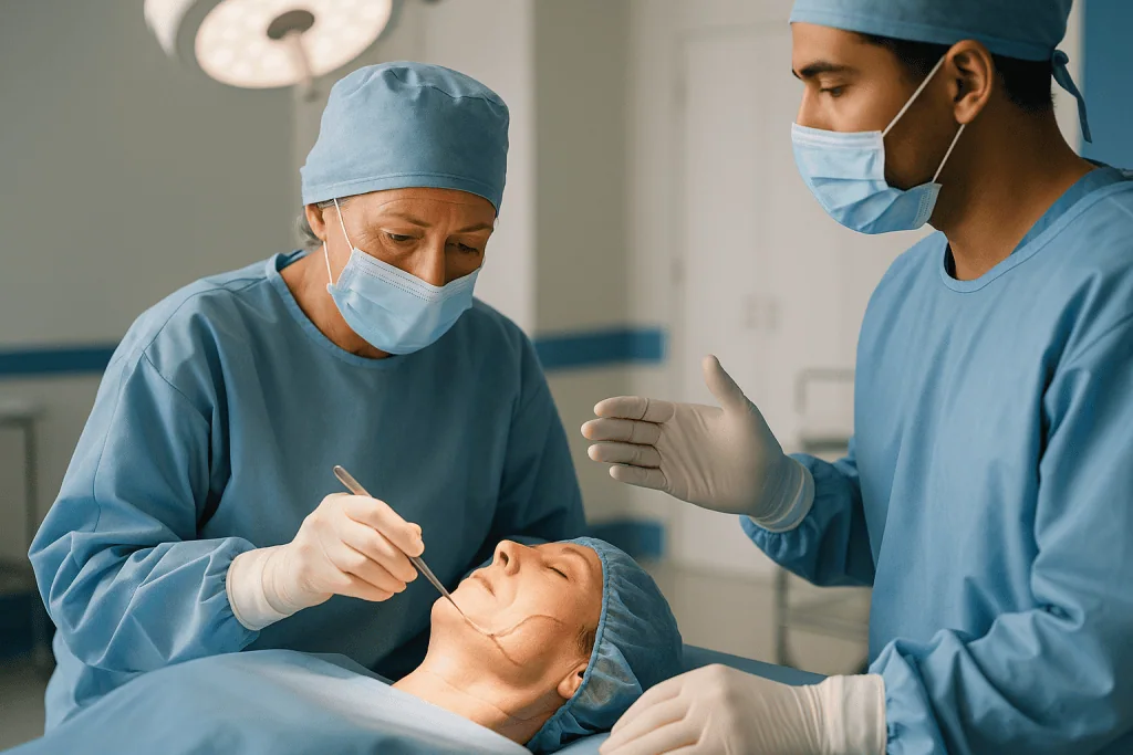
The incision follows a curve that begins in front of the ear, sweeps under the earlobe, and continues either behind the ear into the hairline or down into a neck crease. The skin and a thin layer of tissue are lifted forward as a flap, exposing the gland.
Next comes the part that takes the most time: finding the nerve. The surgeon works from the back edge of the gland toward the ear canal, separating the gland from the cartilage of the ear and the muscle behind it, and identifying the main trunk of the facial nerve where it emerges from the skull. The next section explains the landmarks used.
With the trunk identified, the surgeon follows it forward into the gland, gently lifting gland tissue away from each branch with fine instruments. The tumor and the surrounding portion of gland come away in one piece as this dissection advances. Throughout, the nerve is handled as little as possible and kept moist, because stretching and drying both impair function.
Once the specimen is out and sent to pathology, the surgeon checks that each branch responds to a gentle electrical stimulus, controls any bleeding, and often places a small drain to prevent fluid collecting. The skin is closed with fine sutures or glue. Johns Hopkins Medicine notes the drain is commonly removed within a day or two, once output has fallen.
How do surgeons find the facial nerve during parotidectomy?
Two broad strategies exist, and experienced surgeons are comfortable with both.
The standard approach is called antegrade dissection: find the main trunk first, then follow it forward. Because the trunk is buried in tissue, surgeons rely on landmarks that do not move. The tragal pointer is the cartilage tip of the ear canal; the nerve typically lies a short distance below and in front of it. The tympanomastoid suture is a groove in the bone between two parts of the skull that points almost directly toward the nerve. The posterior belly of the digastric muscle, a neck muscle running up toward the skull base, marks the depth at which the nerve sits. None of these is perfect on its own, which is why surgeons triangulate with all three, and why some also palpate the styloid process, a spike of bone just deep to the nerve.
The alternative is retrograde dissection: identify a peripheral branch first and trace it backward toward the trunk. The marginal mandibular branch can be found where it crosses a vein below the jaw; the buccal branch runs alongside the parotid duct, the tube that carries saliva into the mouth. Retrograde dissection is useful when a tumor sits directly on top of the trunk, when prior surgery or radiation has distorted the landmarks, or when a small tumor near the front of the gland can be removed with a partial parotidectomy without exposing the whole nerve.
Neither method is proven superior in every situation. What the literature does show is that the surgeon’s familiarity with the technique matters more than the label. Whichever route is taken, the principle is identical: see the nerve before cutting anything near it, and never divide tissue whose contents are uncertain.
Is facial nerve monitoring for parotidectomy necessary?
Intraoperative facial nerve monitoring uses fine electrodes placed in the facial muscles, connected to a machine that sounds an alert when a branch is touched or stimulated. It also lets the surgeon touch a structure with a probe and confirm, before cutting, whether it is nerve or scar.
Is it necessary? The honest answer is that it is widely used, generally considered helpful, and not universally mandated. Guideline-level statements from head and neck surgical bodies describe monitoring as an adjunct to, not a replacement for, anatomical knowledge and careful technique. Published studies and systematic reviews have reported that monitoring shortens operating time in some series and may reduce the rate of temporary weakness, particularly in revision surgery or when the anatomy is distorted by a large or inflamed tumor. The evidence that it reduces permanent paralysis is weaker and inconsistent, partly because permanent injury is uncommon enough that trials struggle to show a difference.
There are also limits worth knowing. A monitor can be falsely reassuring if the anesthesia team has given muscle relaxant, if an electrode has slipped, or if the nerve has been stretched rather than cut, since stretch injuries may not trigger an alarm in real time. A monitor can also produce noise from ordinary electrical equipment in the room.
What this means for you: it is reasonable to ask your surgeon whether monitoring will be used and how they weigh it in your case. A surgeon who says “routinely” and a surgeon who says “selectively, for these reasons” can both be practicing well within the evidence. What you are really listening for is a clear rationale, not a particular answer.
Superficial, total or radical: which parotidectomy puts the facial nerve most at risk?
The names describe how much gland is removed and, implicitly, how much of the nerve must be exposed. Understanding them helps you interpret your operation note and the risk your surgeon quotes.
| Type | What is removed | Facial nerve exposure | Typical situation |
|---|---|---|---|
| Partial superficial (or extracapsular dissection) | Tumor with a margin of gland tissue | Only nearby branches | Small, mobile benign tumor near the surface |
| Superficial parotidectomy | Entire superficial lobe | Trunk and all branches on the surface | Most benign tumors; some low-grade cancers |
| Total parotidectomy | Superficial and deep lobes | Trunk and all branches, with dissection beneath them | Deep-lobe tumors; many cancers |
| Radical parotidectomy | Whole gland plus the facial nerve, partly or wholly | Nerve deliberately divided | Cancer invading the nerve; usually with reconstruction |
The gradient is intuitive. Every additional branch that must be exposed and handled is another branch that can be bruised. Total parotidectomy adds a particular hazard: gland tissue beneath the nerve has to be teased out by lifting the branches gently and working under them, which places tension on the nerve that superficial surgery does not.
A related debate concerns how little to remove. For a small benign tumor, some surgeons favor extracapsular dissection, removing the tumor with a thin cuff of gland without formally identifying the trunk. Advocates argue it reduces nerve handling and Frey syndrome. Critics worry about incomplete removal if the tumor’s capsule is breached. Both approaches have supporting literature, and neither is universally preferred. Your surgeon’s recommendation will depend on tumor size, location, biopsy result and their own experience, all of which are legitimate questions to raise.
How common is facial paralysis after parotidectomy?
This is the question people most want answered with a number, and it deserves a careful one. Two different things are being asked at once: temporary weakness and permanent paralysis.
Temporary weakness is common. Patient information from Johns Hopkins Medicine and Cleveland Clinic describes it as affecting a significant proportion of people after parotidectomy, most often as mild drooping of the corner of the mouth or a slightly slower blink, appearing in the first day or two and improving over weeks to months. It happens because the nerve has been touched, stretched, warmed by cautery nearby, or temporarily deprived of blood supply, not because it has been cut. Published series report a wide range, and the figure is higher after total parotidectomy than after superficial or partial procedures, and higher again in revision surgery and in operations for large or inflamed tumors.
Permanent paralysis, meaning weakness that has not recovered after roughly a year, is uncommon after surgery for benign disease, described by the same sources as occurring in a small percentage of cases. When it happens, it is often confined to a single branch rather than the whole face. The marginal mandibular branch to the lower lip is the most frequently affected, because it is long, thin and runs close to the lower edge of the gland.
Cancer surgery changes the picture. When a tumor invades the nerve, some or all of it may need to be removed deliberately, which is not a complication but a planned part of treatment. Ask your surgeon which category your operation falls into and what their own experience has been with facial paralysis after parotidectomy of the type you are having. Those personal figures are more relevant than any national average.
How long does it take for damaged facial nerves to heal?
It depends entirely on what kind of damage occurred, and surgeons think in three categories.
The mildest is a conduction block, sometimes called neurapraxia: the nerve is bruised or stretched, its insulating sheath is intact, and signals simply stop for a while. Recovery is usually measured in days to a few weeks, and it tends to be complete. Most post-operative weakness after parotidectomy falls into this group.
The middle category is axonotmesis: the fibers inside the nerve are disrupted but the outer sheath survives. The fibers beyond the injury die back and must regrow from the point of damage to the muscle. Nerve regrowth is slow, on the order of a millimeter a day in the figures commonly cited in nerve-injury literature, so a branch injured near the ear may take several months to reach the mouth. Recovery is often good but can be incomplete, and misdirected regrowth can cause synkinesis, an involuntary movement of one part of the face when another moves, such as the eye narrowing when you smile.
The most severe is neurotmesis: the nerve is divided. Without repair, meaningful recovery is unlikely. With repair, regrowth follows the same slow schedule and typically produces partial rather than perfect movement.
What this means in practice: weakness that is improving by the second or third week is reassuring. Weakness that is unchanged at three months is not a verdict, since regrowth may still be under way, but it is the point at which many teams arrange nerve conduction testing or refer to a facial nerve clinic. Information from NIH’s National Institute of Neurological Disorders and Stroke on facial nerve injury describes recovery continuing for many months, which is why surgeons resist calling any weakness permanent before roughly a year.
What happens if the facial nerve has to be sacrificed?
Sometimes the nerve cannot be saved. A cancer may have grown into it, so that leaving it behind means leaving tumor behind. Occasionally a benign tumor has wrapped so tightly around a branch, or scar from previous surgery is so dense, that a branch is divided despite every effort. In either case, the plan shifts from protection to reconstruction, ideally in the same operation.
The most direct repair is to sew the two nerve ends together under a microscope, which is possible only when they meet without tension. More often a gap remains, and the surgeon bridges it with a nerve graft, a segment of a less critical sensory nerve harvested from the neck, arm or leg. The graft acts as a tunnel through which the facial nerve’s fibers regrow. Movement returns slowly over many months, as described in the previous section, and rarely matches the other side exactly.
When the trunk itself is removed, or when the muscles have been without a nerve for a long time, surgeons may borrow a signal from a different nerve. The nerve that moves the chewing muscles, or a branch from the opposite facial nerve, can be connected to the paralyzed side. Newer techniques transfer small pieces of muscle along with their own nerve and blood supply into the face.
Alongside these dynamic procedures, static ones matter enormously for comfort and safety. A small weight placed in the upper eyelid helps it close; a sling of tissue lifts the corner of the mouth; eyebrow lifts correct drooping. Eye protection is the immediate priority after any complete paralysis, because a cornea that cannot be covered dries and scars. Your team will explain lubricants, taping and follow-up before you leave. Whether and how to reconstruct is a decision made with the surgical team, guided by the pathology and your own goals.
What does superficial parotidectomy recovery look like week by week?
Timelines vary, and your team’s instructions override any general description, but patient information from Johns Hopkins Medicine and Cleveland Clinic sketches a recognizable pattern.
The first two days. You wake with a dressing, possibly a thin drain, and a numb ear. The numbness comes from a sensory nerve to the earlobe that is often stretched or divided during the approach; it is expected, and it is separate from the facial nerve. The side of your face may feel tight. If any facial weakness is present, this is when it shows: ask the nurse to check your eye closure and smile so there is a baseline. Many people go home after one night, some the same day.
Days three to seven. The drain, if present, usually comes out within a day or two once output has dropped. Swelling peaks and starts to settle. Chewing on the operated side may feel awkward; soft foods help. Gentle walking is encouraged; heavy lifting and vigorous exercise are usually deferred.
Weeks two to four. Most people return to desk work and driving in this window, once they are off any medicine that impairs alertness and can turn their head comfortably. Sutures are removed or dissolve. Mild facial weakness, if it occurred, often begins improving here. The pathology result is typically discussed at a follow-up visit.
One to three months. The scar softens and fades from pink toward skin tone; sun protection helps. Ear numbness slowly shrinks, although a small patch may persist. A hollow in front of the ear is normal after the gland is removed and usually becomes less noticeable as swelling resolves.
Recovery from total parotidectomy follows the same shape with more swelling and a somewhat longer tail. Cancer surgery may add radiotherapy planning during this period, which your oncology team will schedule.
Frey syndrome after parotidectomy and other side effects to expect
Facial nerve weakness gets the attention, but several other effects are common enough that you should hear about them before, not after.
Frey syndrome, also called gustatory sweating, is the oddest. Months after surgery, some people notice that the skin in front of the ear flushes and sweats when they eat, especially sour or spicy food. The mechanism is a case of mistaken wiring: nerve fibers that once told the parotid gland to make saliva regrow into the skin and connect with sweat glands instead, so the signal to salivate becomes a signal to sweat. Patient sources such as Cleveland Clinic describe it as occurring in a meaningful minority of people, usually mild, and often not bothersome. When it is, treatments exist, including topical preparations and injections that block the misdirected signal; those are decisions for your treating team.
Ear numbness is expected, as described earlier, and is the price of protecting the facial nerve while lifting the skin flap. Some surgeons preserve a branch of the sensory nerve to reduce it.
A salivary collection or fistula occurs when residual gland tissue keeps producing saliva that pools under the skin or leaks through the wound. It typically settles with drainage and dressings over days to a few weeks.
A visible hollow in front of the ear reflects the volume of gland removed. Some surgeons fill it with a flap of nearby tissue or fat at the time of surgery.
First bite syndrome is a sharp pain in the jaw with the first mouthful of a meal, more common after deep-lobe or neck surgery; it usually eases over time.
None of these is dangerous. All are worth knowing about, because an unexplained new symptom months later is far less alarming when someone has told you it might happen.
What people often get wrong about parotidectomy and the facial nerve
“If my face is weak afterward, the nerve was cut.” Almost always false. Most post-operative weakness comes from stretch, bruising or heat near an intact nerve, and it recovers. Surgeons check every branch with a stimulator before closing, and a divided nerve would be recognized at the time.
“A benign tumor doesn’t need surgery.” Often false. Pleomorphic adenomas keep growing, become harder to remove safely as they enlarge, and carry a small long-term risk of malignant change, according to Mayo Clinic. Some slow-growing tumors can reasonably be watched, but that is a decision made with imaging and biopsy in hand, not by default.
“Nerve monitoring guarantees the nerve is safe.” False. Monitoring helps identify the nerve and warns of contact, but it can miss stretch injuries and can be muted by muscle relaxants. Anatomical knowledge remains the primary safeguard.
“The numb ear means the facial nerve was damaged.” False. Ear numbness comes from a separate sensory nerve that is routinely affected during the approach. It has nothing to do with facial movement.
“Weakness at three months is permanent.” Not necessarily. Regrowth is slow, and recovery can continue for many months. Surgeons generally do not label weakness permanent before about a year, and interventions exist at every stage.
“Sweating when I eat means something has gone wrong.” False. It is Frey syndrome, an expected consequence of nerve regrowth into the skin, and it is treatable if it bothers you.
“A smaller incision is always better.” Not for this operation. The incision is designed to give a clear view of the nerve; a view that is too narrow raises the risk of the injury you most want to avoid. Cosmetic results from the standard incision are generally good because it follows natural creases.
Questions to ask your care team before parotidectomy
The consultation goes better when you arrive with specific questions rather than a general sense of worry. These are the ones that tend to unlock the most useful answers.
- Based on my imaging and biopsy, what type of parotidectomy are you planning, and why that one rather than a smaller or larger operation?
- Where does the tumor sit in relation to the facial nerve: on top of it, beneath it, or wrapped around a branch?
- Will you use facial nerve monitoring, and how do you decide?
- In your own practice, how often do people have temporary weakness after this type of operation, and how often is it permanent?
- If a branch has to be divided, will you repair it during the same operation, and how?
- What is the plan for protecting my eye if it does not close fully afterward?
- Should I expect a drain, and how long do you usually leave it?
- How long do you expect me to stay in hospital, and what would make you keep me longer?
- When will the pathology result be available, and how will I hear about it?
- What are the chances the final pathology changes the plan, for example by adding radiotherapy?
- Are there alternatives to surgery in my case, including watching the tumor, and what are the trade-offs?
- Who do I call, at any hour, if something worries me in the first two weeks?
Write the answers down or bring someone who will. It is also fair to ask what the surgeon would want to know if they were the one on the table. Surgeons who do this operation regularly are used to these questions and usually welcome them, because a patient who understands the nerve is a patient who reports early if something changes.
When to call your doctor after parotidectomy
Most recoveries are uneventful. Certain signs, however, should prompt a same-day call to your surgical team or, where indicated, emergency care.
Call your surgical team promptly if you notice:
- Rapidly increasing swelling on the side of the face or neck, especially if it feels tense or the skin looks bruised and stretched, which can indicate bleeding under the flap.
- Difficulty breathing or swallowing, or a feeling of tightness in the throat: this warrants emergency care.
- Fever, spreading redness around the incision, warmth, or pus-like discharge, which may signal infection.
- Clear or watery fluid leaking from the wound or collecting under the skin, particularly around mealtimes, which may be saliva.
- New or worsening facial weakness that was not present when you left hospital, or weakness that was improving and then declines.
- An eye that will not close, feels gritty, is red or painful, or has blurred vision; the cornea needs protection, and a same-day assessment is appropriate.
- Severe pain that is not eased by the plan your team gave you.
- Calf pain, chest pain or sudden breathlessness after any operation, which need emergency evaluation for a blood clot.
Less urgent but still worth reporting at your follow-up: sweating or flushing in front of the ear when you eat, persistent ear numbness beyond what you were told to expect, a sharp pain with the first bite of meals, or a scar that is thickening or itching.
The general rule is simple. You were given a contact number for a reason; use it if something feels wrong. Teams would far rather answer an unnecessary call than learn about a bleed or an unprotected eye a day too late. Every decision about what to do next belongs to the clinicians who know your operation.
Frequently asked questions
How common is facial paralysis after parotidectomy?
Temporary weakness is common and usually mild, most often a slight droop of the mouth corner or a slower blink that improves over weeks to months, while permanent paralysis is uncommon after surgery for benign tumors, according to Johns Hopkins Medicine and Cleveland Clinic. Rates are higher after total parotidectomy, revision surgery and operations for large or inflamed tumors. Your surgeon’s own figures for your type of operation are the most relevant to ask about.
How long does it take for damaged facial nerves to heal?
It depends on the type of injury. A stretched or bruised nerve with an intact sheath often recovers within days to a few weeks. When fibers inside the nerve are disrupted, they regrow slowly over months, and recovery may be partial. A divided nerve needs repair, after which movement returns gradually over many months. Surgeons generally wait about a year before calling any weakness permanent.
Is facial nerve monitoring necessary for parotidectomy surgery?
It is widely used and generally considered helpful, but not universally mandated. Monitoring helps the surgeon confirm nerve branches and may reduce operating time and temporary weakness, particularly in difficult cases. Evidence that it lowers permanent paralysis is inconsistent, and it can miss stretch injuries or be muted by muscle relaxants. Anatomical knowledge remains the primary safeguard; monitoring is an adjunct. Ask your surgeon how they decide.
Is a parotidectomy considered major surgery?
Yes, in terms of anesthesia, duration and skill required, since it involves fine dissection around a nerve with no backup and typically lasts two to four hours according to Johns Hopkins Medicine and Cleveland Clinic. Physically, however, recovery is often gentler than other major operations, with many people home within a day and back to desk work within a few weeks.
What does superficial parotidectomy recovery involve?
Most people stay one night or go home the same day, have any drain removed within a day or two, and return to light activity and desk work within about two weeks, based on Johns Hopkins Medicine and Cleveland Clinic patient information. Ear numbness and a hollow in front of the ear are expected. Swelling settles over weeks, and the scar fades over months. Your team’s specific instructions take precedence.
Why is my ear numb after parotidectomy?
Because a sensory nerve supplying the earlobe, separate from the facial nerve, runs across the surgical field and is often stretched or divided while lifting the skin flap. This numbness is expected, does not affect facial movement, and typically shrinks over months, although a small permanent patch is common. Some surgeons preserve a branch of this nerve to reduce it.
What is Frey syndrome after parotidectomy?
Frey syndrome is flushing and sweating of the skin in front of the ear when you eat, appearing months after surgery. It happens because nerve fibers that once stimulated saliva production regrow into the skin and connect with sweat glands instead. Cleveland Clinic describes it as affecting a meaningful minority of people, usually mildly. Treatments exist if it bothers you, and your treating team can advise.
Can a surgeon repair the facial nerve during a parotidectomy?
Yes, when a branch or the trunk must be divided, surgeons can sew the ends together under a microscope or bridge a gap with a nerve graft taken from elsewhere in the body, usually during the same operation. Movement returns slowly over many months as fibers regrow, and rarely matches the other side perfectly. Additional procedures to protect the eye and lift the mouth are available.
Does every parotid tumor need surgery?
No, but most do. Benign pleomorphic adenomas are generally removed because they keep growing and carry a small long-term risk of malignant change, according to Mayo Clinic. Some slow-growing tumors with very low malignant potential may be watched with repeat imaging, particularly in older adults. That decision depends on imaging, biopsy and your health, and belongs with you and your surgeon.
What should I do if my eye will not close after parotidectomy?
Contact your surgical team the same day. An eye that cannot close fully is at risk of drying and corneal damage, so protection with lubrication and taping is the immediate priority while the nerve recovers. Your team will assess whether the weakness is expected to improve and arrange eye care and follow-up. Do not wait for a scheduled appointment if the eye is red, gritty or painful.
References
This article is for general information only and is not a substitute for professional medical advice. Please consult a qualified doctor about your individual situation.

More from the Blog
Ablation or Surgery for Liver Cancer? What Makes Each Option Suitable
Ablation and surgery are both used to treat liver cancer that has not spread beyond the liver. Ablation destroys a small tumor in place…
How Doctors Choose Between HDR and LDR Brachytherapy: Temporary Source or Permanent Seeds
Doctors choose between HDR and LDR brachytherapy based on the cancer type and size, how the tumor sits next to healthy organs, the patient's…

Immunotherapy Cycles Explained: Why Infusions Repeat Every Few Weeks and for How Long
Immunotherapy is usually given in cycles because the drugs are cleared from the body over days to weeks and the immune response they trigger…
How to Support Someone Through Radiation Therapy: Rides, Meals and Watching for Fatigue
To support someone going through radiation, take on the repeatable jobs: a shared ride rota for daily weekday appointments, small frequent meals matched to…

Recovery After Melanoma Surgery: Stitches, Skin Grafts and Getting Back to Activity
Recovery after melanoma surgery usually takes about one to three weeks for a wound closed with stitches, and several weeks longer when a skin…
Who Sits on a Head Cancer Tumor Board and How the Group Reaches a Recommendation
A head and neck cancer tumor board is a scheduled meeting where surgeons, radiation oncologists, medical oncologists, radiologists, pathologists, and supportive specialists review one…