

Breast Imaging Around Fat Transfer: Why Scans Are Taken First and Reviewed Later
Key Takeaways
- A mammogram taken before fat transfer serves as the comparison film that lets radiologists judge whether later calcifications and cysts are stable or new.
- Grafted fat has no blood supply on the day it is placed, which is why surgeons inject it in fine threads and why part of every graft is reabsorbed.
- Fat necrosis after fat transfer forms oil cysts and rim calcifications that are well recognized on imaging, especially when the graft history is on the request form.
- Transferred fat keeps the behavior of its donor site, so substantial weight gain or loss changes both the result and the appearance of the mammogram.
- Routine screening intervals set by age and risk do not change because of a fat graft; an additional settled-breast scan is often added as a new baseline.
- After a mastectomy with reconstruction, surveillance of that side relies more on examination, ultrasound and MRI than on mammography, while the other breast keeps its own schedule.
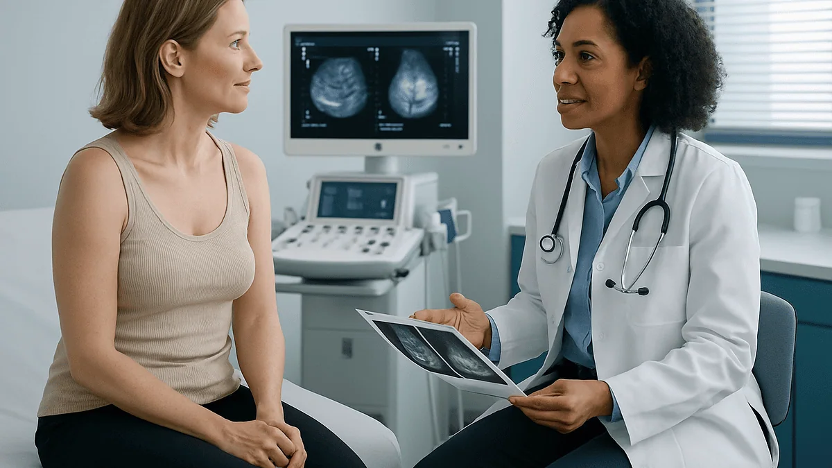
A mammogram after fat transfer is usually arranged so radiologists can compare the new images with a baseline scan taken before surgery. Grafted fat can form oil cysts, calcifications or firm areas of fat necrosis that resemble other findings, so the before-and-after comparison helps the imaging team interpret changes. Timing and follow-up are decided by the surgeon and radiologist together.
The folder on her lap was thicker than she expected. She had come to talk about reshaping her breasts with her own fat, and the first thing the surgeon asked for was not a photograph of the result she hoped for, but a scan of what was already there. A mammogram, before anything else. She left the consultation with an imaging appointment instead of a surgery date, and a question she had not thought to ask: why look so carefully at a breast nobody had touched yet?
The answer is less about suspicion and more about reading. A mammogram after fat transfer tells a different story from one taken before, and the two are read side by side, sometimes years apart. Grafted fat settles, scars, calcifies, occasionally forms a lump. None of that is unusual, but all of it can look like something else on a screen.
This explainer walks through what imaging teams are actually looking for, why the baseline matters so much, and what the weeks and years after a graft typically involve.
Why a mammogram before fat transfer is taken first
A mammogram is a low-dose X-ray of the breast that shows glandular tissue, fat, cysts and calcifications, the tiny mineral deposits that radiologists examine closely. Mayo Clinic describes it as the standard tool for both routine screening and investigating a specific concern. Before fat transfer, a surgeon typically wants that picture for two reasons that have nothing to do with the cosmetic result.
The first is to confirm the breast is suitable for surgery at all. If the baseline scan shows an area that needs further work-up, that work-up comes first; moving fat into a breast with an uninvestigated finding would muddy any later assessment. The second reason is comparison. Every mammogram is read against earlier films when they exist. Once fat has been grafted, the breast will contain new structures that were not there before, and the only way to say with confidence that a finding is new, old, or changed is to have the old image to hand.
Think of it as a before photo for the radiologist rather than the patient. Without it, every post-graft calcification has to be judged on its own, which raises the chance of an additional scan or a biopsy that a comparison film might have made unnecessary. Guidance from the NHS and the CDC emphasizes that stable findings over time are one of the strongest reassurances in breast imaging, and stability can only be measured from a starting point.
Who orders the baseline, at what age, and whether an ultrasound is added depends on the person’s screening history and risk profile. That judgement sits with the surgical and radiology teams, not with a blanket rule.
What actually happens to grafted fat inside the breast
Fat transfer, also called autologous fat grafting, means fat is removed from one part of the body by liposuction, processed to separate it from fluid and oil, and injected in small amounts into another area. Autologous simply means the tissue comes from the same person. In the breast, the fat is placed in many fine threads through small entry points rather than as a single mass.
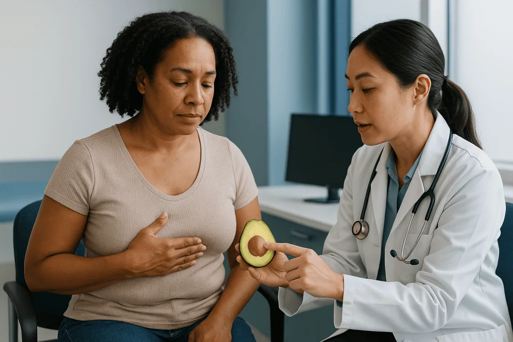
Those threads matter, because grafted fat has no blood supply of its own on the day it is placed. Each fat cell survives the first days by absorbing nutrients from surrounding tissue, then depends on new capillaries growing in. Cells that sit too far from a blood vessel do not make it. That is why surgeons spread the graft thinly and why a portion of any graft is expected to be reabsorbed in the months after surgery. The final volume is usually smaller than the volume injected, which is one reason some people are offered more than one session.
Fat that dies but is not fully cleared away becomes fat necrosis. Necrosis is the medical word for tissue death. The body walls it off, sometimes into a pocket of oil (an oil cyst), sometimes into firm scar, and over time calcium can settle at the edges. On a mammogram these show up as round lucent spots, rim-shaped calcifications, or denser scar-like areas.
Nothing about this process is unique to breasts. Fat necrosis happens after any breast surgery, after injury, even after a seatbelt bruise. What fat transfer changes is the quantity of fat exposed to the risk, and therefore how often these findings appear on later scans.
Does fat transfer affect mammograms? What the evidence shows
This is the question that worries most people, and the honest answer has two halves. Fat transfer does change what a mammogram shows. It does not, on current evidence, stop a mammogram from doing its job, provided the imaging team knows the history and has a baseline to compare with.
The changes are predictable. Oil cysts appear as smooth, round, fat-density spots, often with a thin calcified rim as they age. Scar from fat necrosis can look like an irregular dense area. Microcalcifications, the fine speckles radiologists scrutinize most carefully, can cluster around dying fat. In isolation, some of these patterns overlap with the appearances that prompt a biopsy. Reviews of imaging after breast fat grafting consistently describe these findings as common and usually recognizable, especially when the radiologist knows a graft has been placed and when earlier films exist.
Radiologists have decades of experience with the same appearances from other causes. Fat necrosis after reduction surgery, lumpectomy or trauma has long been part of routine reading, and the features that distinguish a benign oil cyst from a suspicious lesion are well described in standard teaching. Where ambiguity remains, the next step is additional imaging such as targeted ultrasound, and if still unclear, a needle biopsy. That pathway is the same one used for any indeterminate finding.
What the evidence does not support is any claim that fat transfer makes breast cancer harder to find in a way that should change screening intervals on its own. Screening recommendations from the CDC and NHS are based on age and personal risk, and a graft history is handled as additional context, not as a reason to scan less often.
Fat necrosis after fat transfer: what causes lumps to form
Someone who feels a firm area a few months after surgery often assumes the worst, or the opposite, assumes it must be the graft and ignores it. Neither reflex is helpful, so it is worth understanding the mechanism.

A lump after fat transfer most often comes from fat that did not establish a blood supply. The body’s clean-up cells move in, the dead fat liquefies into oil, and a fibrous wall forms around it. The result can feel smooth and mobile, like a small marble, or firmer and less distinct if scar predominates. Larger grafts placed as a single bolus rather than in fine threads carry a higher chance of central necrosis, because the center of a big deposit sits farthest from any vessel.
Other causes of a post-operative lump include a hematoma, a collection of blood that forms in the first days, and a seroma, a pocket of clear fluid. Both usually resolve, though some are drained. Infection can also produce a tender, warm swelling and is treated as a separate problem. Finally, and this is the reason imaging exists, a new lump can be unrelated to the graft at all.
Because touch cannot reliably tell these apart, a new or changing lump is assessed clinically and, where appropriate, with imaging. Many fat necrosis lumps soften or shrink over months as the body absorbs the oil; some persist indefinitely and are simply documented and watched. Whether a lump is monitored, imaged again or sampled is a decision for the surgeon and radiologist, informed by its appearance, its timing and the baseline films.
Who fat transfer to the breast is usually for, and who is asked to wait
Fat grafting to the breast is used in two broad settings. In reconstruction, it fills contour gaps after mastectomy or lumpectomy, softens the edge of an implant, or builds volume over several sessions. In cosmetic breast augmentation, it offers a modest increase in size and shape for people who want to avoid an implant and who have enough donor fat elsewhere. Mayo Clinic notes that fat grafting generally provides a smaller change than implants, which is a central part of any candid consultation.
People commonly asked to wait, or offered a different route, include those with an unresolved finding on the baseline mammogram, which needs investigation first. A personal history of breast cancer does not rule out grafting; it is used in reconstruction every day, but the oncology team is usually involved in timing and in follow-up imaging. Active smoking, uncontrolled diabetes and other conditions that impair wound healing and blood supply reduce graft survival and raise complication rates, so surgeons often ask for these to be addressed first. Significant planned weight loss is another reason to pause, because the grafted fat behaves like the fat it came from and will shrink with it.
Very low body fat limits what can be harvested. Pregnancy and breastfeeding are reasons to postpone. People with strong family histories or known genetic risk are typically advised to discuss the plan with a breast specialist, not because grafting is forbidden, but because their imaging schedule is already more intensive and should be built into the decision.
None of these are rules a patient applies alone. They are the questions a surgical team works through, and the outcome of that conversation varies from person to person.
Breast fat grafting recovery time: what the first weeks look like
Recovery involves two sites, the donor area and the breast, and the donor area is frequently the sorer of the two. Liposuction leaves bruising and swelling that peak within the first several days, and a compression garment is commonly worn to limit fluid build-up. The breasts themselves tend to feel full, tender and tight, with bruising around the small entry points.
NHS guidance on breast enlargement surgery describes a recovery measured in weeks rather than days, with most people needing time off work in the early phase and avoiding strenuous activity until the surgeon clears it. Individual plans differ, so the specific timeline is set by the treating team. Early walking is encouraged in almost every protocol because it supports circulation and lowers the risk of blood clots after any surgery.
A few points surprise people. The breasts look larger in the first weeks than they will later, because swelling adds volume that disappears and because some grafted fat is reabsorbed. Surgeons generally advise against pressure on the chest during the early period, which affects sleeping position and bras. Numbness or odd sensations around the entry points and donor area are common and usually settle over months as small nerves recover.
The graft itself is judged over a longer horizon. Volume continues to change for several months while the surviving fat integrates and the dead fraction is cleared. Follow-up visits in that window check healing, note any firm areas, and decide whether imaging is needed sooner than the routine schedule. Patients who keep a simple diary of changes, including anything they feel and when it appeared, give their team far more to work with than memory alone.
How radiologists read a mammogram after fat transfer
Reading post-graft films is a matter of pattern recognition against context. The radiologist wants three things: the knowledge that a graft was placed, the baseline images, and any prior post-operative scans. With those, the typical appearances of grafted fat are usually straightforward. Without them, the same pictures can prompt extra tests.
The table below summarizes features that imaging teams weigh. It is written to explain the radiologist’s lens, not to help anyone grade their own scan.
| Finding | Typical appearance after fat transfer | What raises the radiologist’s attention |
|---|---|---|
| Oil cyst | Round, smooth, fat-density; may develop a thin calcified rim over time | Irregular margin or growth between scans |
| Calcifications | Coarse or rim-shaped, scattered, often stable over years | Fine, clustered, linear or branching patterns |
| Fat necrosis scar | Dense area that stabilizes or shrinks on later films | New or enlarging mass with spiculated edges |
| Grafted fat | Increased fat-density tissue in the treated zones | No concern on its own; noted for comparison |
Stability is the recurring theme. A rim-calcified oil cyst that looks identical on two scans a year apart is reassuring in a way no single image can be. Mayo Clinic’s description of mammography stresses that comparison with earlier images is a routine part of interpretation, and that is exactly why the pre-surgery scan was taken.
When a feature is indeterminate, the standard sequence applies: additional views, targeted ultrasound, and if needed a biopsy. A biopsy in this setting is not a sign that something went wrong; it is the imaging pathway working as designed.
Can weight gain affect mammogram results and fat grafts?
Yes, in two different ways, and the distinction is useful. Weight change alters the mammogram itself, and it alters the graft.
Breast tissue is a mix of glandular and fatty components. Mayo Clinic’s guidance on dense breast tissue explains that denser, more glandular breasts appear whiter on a mammogram and can make some findings harder to see, while fatty tissue appears darker and is easier to read through. Weight gain generally adds fat to the breast and can lower density; weight loss can do the reverse. Either shift changes the look of the film, which is one more reason radiologists lean on serial comparison rather than a single image.
The second effect concerns the graft. Transferred fat keeps the biology of the place it came from. If it was harvested from the abdomen, it will enlarge and shrink with the abdomen as body weight moves. That means a result that looks settled at one weight can change noticeably after a substantial gain or loss. Surgeons often ask people to be at a stable weight before the procedure for this reason, and it is why large planned weight changes are a common reason to postpone.
Hormonal changes act on the breast too. Pregnancy, breastfeeding and menopause each shift the balance of glandular and fatty tissue, and some people notice their grafted breasts respond differently from how they expected. None of this makes imaging unreliable; it makes the history on the request form more important. Telling the mammographer about weight change, hormonal events and the date of the graft is a small act that materially helps whoever reads the scan.
When is the first mammogram after fat transfer usually scheduled?
There is no single interval written into guidelines, and anyone who promises one is simplifying. What exists is a logic that most teams follow.
Routine screening continues on the schedule that applies to the person’s age and risk. The CDC summarizes US recommendations that screening is offered on a regular cycle through midlife and beyond, and the NHS breast screening program invites eligible women at fixed intervals. A fat graft does not reset that calendar. If someone is already in a screening program, the next invitation usually stands; if they are younger than screening age, imaging is driven by clinical need rather than routine.
Beyond routine screening, many surgeons arrange a post-operative scan once the breast has settled, after the swelling has gone and the graft has stabilized. The purpose is to document the new baseline: where the oil cysts are, which calcifications belong to the graft, what the scar looks like. That image becomes the reference for everything that follows. Timing is set individually, and some teams choose ultrasound rather than mammography for this early check, particularly in younger people with dense tissue.
Imaging may also be brought forward for a reason: a new lump, a change in shape, pain that persists, or a finding on examination at follow-up. In reconstruction after cancer, the oncology team’s surveillance plan takes precedence and the graft history is folded into it.
The practical advice is simple. Ask the surgical team when they expect the next scan, who will order it, and how the imaging department will be told about the graft. Then keep a copy of the reports, because people move, and the baseline is only useful if it travels with them.
Fat grafting after mastectomy and reconstruction: imaging and recovery
In reconstruction, fat transfer is rarely the main event. It is usually a refinement, added after an implant or a tissue flap to smooth hollows, soften edges and improve symmetry, often across more than one session. Recovery from the grafting sessions themselves is generally lighter than from the original reconstruction, which involves a larger operation and a longer healing period.
How long recovery from a mastectomy with reconstruction takes depends heavily on the method. Implant-based reconstruction and flap reconstruction, where tissue is moved from the abdomen or back, have different timelines, and both are longer than a fat grafting session. The NHS and Mayo Clinic describe recovery from major breast surgery in terms of weeks before normal activity and months before the final result is seen. The treating team gives the specific plan, and it is adjusted to how healing progresses.
Imaging after reconstruction follows a different path from screening a native breast. After a mastectomy there is little or no breast tissue left to screen, so routine mammography of that side is often not performed; surveillance relies on examination, with imaging used for specific concerns. Fat necrosis is still common in reconstructed breasts and can produce lumps that worry people, particularly those who have already been through cancer. The same principles apply: new lumps are assessed, not assumed, and ultrasound or MRI are frequently chosen because they handle implants and flaps well.
The remaining breast, if there is one, continues on its own screening schedule, which may be more intensive after a cancer diagnosis. Coordination between the oncology team, the reconstructive surgeon and radiology is what keeps that picture coherent, and patients are entitled to ask how that coordination is being managed.
Ultrasound and MRI: when a mammogram is not the whole story
Mammography is the backbone of breast imaging, but it is not the only tool, and after fat transfer the others earn their place.
Ultrasound uses sound waves rather than X-rays and is particularly good at telling a fluid-filled structure from a solid one. An oil cyst, which is essentially a tiny pocket of liquefied fat, has a characteristic look on ultrasound that often settles a question the mammogram left open. It is also the usual first step when a specific lump can be felt, because the probe can be placed directly over it. Ultrasound involves no radiation, which makes it the common choice in younger people and in pregnancy.
MRI, magnetic resonance imaging, builds detailed cross-sectional pictures using a magnetic field and usually a contrast dye. It is the most sensitive of the three for picking up areas of unusual blood flow, and it is used in high-risk screening programs and in some reconstruction follow-up. The trade-off is that it also picks up more benign changes, including the inflammatory activity around healing fat, which can lead to additional tests. For that reason MRI is used selectively rather than routinely after grafting, and its timing relative to surgery is something radiologists consider carefully.
None of these replaces the others. The pattern that emerges from mainstream guidance is layered: mammography for the overview, ultrasound for a targeted question, MRI when risk or ambiguity justifies it. Which layers apply to a given person is a radiology decision made with the surgical team, and the patient’s role is to make sure each report is filed with the rest of the history.
What people often get wrong
Fat transfer attracts a particular set of myths, and most of them run in opposite directions.
The first is that fat grafting makes cancer impossible to detect. The evidence does not support this. It changes the images, and those changes are well characterized; with history and comparison films, radiologists read them routinely. The opposite myth, that fat grafting has no effect on imaging at all, is equally unhelpful. It does, which is why the pre-surgery mammogram exists.
The second is that any lump after a graft is just fat. Most are, but a lump is assessed on its features and its behavior, not on the assumption that surgery explains it. The reverse error, treating every firm area as an emergency, causes needless distress. The balanced position is that new or changing lumps are reported and examined, and the team decides what, if anything, comes next.
The third concerns permanence. Grafted fat is living tissue. It survives, but it also responds to weight change and hormones, and a portion is reabsorbed early on. The result is durable rather than fixed, and the breasts will continue to age and change like any others.
The fourth is that fat grafting is a lighter version of an implant with the same outcome. Mayo Clinic notes that the volume gain is typically modest, and some people need more than one session to approach what they had in mind. That is a feature of the biology, not a failure.
The last is the belief that screening can be relaxed because the breasts have been checked for surgery. The baseline is one scan on one day. Screening works through repetition over years, and a graft is a reason to keep that rhythm, never to abandon it.
Questions to ask your care team
A good consultation leaves someone with answers that are specific to them, and the right questions make that more likely. These are the ones that matter most for imaging around fat transfer.
- Which imaging do you want before surgery, and why that modality for me?
- Will you review the baseline scan yourself, and will the radiologist’s report be in my surgical notes?
- How will the imaging department be told that I have had a fat graft when I attend future scans?
- When do you expect the first scan after surgery, and who orders it?
- If I already attend a screening program, does anything change?
- How much of the grafted volume do you typically expect to be reabsorbed, and how would you know whether a second session is appropriate for me?
- What would make you want to image sooner than planned?
- If a lump appears, who do I contact, and what is the usual pathway from there?
- How should I describe this procedure to a radiologist or doctor who does not know my history?
- Are there features of my personal or family history that change the imaging plan?
Write the answers down. The value of these questions is not the reassurance in the room but the record afterward, because imaging teams years from now will rely on what was documented, not on what was said.
People in reconstruction should add one more question: how does the oncology surveillance plan and the reconstruction plan fit together, and who holds the overview? When several teams are involved, knowing which clinician coordinates the imaging prevents scans being duplicated or, worse, missed.
When to call your doctor
Most of what people notice after fat transfer is expected healing, and the follow-up schedule is built to catch the rest. Some signs, though, should prompt a same-day call to the surgical team or, out of hours, urgent care, rather than waiting for the next appointment.
Contact the team promptly for a breast or donor site that becomes increasingly red, hot, swollen or painful, particularly with fever or chills, because these can indicate infection or a collection of blood or fluid that needs assessment. Rapid swelling of one breast in the first days, a wound that opens or leaks persistently, or a spreading rash around the entry points also need review.
Seek emergency care immediately for chest pain, sudden shortness of breath, coughing up blood, or a calf that becomes painful, swollen or warm, since any surgery raises the risk of a blood clot and these are the signs that one may have formed or traveled to the lung. Fainting, confusion or a very fast heartbeat are likewise emergencies.
In the months and years after, call about any new lump that is growing, hardening or changing shape, skin that dimples or thickens, a nipple that newly inverts or discharges, or a change in the size or outline of one breast that was not there before. Those are the same signs that warrant assessment in anyone, and the fact that a graft was placed does not alter the advice; it only adds context for whoever examines the breast.
The team may examine, reassure, arrange imaging or sample an area. Each of those is a normal step on a well-worn pathway, and the decision about which applies belongs to them.
Frequently asked questions
Does fat transfer affect mammograms?
It changes what the images show without preventing them from working. Oil cysts, calcifications and scar from fat necrosis are common after grafting and are familiar to radiologists from other breast surgery. With the pre-operative baseline and a note of the graft on the request form, these findings are usually recognized; where doubt remains, ultrasound or biopsy follows the standard pathway.
How long does it take to recover from fat grafting to the breasts?
Recovery is measured in weeks, with the liposuction donor site often sorer than the breasts. NHS guidance on breast surgery describes time off work early on and avoiding strenuous activity until cleared by the surgeon. Swelling settles over the first weeks, while the graft volume continues to change for months as surviving fat integrates and the rest is reabsorbed. The treating team sets the individual timeline.
What causes lumps to form on the breast after fat transfer?
The most frequent cause is fat necrosis: grafted fat that failed to gain a blood supply liquefies, is walled off, and may calcify, producing a smooth or firm lump. Blood or fluid collections and infection are other early causes. Because a lump can also be unrelated to the graft, new or changing lumps are examined and imaged rather than assumed to be benign.
Can weight gain affect mammogram results?
Yes. Weight gain generally adds fat to the breast and can reduce density, altering how the mammogram looks, while weight loss can increase density. Mayo Clinic notes that denser tissue appears whiter and can hide some findings. Grafted fat also enlarges or shrinks with body weight, so surgeons usually prefer a stable weight before surgery and radiologists want to know about major changes.
When should I have my first mammogram after fat transfer?
There is no fixed interval in guidelines. Routine screening continues on the schedule set by age and risk, and many surgeons add a scan once swelling has settled to document the new baseline. Some teams use ultrasound for that early check, especially in younger people with dense breasts. Ask the surgical team when they expect the next scan and who will order it.
Will fat grafting breast lumps need a biopsy?
Not usually, but sometimes. Many lumps from fat necrosis have characteristic features on mammography or ultrasound and are simply documented and followed. When the appearance is indeterminate, a needle biopsy is the standard way to settle the question. A biopsy in this setting means the imaging pathway is doing its job, not that something has gone wrong with the graft.
How long does it take to recover from a mastectomy with reconstruction?
Longer than from a fat grafting session, and it depends on the method. Implant-based and flap-based reconstruction have different timelines, with weeks before normal activity and months before the final shape is seen according to NHS and Mayo Clinic descriptions. Fat transfer is often added later as a refinement, and each of those sessions has a lighter recovery than the original surgery.
Is fat necrosis after fat transfer dangerous?
Fat necrosis itself is a benign process: dead fat that the body has walled off. It can cause a lump, tenderness or a visible change in contour, and some areas soften over months while others persist. Its importance lies in imaging, where it can resemble other findings, which is why it is documented carefully rather than treated as a danger in its own right.
Should I tell the mammographer that I have had fat transfer?
Yes, every time. The person performing the scan records the history on the request form, and the radiologist relies on it when interpreting oil cysts, calcifications and scar. It also helps to mention the approximate timing of the surgery, any weight change, and where previous films were taken so they can be retrieved for comparison.
Does a fat graft mean I can skip routine breast screening?
No. The baseline scan before surgery is a single image on a single day, while screening works through repeated comparison over years. The CDC and NHS screening recommendations are based on age and risk and are not changed by a fat graft. If anything, the graft is a reason to keep the schedule, because stability over time is what makes post-graft findings easy to read.
References
This article is for general information only and is not a substitute for professional medical advice. Please consult a qualified doctor about your individual situation.

More from the Blog
Liposuction Alone or Gland Excision? How Gynecomastia Surgery Is Matched to Tissue Type
Liposuction removes fatty tissue, while surgical excision removes the firmer glandular disc behind the nipple, so surgeons choose based on what the chest actually…

Gynecomastia Surgery Prep: Smoking, Supplements and the Tests Booked Beforehand
Preparing for gynecomastia surgery usually means stopping all nicotine for the period your surgeon sets, giving the team a complete list of medicines and…
How Does the Chest Settle After Gynaecomastia Treatment? Results Over the First Months
After gynaecomastia treatment, the chest settles in stages rather than all at once. Swelling and bruising are most noticeable in the first one to…
Combined Procedures in Breast Aesthetics: When a Lift and Implants Happen in One Operation
A combined breast lift and augmentation, also called augmentation mastopexy, reshapes and raises sagging breast tissue while placing an implant for volume, all under…

Reading a Breast Aesthetics Surgical Plan: Measurements, Options and Questions to Raise
A breast aesthetics surgical plan usually lists your chest and breast measurements, the surgeon's proposed technique, and the alternatives that were considered. Reading it…

Preparing for Breast Augmentation: Imaging, Medication Review and the Final Sizing Visit
Preparing for breast augmentation usually involves three steps spread over several weeks: a baseline breast imaging review if you are of screening age or…