

Bringing Old X-Rays and Records to a Dental Anatomy Assessment: Why History Helps Planning
Key Takeaways
- A single dental X-ray shows where things stand; a pair taken years apart shows whether a shadow, bone level, or root is stable or changing, which is the information planning depends on.
- Periodontal charts and treatment notes are often more decisive for planning than images alone, because gum disease and previous work are judged by trend and detail rather than appearance.
- Patients in the United States have a legal right to copies of their dental records, but practices do not send them to a new dentist automatically; the request has to come from you.
- Digital image files in standard formats preserve the margin detail where diagnosis happens; photographs of a screen or photocopies of film lose it.
- Bringing recent images may allow the team to skip or narrow new X-rays, though a current image is still needed when the clinical question is current, and the decision sits with the treating team.
- When records are lost, insurance statements listing procedures by tooth number, receipts, and an honest personal timeline still help reconstruct history for the clinician.
Bringing old X-rays and records to a dental anatomy assessment gives the clinician a timeline rather than a single snapshot. Prior radiographs, periodontal charts, and treatment notes show whether bone levels, cavities, or root changes are stable or progressing, may reduce the need for repeat imaging, and help the team plan around existing work. Request copies from previous dentists in advance; the treating team decides what still needs imaging.
The envelope had been in a kitchen drawer for years: three small films from a dentist two cities ago, a panoramic image in a paper sleeve, and a folded printout of a gum chart nobody had ever explained. When a new dental assessment was booked, the question came up almost as an afterthought. Is any of this worth carrying across town, or is it just clutter?
It turns out to be one of the more useful things a patient can do. If you plan to bring dental records to appointment day, you hand the clinician something a brand-new X-ray cannot provide on its own: time. A single image shows where things stand. Two images taken years apart show where things are going.
This explainer walks through what a dental anatomy assessment involves, which records genuinely help, how to request and transfer them, what to do when old films are lost, and how history changes the way a plan is built.
Why bring dental records to appointment for an anatomy assessment?
A dental anatomy assessment is a structured look at the shape, position, and condition of the teeth, the bone that holds them, the gums, and the way the upper and lower jaws meet. Clinicians describe it as building a baseline. The trouble with a baseline is that it has no past. On its own, a slightly shortened root, a small dark shadow near a filling, or a millimeter of lost bone raises a question it cannot answer: was this always here, or is it new?
Old records answer that question. A radiograph, which is simply the medical term for an X-ray image, taken four years earlier can show the same shadow at the same size, which changes it from a suspected problem into a known, stable feature. The reverse is also true. A change that looks trivial today becomes meaningful when the earlier image shows nothing at all in that spot.
Mayo Clinic notes that a dental exam typically combines a visual and manual check of teeth, gums, and jaw with X-rays chosen according to individual risk and history. History is not decoration in that sentence; it is one of the inputs. Records also carry information that no camera captures, such as why a tooth was extracted, whether a root canal was completed or abandoned, and which materials were used in a crown.
The practical upshot is straightforward. When you bring dental records to appointment, the clinician spends less time reconstructing your story from clues and more time interpreting it. For people with extensive past work, several previous dentists, or a history of gum disease, the difference between a snapshot and a timeline can shape the entire plan.
What actually happens during a dental anatomy assessment
Expect the appointment to move through predictable stages, though the order varies between practices. Most begin with a conversation. The clinician asks about pain, sensitivity, previous treatment, medical conditions, and medicines, because conditions such as diabetes and some medicines affecting saliva or bone can change how the mouth behaves.
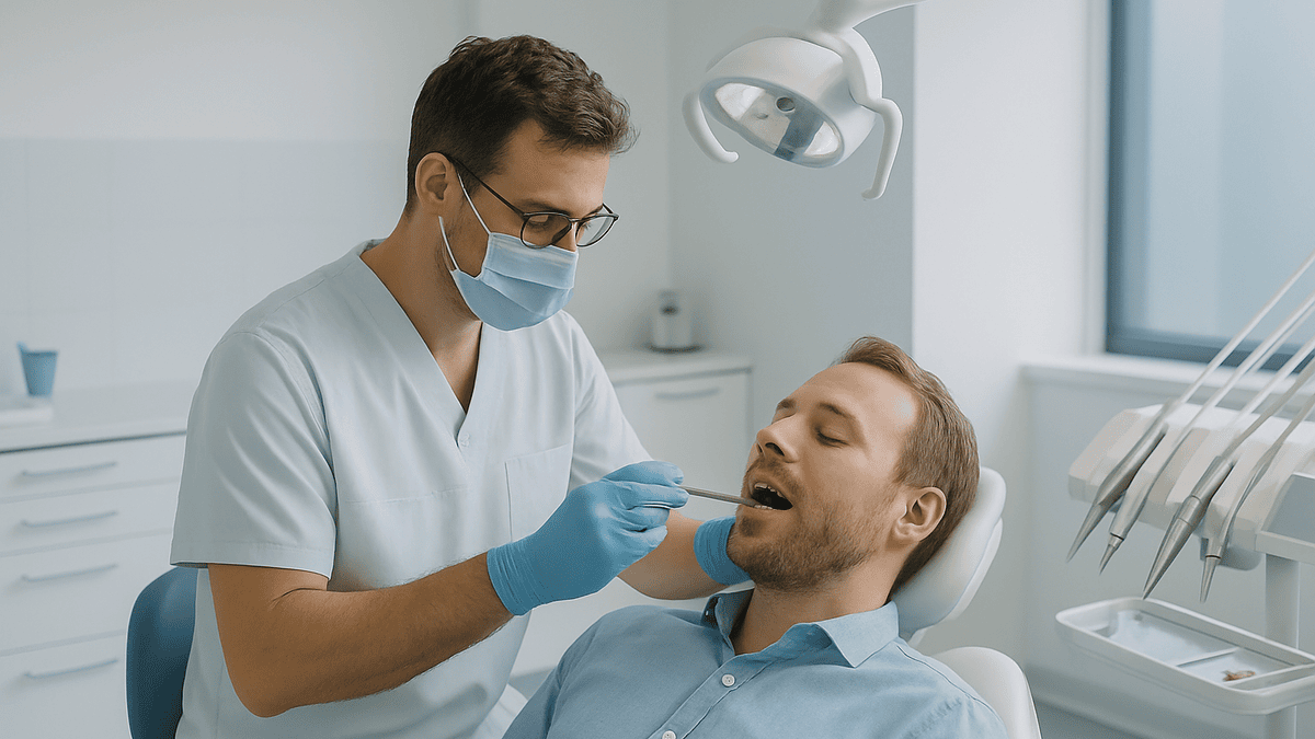
The hands-on part follows. Each tooth is checked for cavities, cracks, worn edges, and the condition of existing fillings or crowns. The gums are examined with a slim probe, a blunt measuring instrument that gauges how deep the small pocket between gum and tooth runs. Those measurements are recorded on a periodontal chart, a tooth-by-tooth map of gum health. The clinician will also look at how the teeth come together when the jaw closes, called occlusion, and check the jaw joints, the soft tissue of the cheeks, tongue, and palate, and the lymph nodes of the neck.
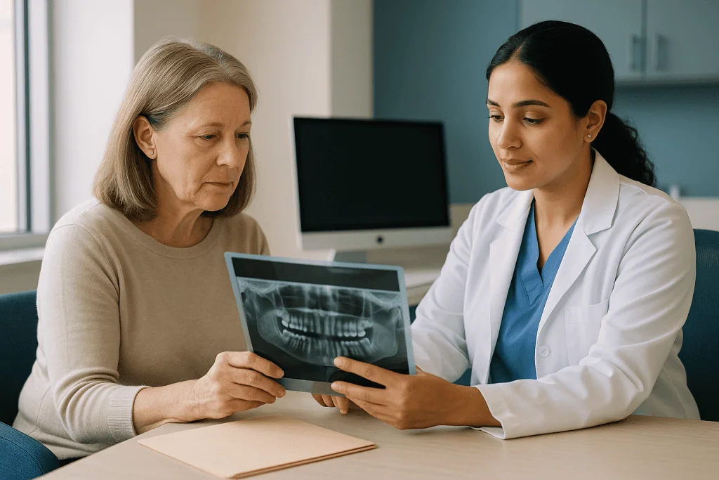
Imaging comes next, if needed. According to MedlinePlus, dental X-rays help find problems not visible during a regular exam, including decay between teeth, bone loss, abscesses, and changes in the bone around the roots. Which images are taken depends on what the exam and your history suggest. This is precisely where old records earn their place: a clinician who can see last year’s images may choose fewer new ones, or target them more narrowly.
The visit ends with a summary. Findings are placed in context, options are outlined, and any decisions about treatment or monitoring are left to you and the treating team. Nothing in a good assessment requires a same-day commitment.
What dental records include, and which ones matter most
Patients often imagine dental records as a stack of X-rays and nothing else. The complete file is broader, and several of the least glamorous items carry the most weight.
- Radiographs. Bitewings, periapicals, panoramic images, and sometimes three-dimensional scans. Each type is explained in the table further down.
- Periodontal charts. The pocket-depth measurements around every tooth, along with notes on bleeding and gum recession. These are the only reliable way to judge whether gum disease is stable or advancing.
- Treatment notes. Dated entries describing what was done, to which tooth, with what materials, and any complications.
- Diagnostic notes and diagrams. Tooth charts marking existing fillings, crowns, missing teeth, and implants.
- Medical history forms. Conditions, medicines, allergies, and any recorded reactions to local anesthetic.
- Laboratory prescriptions. Specifications for crowns, bridges, or dentures, including shade and material.
- Orthodontic records. Models, photographs, and cephalometric images, which are side-view skull X-rays used to plan tooth movement.
If you can only obtain part of the file, prioritize the radiographs and the periodontal charts. Together they describe the two things most likely to change over time: the teeth themselves and the bone supporting them. Treatment notes come a close third, particularly for teeth that have had root canals, because the clinician will want to know whether the procedure was completed and how the tooth was restored afterward.
MedlinePlus, in its guidance on personal health records, encourages patients to keep copies of test results and treatment summaries in one place. Dental information belongs in that same folder. A mouth is not separate from the rest of the body, and a clinician assessing it benefits from the same organized history any other specialist would.
Why old X-rays tell a story a new one cannot
Consider a small dark area on a bitewing near the edge of an old filling. Dark on a dental X-ray usually means less dense tissue, which can indicate decay. It can also indicate a normal anatomical feature, a slight gap in the filling that has been there for years, or a shadow created by the angle of the beam. Faced with that single image, a careful clinician has three choices: monitor, investigate, or treat. Each has a cost in time, comfort, or tooth structure.
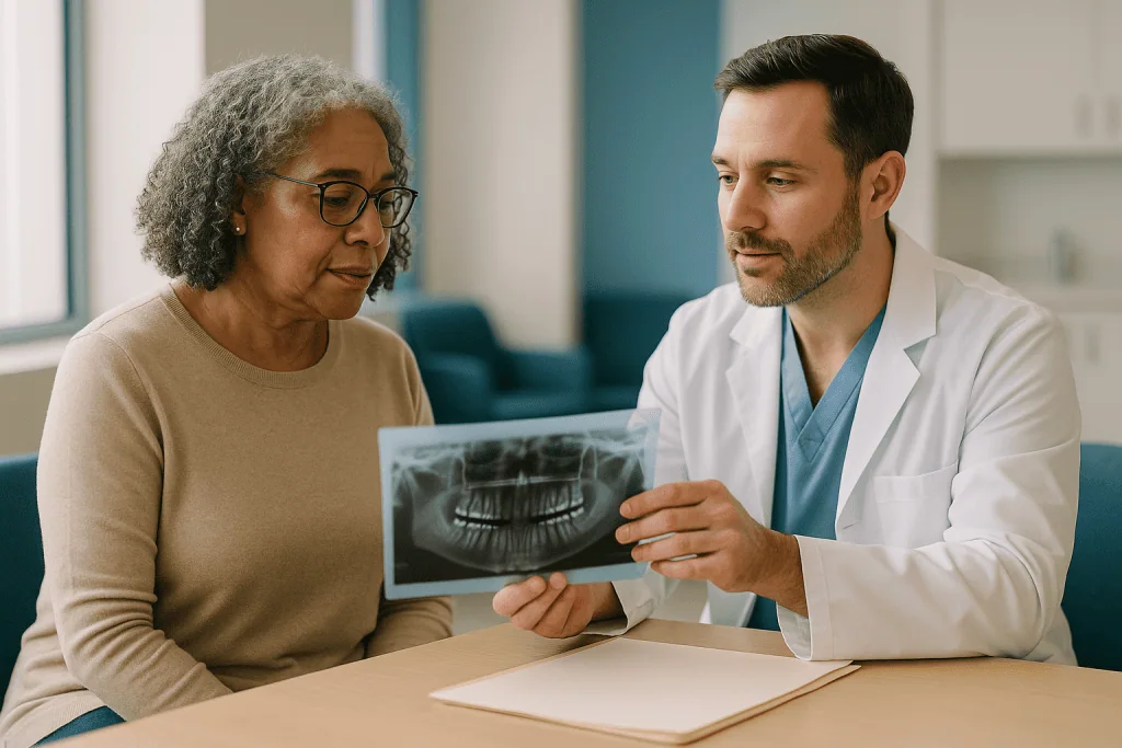
Now place a bitewing from three years earlier beside it. If the dark area is identical, the picture changes from ambiguous to reassuring. The team can reasonably choose to monitor. If the area has grown, the decision to intervene rests on evidence of progression rather than suspicion. Either way, the old image has done work that no amount of squinting at the new one could.
Bone tells the same kind of story. Cleveland Clinic describes how dental X-rays reveal bone loss associated with gum disease, but a single measurement of bone height has limited meaning. Some people have naturally shorter bone crests; others have lost bone slowly and stopped. Serial images separate the two. The same logic applies to root shortening after orthodontic treatment, the slow expansion of a cyst, the position of a wisdom tooth relative to the nerve canal, and the fit of a crown margin.
There is an honest limit here. Older images taken with different equipment, angles, and exposure settings cannot be compared millimeter for millimeter. Clinicians account for that by looking at trends rather than exact figures. Even an imperfect earlier image usually narrows the range of possibilities, which is the whole point of planning.
Types of dental X-rays and what each reveals over time
Not every image in an old envelope is equally useful for every question. The table below summarizes the common types, what they show, and how they help when compared with a newer image. Definitions follow the conventions used by MedlinePlus and Cleveland Clinic.
| Image type | What it shows | Why an old copy helps |
|---|---|---|
| Bitewing | Crowns of upper and lower back teeth in one view, plus the bone crest between them | Tracks decay between teeth and early bone changes over time |
| Periapical | One or two whole teeth from crown to root tip and surrounding bone | Compares root health, healing after root canal, and changes around the root tip |
| Panoramic | Entire upper and lower jaws, all teeth, sinuses, and jaw joints in a single wide image | Shows position of unerupted teeth, cysts, and jaw structure; useful for long-range comparison |
| Occlusal | Floor or roof of the mouth in one broad view | Locates extra teeth or objects; less often used for follow-up |
| Cephalometric | Side view of the skull and jaws | Orthodontic growth and jaw relationship over years |
| Cone-beam CT | Three-dimensional scan of a region of the jaw | Detailed bone volume and nerve position; prior scans help avoid repeat scanning |
A cone-beam CT, sometimes written CBCT, is a specialized three-dimensional X-ray that captures a cylinder of tissue rather than a flat picture. It carries a higher dose than a bitewing and is reserved for specific questions such as implant planning or complex root anatomy. Because of that, a previous scan is especially worth retrieving; the treating team can decide whether it still answers the current question.
Film images and digital images differ in handling but not in value. Digital files can be enlarged and adjusted for contrast, which makes side-by-side comparison easier, but a clear film held to a light box still tells its story.
How to get dental records from a previous dentist
In the United States, patients have a legal right under federal privacy law to access their health records, and dental records are included. Exercising that right is usually simpler than people expect, though it works best with a little lead time.
Start by contacting the former practice directly, by phone or through its patient portal. Ask for a copy of your complete record, and name the items specifically: radiographs, periodontal charts, treatment notes, and any laboratory prescriptions. Many practices have a short authorization form to sign; some accept a signed letter. If you prefer, you can ask that the copies be sent straight to your new provider, which avoids handling the files yourself.
Timing matters. Practices may take days to a few weeks to fulfill a request, depending on whether records are digital or in paper archives, so make the call as soon as the assessment is booked. If the practice has closed, records are typically transferred to another practice or a custodian, and the state dental board can often say where. If a dentist has retired, a forwarding notice is frequently posted online or at the former address.
Ask about format. Digital radiographs are best supplied as standard image files or in the format dentists commonly exchange, so they open on the new team’s software. Screenshots and photographs of a monitor lose detail and should be a last resort. For film, request the original films or high-quality scans, not photocopies.
A short, polite note describing why you need the records, that is, an upcoming anatomy assessment at a new practice, often speeds things along. Staff handle these requests routinely and generally understand that continuity of care depends on them.
How to transfer dental records to a new dentist without losing detail
Requesting records is half the task. Getting them to the new practice in a usable form is the other half, and it is where information most often goes missing.
The cleanest route is practice to practice. When the previous office sends files directly to the new one, images stay in their native digital format, the periodontal chart arrives as the clinician’s own record rather than a photo of it, and nothing sits on a phone waiting to be forgotten. Ask both offices whether they use compatible systems; if not, most can export standard image files with a short accompanying summary.
If you handle the transfer yourself, keep three principles in mind. First, preserve originals. Do not crop, filter, or compress image files, and do not laminate or tape film. Second, keep dates attached. A radiograph without a date is a puzzle with a missing piece; if the file name does not include one, write it on a separate sheet. Third, keep the set together. A partial periodontal chart or a single bitewing separated from its pair loses much of its comparative value.
Bring the files on the day even if you have already sent them ahead. Uploads fail, emails land in spam folders, and a memory stick or a paper sleeve in your bag is a reliable backup. Tell the receptionist you have records with you when you check in, so the clinician can review them before the exam begins rather than at the end.
MedlinePlus recommends keeping your own copy of important health records and updating them after each visit. Once the new team has scanned your files, ask for the updated set back. The folder you began with becomes the seed of a record that follows you to every future appointment.
How long are dental X-rays kept, and what if they are gone?
Retention rules for dental records in the United States are set by state law and professional regulation rather than a single national standard, and the required period varies from state to state. Many practices keep records longer than the minimum, and digital storage has made it easier to retain images indefinitely. The realistic answer to how long your X-rays exist is: probably longer than you assume, but not forever, and it depends where you were treated.
When records genuinely cannot be found, the assessment does not fail. The clinician simply proceeds with the exam and, where the findings justify it, takes the images needed to establish a fresh baseline. Mayo Clinic notes that the frequency and type of dental X-rays depend on individual risk factors, age, and dental history, so a person without prior images is assessed on the same principles as anyone else; the team just has less past to draw on.
You can still supply history without films. Old appointment cards, insurance statements listing procedures by tooth number, receipts for crowns or root canals, and your own memory of when and where work was done all help reconstruct a timeline. Photographs of your smile from different years, though not diagnostic, can show tooth movement, wear, or gum changes.
Be candid about gaps. A clinician who knows you cannot recall whether a root canal was finished will plan accordingly, perhaps with a targeted periapical image, rather than assuming it was. What undermines planning is not missing information but unknown missing information.
Going forward, request a copy of every new set of images at the end of each visit. Storage is cheap, and the next time someone asks whether you have old X-rays, the answer will be yes.
Who benefits most from bringing history, and when a fresh baseline is preferred
Almost everyone benefits from bringing records, but the value is not evenly distributed. Some situations make history close to essential.
People with a diagnosed history of gum disease gain the most, because periodontal management is fundamentally about trends. A pocket that measured a certain depth two years ago and measures the same today is a different clinical situation from one that has deepened, even if the number is identical. Anyone with multiple crowns, bridges, implants, or root-canal-treated teeth also benefits, since the notes describing that work guide how it is examined and protected. Adults who moved between several dentists, people planning orthodontic or implant treatment, and those with a history of jaw joint problems or cysts all fall into the high-value group.
Children and adolescents are a special case. Their jaws are growing and teeth are erupting, so older images document developmental stages that cannot be recreated. Panoramic images from early adolescence are often the clearest record of where wisdom teeth started.
There are also situations where the team will ask for a fresh baseline regardless of what you bring. Images that are badly underexposed, undated, or taken at angles that make comparison impossible add little. Very old films may show teeth that have since been treated or removed. If a new symptom has appeared, a current image of that specific area is usually needed whatever the archive shows. Pregnancy is a common reason to postpone non-urgent imaging; the NHS advises telling your dentist if you are or might be pregnant so that imaging decisions can be made with that in mind.
None of this means old records were wasted. They still orient the clinician, and the decision about what to image now remains with the treating team.
Does bringing old X-rays mean fewer new ones? What the evidence supports
The honest answer is: often, but not automatically. Radiation from dental imaging is low. MedlinePlus states that the amount of radiation from dental X-rays is very low, and Cleveland Clinic describes it as a small fraction of the background radiation people receive from the environment each year. Low is not zero, though, and the guiding principle across health systems is that images should be taken when they are expected to change management.
Old images support that principle in two ways. First, they can make a planned image unnecessary. If a recent set of bitewings from the previous practice is available and the clinical exam raises no new concerns, the team may reasonably defer repeating them. Second, they can shrink the field. Instead of a full-mouth series, a single periapical of the one tooth that has changed may be enough.
What old images cannot do is substitute for a current picture when a current picture is needed. A film from several years ago says nothing about what has happened since. Mayo Clinic and the NHS both frame X-ray frequency as a function of individual risk rather than a fixed calendar; the NHS notes that recommended check-up intervals range from every three months to every two years depending on oral health, and imaging follows similar risk-based reasoning. Someone with a high cavity rate or active gum disease will generally need more recent images than someone whose mouth has been stable for a decade.
It is fair to ask the clinician which new images are proposed and what each is expected to show. A good team welcomes that conversation, and bringing history makes it more specific. The decision, as always, sits with the treating team.
What the following days and weeks usually look like after the assessment
An anatomy assessment is diagnostic, so there is rarely any physical recovery to speak of. Some people notice mild gum tenderness for a day or so after periodontal probing, particularly where gums were already inflamed, and a little bleeding during brushing that evening is common. Neither needs treatment; both typically settle with ordinary brushing and flossing.
The more meaningful aftermath is informational. Within days to a couple of weeks, depending on the practice, you should receive a summary of findings and, where relevant, a written plan. Ask for it if it does not arrive. The plan may propose immediate treatment, a period of monitoring, referral to a specialist such as a periodontist (a dentist who focuses on gums and supporting bone) or an endodontist (a dentist who focuses on root canals), or simply a recall interval.
This is also when history pays a second dividend. If the team decided that a suspicious area could be watched rather than treated because old images showed stability, the plan will specify when to re-image and what change would prompt action. That schedule is a shared agreement, not a promise about outcomes. Mouths change, and a monitoring plan exists precisely to catch change early.
Use the interval to consolidate your records. Add the new images and chart to your folder, note the date and the practice, and keep a copy of the plan alongside them. If you were referred to a specialist, send or bring the same set; specialists rely on prior imaging as heavily as general dentists do, and a referral with history attached is a referral that starts on the right foot.
What people often get wrong about dental records and X-rays
A handful of misunderstandings surface repeatedly in waiting rooms and online forums. Correcting them helps patients use their records well.
Old X-rays are useless because they are out of date. This reverses the logic. Their age is the asset. A five-year-old image is valuable precisely because it shows what your mouth looked like five years ago, allowing today’s image to be read as change or stability rather than as an isolated finding.
The new dentist will get the records automatically. Practices do not routinely exchange files without a patient’s request and authorization. You have to ask, and you may have to ask twice.
A photograph of the screen is as good as the file. It is not. Detail at the margins of fillings and the crest of bone, exactly where diagnosis happens, is lost in a phone photo of a monitor. Request the actual image files.
Bringing records means no new X-rays. It may reduce or narrow them, but a current image is still needed when the clinical question is current. The team decides.
Dental radiation is dangerous enough that skipping all imaging is safer. MedlinePlus and Cleveland Clinic describe the dose from dental X-rays as very low. The greater risk for most people is decay or bone loss developing unseen. The sensible position is risk-based imaging, not avoidance.
Records belong to the dentist. The physical files are the practice’s property, but the information is yours, and federal law gives you the right to obtain copies.
Only X-rays matter. Periodontal charts and treatment notes are frequently more decisive than images for planning around gum health and previous work.
Bring dental records to appointment: a practical checklist for the day
Preparation is mostly about gathering and labeling. A little order on your side saves the clinician a great deal of interpretation on theirs.
- Request records from every previous dentist as soon as the appointment is booked, and name radiographs, periodontal charts, and treatment notes specifically.
- Ask for digital images in standard file formats, or original films rather than photocopies.
- Confirm each image and chart carries a date; add one on a separate sheet if it does not.
- Write a one-page timeline in your own words: where you were treated, roughly when, and what you remember being done.
- List current medicines, medical conditions, and allergies, including any past reactions to dental anesthetic.
- Bring insurance statements or receipts that list procedures by tooth number if formal records are missing.
- Pack a physical or electronic backup even if files were sent ahead.
- Tell reception on arrival that you have records so they reach the clinician before the exam.
The timeline deserves a word. Patients sometimes feel embarrassed that they cannot remember whether a tooth was crowned eight years ago or twelve. Approximate dates are still useful, and honesty about uncertainty is more helpful than a confident guess. A clinician reading a note that says a lower molar was treated sometime after a move will search that region with appropriate care.
Do not worry about presentation. A neatly labeled paper folder works as well as a formatted digital archive. What matters is that the images, charts, and notes are together, dated, and legible, and that they arrive in the room at the same time you do. When you bring dental records to appointment in that state, you have done the part of planning that only you can do.
Questions to ask your care team
An assessment goes better as a dialogue. These questions help you understand how your history shaped the findings and what happens next; none requires a same-day answer.
- Which of the old images and charts were useful, and which were too old or unclear to help?
- Did any earlier image change how you interpreted something you saw today?
- Which new images do you recommend, what will each one show, and why is it needed now?
- Are there areas you have decided to monitor rather than treat, and what change would prompt action?
- How does my gum chart today compare with the earlier one, tooth by tooth?
- Is there any previous work, such as a root canal or crown, that you would like more information about?
- What recall interval do you suggest, and what is that recommendation based on?
- Can I have a copy of today’s images and chart before I leave?
- If I am being referred, will you send my full history to the specialist, or should I bring it?
- Is there anything in my medical history or medicines that affects how you plan around my teeth or gums?
The last question matters more than people expect. Conditions such as diabetes, some heart conditions, and medicines that affect bone or saliva can alter how the mouth responds to treatment and how closely it should be watched. The dental team will not change those medicines and should not be asked to; that decision belongs with the prescribing clinician. Knowing about them, however, allows the dental plan to be built realistically.
Write the answers down or ask for them in the written summary. A plan you can reread is a plan you can follow.
When to call your doctor or dentist
A dental anatomy assessment is not a treatment, and most people leave with nothing more than a plan and perhaps briefly tender gums. Some signs, however, should not wait for a scheduled recall and warrant a prompt call to your dental team or, in certain cases, urgent medical care.
Contact your dentist promptly if you develop toothache that keeps you awake or does not ease with over-the-counter measures your pharmacist or clinician considers appropriate for you; swelling of the gum, face, or jaw; a tooth that suddenly feels loose or has changed position; bleeding from the gums that does not stop with gentle pressure; a broken tooth or lost filling exposing sensitive tissue; or a sore, patch, or lump in the mouth that has not healed within a few weeks.
Seek urgent medical care, through an emergency department or emergency services, if facial swelling spreads toward the eye or down the neck, if you have difficulty swallowing or breathing, if swelling is accompanied by a high fever or feeling generally very unwell, or if you cannot open your mouth normally. These can indicate a spreading infection that needs treatment beyond what a dental office can provide.
The NHS advises that anyone with severe dental pain, swelling, or bleeding should contact a dentist urgently rather than waiting for a routine check-up. If you are unsure whether a symptom is urgent, a phone call is always reasonable; dental teams triage these questions daily.
Whatever the situation, bring or send your records again. History is as useful in an urgent visit as in a planned one, and the team treating you will want to know what has been done before. Every decision about investigation and treatment remains with the clinicians examining you.
Frequently asked questions
Should I bring dental records to an appointment even if they are several years old?
Yes. Older images are valuable precisely because they show an earlier point in time, letting the clinician read today’s findings as change or stability rather than as isolated observations. A five-year-old bitewing beside a new one can turn an ambiguous shadow into a known stable feature or reveal slow progression. Very old, undated, or badly exposed images help less, but the team can judge that quickly once they see them.
How do I get dental records from a dentist I saw years ago?
Contact the practice directly and ask for a copy of your complete record, naming radiographs, periodontal charts, and treatment notes. You may need to sign a short authorization. If the practice has closed or the dentist retired, records are usually held by a successor practice or custodian, and the state dental board can often tell you where. Allow days to a few weeks, so start as soon as the assessment is booked.
What do dental records include besides X-rays?
A complete dental record contains radiographs, periodontal charts recording gum pocket depths around each tooth, dated treatment notes describing procedures and materials, tooth charts marking existing work, medical history forms listing conditions and medicines, laboratory prescriptions for crowns or dentures, and orthodontic records where relevant. If you can only obtain part of the file, prioritize the X-rays and gum charts, followed by treatment notes for any root-canal-treated teeth.
How long are dental X-rays kept by a practice?
Retention periods for dental records in the United States are set by state law and professional regulation and vary between states, so there is no single national figure. Many practices keep records longer than the minimum, and digital storage has made long-term retention easier. If your films have been discarded, the assessment still proceeds; the clinician establishes a fresh baseline and uses whatever other history you can supply.
Can I transfer dental records to a new dentist myself, or should the practices do it?
Either works, but practice-to-practice transfer is cleaner because images stay in their native digital format and nothing is lost in handling. If you carry the files yourself, keep originals uncropped and uncompressed, make sure every image and chart carries a date, and keep sets together. Bring a physical or electronic backup on the day even if you sent files ahead, since uploads and emails do fail.
Will bringing old X-rays mean I will not need new ones?
Sometimes, but not automatically. Recent images may allow the team to defer repeating them or to take a single targeted image instead of a full series. A current picture is still needed when the clinical question is current, such as a new symptom or a change found on examination. Mayo Clinic notes that X-ray frequency depends on individual risk and history; the treating team makes the final decision.
Is it safe to have new dental X-rays if I already had some recently elsewhere?
The radiation from dental X-rays is described by MedlinePlus as very low and by Cleveland Clinic as a small fraction of everyday background radiation. Even so, images should be taken only when they are expected to change management. Tell the team about recent imaging and bring the files if you can; that information helps them decide whether a repeat is genuinely needed. Mention pregnancy or the possibility of pregnancy so imaging can be planned accordingly.
What if my old X-rays are on film rather than digital?
Film is still useful. Ask the previous practice for the original films or high-quality scans rather than photocopies, and protect them from bending, heat, tape, and lamination. Digital images are easier to enlarge and compare side by side, but a clear film viewed on a light box reveals the same anatomy. Many practices can scan film into their system so it sits alongside your new images for future comparison.
Does a dental anatomy assessment hurt or need recovery time?
An anatomy assessment is diagnostic, so there is rarely any recovery. Some people notice mild gum tenderness or slight bleeding when brushing for a day or so after periodontal probing, particularly where gums were already inflamed. This usually settles with normal brushing and flossing. The meaningful follow-up is informational: expect a written summary of findings and a plan for treatment, monitoring, or referral within days to a couple of weeks.
What should I do if I cannot remember what dental work I have had?
Gather what you can: insurance statements listing procedures by tooth number, receipts for crowns or root canals, old appointment cards, and photographs from different years showing your teeth. Write a short timeline with approximate dates and be honest about gaps. Clinicians plan well around known uncertainty; what undermines planning is information nobody realizes is missing. The examination and any needed images will fill in the rest.
References
This article is for general information only and is not a substitute for professional medical advice. Please consult a qualified doctor about your individual situation.

More from the Blog

Tooth Extraction Healing Timeline: The Socket From the First Day to the Fourth Week
After a tooth extraction, a blood clot forms in the socket within the first day, soft granulation tissue replaces it over the following week,…
How Periodontal Disease Develops Below the Gumline and Why SRP Deep Cleaning Is Planned
Periodontal disease begins when bacterial plaque collects along the gumline, inflames the gums (gingivitis) and, if it persists, spreads below the gum to destroy…
Before Clear Aligners Start: Digital Scans, Cavities to Treat and the Aligner Plan
Before starting clear aligners, an orthodontist or dentist usually completes a full dental exam, X-rays, photographs and a digital scan of both jaws. Any…

Canker Sore vs Cold Sore: How to Tell Them Apart in Ten Seconds
Location is the fastest clue. Canker sores are shallow, round ulcers with a white or yellow center that form inside the mouth, on the…
Is a Dental Spa Right for Anxious Patients? Comfort Care Without Cutting Corners
A dental spa can suit many anxious patients when the comfort features sit on top of standard clinical care rather than replacing it. Warm…
Simple vs Surgical Tooth Extraction: How the Two Procedures and Recoveries Differ
A simple extraction removes a tooth that is fully visible above the gum, using an elevator and forceps, and usually heals within days. A…