

CBCT Scan: Why 3D Imaging Comes Before Every Serious Implant Plan
Key Takeaways
- A CBCT scanner captures hundreds of images in one 10–40 second rotation and rebuilds them into a 3D volume with detail as fine as 0.075–0.4 millimeters.
- Small-field dental CBCT typically delivers about 20–100 microsieverts, roughly one to two weeks of natural background radiation, versus around 2,000 microsieverts for a medical head CT.
- Surgeons conventionally keep at least 2 millimeters of clearance between an implant tip and the lower jaw's nerve canal, a margin only cross-sectional 3D imaging can verify.
- Panoramic X-rays can magnify structures by roughly 20–30 percent unevenly across the image, which is why their measurements are estimates rather than surgical numbers.
- In the US, a standalone dental CBCT commonly costs about $150–$750, driven mainly by field-of-view size, geography, and whether a radiologist's report is included.
- Studies of dental CBCT volumes report incidental findings, sinus changes among the most frequent, in a substantial share of scans, which is why the entire volume, not just the implant site, deserves a full read.
Quick Answer
A CBCT (cone beam computed tomography) scan builds a detailed 3D image of the jaws, teeth, nerve canals, and sinuses in a single quick rotation around the head. Before dental implant surgery, it lets clinicians measure bone height, width, and density and map nerve and sinus positions with millimeter precision, information flat X-rays cannot reliably provide, which is why most serious implant plans start with one.
The first time most patients see their own jaw in three dimensions, they go quiet. On the screen, the dentist rotates a ghostly gray skull, slices through it like a loaf of bread, and points to a thin white tunnel running through the lower jaw. That tunnel carries the nerve that gives feeling to your lip and chin. An implant placed a couple of millimeters too deep can find it.
For decades, dentists planned surgery from flat films, shadows, essentially, of a structure that is anything but flat. A ridge of bone can look tall and generous on a panoramic X-ray and turn out to be thin as a knife edge when viewed from the side.
That gap between what a flat image suggests and what the anatomy actually holds is exactly the problem cone beam imaging was built to close.
What is a CBCT scan, exactly?
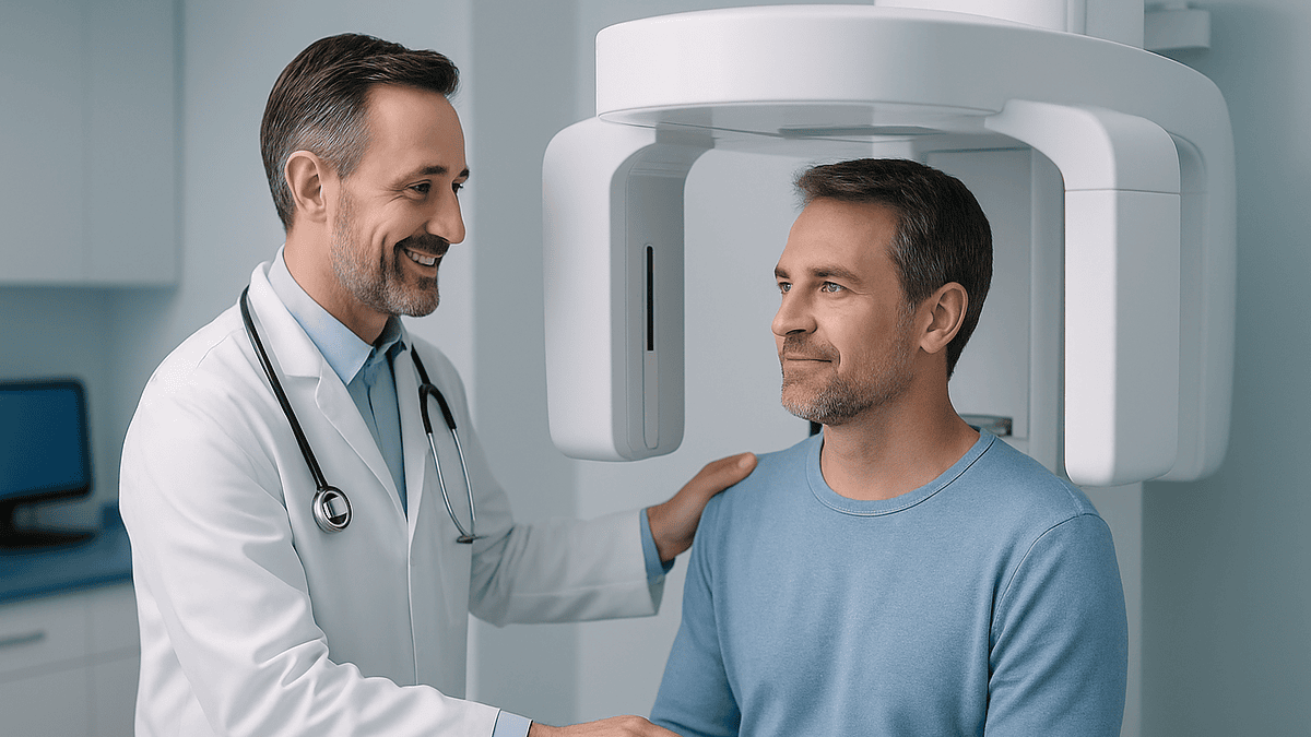
CBCT stands for cone beam computed tomography. Instead of the fan-shaped beam a hospital CT uses to capture the body slice by slice, a CBCT machine sends out a cone-shaped X-ray beam that covers the whole region of interest at once. The scanner arm makes a single rotation around your head, usually while you sit or stand, and a flat-panel detector captures anywhere from roughly 150 to 600 individual projection images along the way.
Software then reconstructs those projections into a three-dimensional volume made of tiny cubes called voxels. Dental CBCT voxels typically measure between 0.075 and 0.4 millimeters on each side, fine enough to trace the wall of a tooth socket or the boundary of a nerve canal. Because the voxels are the same size in every direction, a clinician can slice the volume horizontally, vertically, or at any oblique angle and trust the measurements in all of them.
The practical differences from a medical CT matter to patients, too. A hospital CT generally requires lying inside a gantry and delivers a substantially higher radiation dose; head CT scans commonly run around 2 millisieverts. A dental CBCT is an open, upright design, finishes its rotation in about 10 to 40 seconds, and, depending on the field of view and settings, delivers a small fraction of that dose. The trade-off is that CBCT shows bone and teeth beautifully but has poor soft-tissue contrast, so it is not a substitute for medical CT or MRI when soft tissue is the question.
What is a CBCT scan used for?
Implant planning is the headline use, and for good reason: placing a titanium post in the jaw is a three-dimensional problem with three-dimensional risks. But the same technology earns its keep across dentistry and oral surgery.
- Dental implant planningmeasuring bone height, width, and density; locating the inferior alveolar nerve, the sinus floor, and the nasal cavity; and deciding whether bone grafting is needed before surgery, not during it.
- Impacted teethpinpointing exactly where a wisdom tooth or unerupted canine sits relative to nerves, roots, and sinuses before extraction or orthodontic exposure.
- Complex root canal casesfinding extra canals, curved roots, or areas of infection at root tips that overlap and hide on flat films.
- Jaw pathologymapping the size and borders of cysts and other bony lesions.
- TMJ evaluationassessing bony changes in the jaw joint, such as flattening or erosion of the condyle.
- Sinus assessment before graftingchecking sinus anatomy and health before a sinus lift procedure.
- Trauma and orthodontic planningevaluating fractures and skeletal relationships in selected cases.
Notice what is missing from that list: routine checkups and cavity detection. Ordinary bitewing X-rays remain better suited, and lower dose, for finding decay between teeth. A CBCT scan is a targeted tool for a specific question, and responsible practices treat it that way.
Why a flat X-ray can’t answer a three-dimensional question
Every conventional dental X-ray is a shadow. The beam passes through cheek, bone, teeth, and tongue, and everything in its path collapses onto a single flat image. Structures in front hide structures behind. A nerve canal that sits toward the tongue side of the jaw can appear, on film, to run straight through the spot where an implant is planned, or the reverse, which is far more dangerous.
Panoramic X-rays add a second problem: distortion. Because the machine sweeps around the head in an arc, magnification is uneven across the image and commonly runs in the range of 20 to 30 percent, varying by region and by how the patient was positioned. A measurement taken off a panoramic film is an estimate, not a number a surgeon should drill by.
The dimension flat films miss entirely is width. An implant needs adequate bone on its cheek side and its tongue side, clinicians generally want at least a millimeter or more of intact bone hugging each surface. No two-dimensional image, however sharp, can tell you whether a ridge that looks tall is six millimeters wide or two. Cross-sectional CBCT slices answer that question directly, one slice at a time, along the entire span where implants will go.
This is why the shift to 3D planning was not a luxury upgrade. It replaced educated guessing about the most consequential measurements in the procedure with actual measurements.
What conditions does a CBCT scan diagnose?
CBCT is a bone-and-teeth instrument, and within that territory it identifies a wide range of conditions that flat imaging misses or muddles.
- Impacted and extra teeth, including their exact position relative to nerves and neighboring roots.
- Infections at root tips (periapical lesions) that hide behind overlapping anatomy on standard films.
- Cysts and benign bony lesions of the jaws, with clear borders and true dimensions.
- Bone loss patterns from periodontal disease, seen in three dimensions rather than as a flat silhouette.
- Sinus problems such as mucosal thickening or fluid, which matter before any implant work near the upper back teeth.
- Degenerative changes in the TMJ, including erosion and remodeling of the joint surfaces.
- Root fractureswith an honest caveat: metal fillings and posts create streaky artifacts on CBCT, so hairline fractures near metal can still be genuinely hard to confirm.
Equally important is what CBCT does not diagnose well. Its soft-tissue contrast is poor, so it cannot evaluate gum tissue, muscles, discs inside the jaw joint, or tumors extending into soft tissue; those questions belong to MRI or medical CT. It is also not the right tool for routine cavity screening, smaller, cheaper, lower-dose bitewings do that job better. A good clinician chooses the image to fit the question, and the evidence supports exactly that kind of selectivity.
What happens during a CBCT scan?
From the patient’s side, this is one of the gentler imaging experiences in medicine. There are no needles, no contrast dye, and no enclosed tube.
You will be asked to remove anything metallic from the head and neck, glasses, earrings, hearing aids, removable dentures, hair clips, because metal scatters the X-ray beam and streaks the image. A technician positions you standing or seated at the machine, resting your chin on a support and often biting gently on a small stabilizer so your head stays perfectly still. Some machines add a light forehead strap.
Then the arm sweeps once around your head. The rotation itself takes roughly 10 to 40 seconds depending on the machine and the resolution setting; the whole appointment, positioning included, rarely exceeds fifteen minutes. Movement is the enemy of sharpness, so the only real instruction is to hold still and breathe normally, swallowing mid-scan is the most common reason a scan needs repeating.
Because the design is open, people who find MRI machines claustrophobic generally have no trouble at all. Children and older adults tolerate it well. The reconstructed volume is available within minutes, and many practices review it with you the same day, rotating the model on screen and walking through what the anatomy allows. If an outside radiologist will read the full volume, a written report may follow within days.
How much radiation does a CBCT scan involve?
Less than most people fear, and more than a routine dental film, both halves of that sentence deserve numbers. Effective dose varies widely with the machine, the field of view (how much anatomy is captured), and the exposure settings, but published ranges from the imaging literature cluster as follows:
| Imaging exposure | Approximate effective dose |
|---|---|
| Single bitewing dental X-ray | ~5 microsieverts |
| Panoramic X-ray | ~10–25 microsieverts |
| Small-field dental CBCT | ~20–100 microsieverts |
| Large-field dental CBCT | ~100–200 microsieverts |
| Medical head CT | ~2,000 microsieverts |
| Average US background radiation, per year | ~3,000 microsieverts |
Put in everyday terms: a small-field CBCT scan sits in the same general neighborhood as a week or two of the natural background radiation everyone receives from soil, air, and cosmic rays, and roughly the exposure of one or two cross-country flights. A medical head CT delivers on the order of ten to a hundred times more than a dental cone beam study of comparable anatomy.
None of this makes the dose zero, and Harvard Health and other mainstream sources are consistent on the principle: every X-ray exposure should be justified by information that changes care. Implant planning clears that bar comfortably. Scanning out of curiosity does not, which is the subject of the next section.
Is a CBCT scan safe, and how often is too often?
The governing principle in radiology goes by the acronym ALARA: as low as reasonably achievable. Applied to CBCT, it produces three practical rules that patients can and should hold their providers to.
First, every scan needs a specific clinical question. “We scan everyone” is not a clinical question. Planning implant surgery, locating an impacted tooth against a nerve, investigating a lesion seen on a flat film: those are.
Second, the field of view should match the question. If the surgeon needs to see one quadrant of the lower jaw, a small-field scan at 20 to 100 microsieverts answers it; capturing the entire skull multiplies the dose without adding useful information. Modern machines also offer lower-dose protocols when fine detail is not essential.
Third, extra caution applies to children and adolescents, whose tissues are more radiosensitive and who have more years ahead for any theoretical risk to play out. Professional imaging guidance calls for tighter justification and smaller fields in younger patients.
As for frequency: there is no fixed legal limit, but the honest answer is that most implant patients need one planning scan, occasionally a follow-up if grafting was performed or anatomy changed. If you are pregnant, tell the office: the dose to the abdomen from a head scan is extremely small, but the justification conversation should still happen, and non-urgent imaging is often simply deferred. Asked plainly, “What question is this scan answering?” is a question any good practice will welcome.
How surgeons turn a CBCT scan into an implant plan
The scan itself is only raw material. What happens next is where 3D imaging actually earns its place at the front of every serious implant plan.
Planning software slices the volume into cross-sections a fraction of a millimeter apart along the arch of the jaw. In each slice, the surgeon can see the exact height of usable bone, its width from cheek side to tongue side, and its density, cortical bone reads bright and dense, spongy bone grayer. The inferior alveolar nerve canal in the lower jaw is traced and highlighted; a widely used safety convention keeps the implant tip at least 2 millimeters above it. In the upper jaw, the sinus floor and nasal cavity get the same treatment.
Then the plan runs backward from the end result. A virtual implant, correct length, correct diameter, is positioned not just where bone happens to exist, but where the future crown needs support. Clinicians call this prosthetically driven planning: the tooth dictates the implant position, and the scan reveals whether the bone can cooperate or whether grafting must come first.
Many practices go one step further and convert the digital plan into a printed surgical guide, a custom template that fits over the teeth or gums and constrains the drill to the planned angle and depth. Research on guided surgery consistently shows placement closer to the plan than freehand technique. Discovering a thin ridge or a low sinus on a screen, weeks before surgery, is a far better experience than discovering it mid-procedure.
How much does a CBCT scan cost?
Prices vary enough that any single figure would mislead, so here is the honest landscape. In the United States, a dental CBCT scan billed as a standalone service commonly runs from roughly $150 to $750, with most patients landing somewhere in the middle of that range. Several factors move the number:
- Field of view. A limited scan of one region costs less than a full-jaw or full-skull capture, and often needs less interpretation time.
- Geography. Urban markets and coastal regions tend to price higher, as with most healthcare services.
- Setting. Dental school clinics and dedicated imaging centers frequently charge less than private surgical practices; some offices own their own machine and bundle the scan into the overall implant fee rather than itemizing it.
- Interpretation. If the full volume is sent to an oral and maxillofacial radiologist for a written report, a genuinely valuable step, that reading may be billed separately.
Two practical suggestions. First, ask for an itemized treatment estimate; a scan folded invisibly into a large implant quote is hard to compare across offices. Second, if you already have a recent CBCT from another provider, ask whether it can be transferred, scans are digital files, and a study taken within the past several months is often perfectly usable, sparing you both the cost and the dose of a repeat.
Is a CBCT scan covered by insurance?
Sometimes, and the answer depends less on the scan than on why it was taken and which policy is being asked.
Dental insurance treats CBCT inconsistently. Many plans that cover implants at all will contribute toward the diagnostic imaging that precedes them, though implant benefits themselves are frequently capped or excluded, and annual maximums on dental plans are modest to begin with. Plans that exclude implants often exclude the planning scan by extension. The only reliable move is a pre-treatment estimate: your dental office submits the imaging and procedure codes, and the insurer responds in writing with what it will pay.
Medical insurance occasionally enters the picture when the scan serves a medical rather than purely dental purpose, evaluating a jaw cyst or tumor, facial trauma, or certain TMJ and airway questions. Coverage there hinges on documented medical necessity and usually on prior authorization, and denials are common when the underlying purpose is implant planning.
A few consolations regardless of coverage. Funds in an HSA or FSA can generally be applied to dental imaging, since it qualifies as medical care. Costs that insurance declines still count toward the itemized medical-expense deduction for those who qualify. And because prices vary substantially between settings, a phone call comparing the standalone scan fee at two or three providers is fifteen minutes well spent. Ask specifically: is the scan itemized, is a radiologist’s report included, and will the office submit a pre-authorization on your behalf?
When a CBCT scan isn’t necessary
A magazine article arguing for 3D imaging owes you the other half of the truth: plenty of dental care neither needs nor benefits from it, and professional imaging guidance says so explicitly.
Routine checkups top the list. Bitewing X-rays detect decay between teeth better than CBCT does, at a small fraction of the dose and cost. There is no evidence-based role for cone beam scanning as a screening tool in patients with no symptoms and no planned surgery, “scanning everyone” is a practice-pattern choice, not a clinical necessity.
Straightforward extractions often qualify, too. When a tooth’s roots and their relationship to nearby structures are clearly visible on a conventional film, adding a 3D scan adds dose without changing the plan. The same logic applies to many orthodontic cases, most routine fillings and crowns, and initial evaluation of simple toothaches.
Even within implant dentistry, judgment matters. Virtually all surgeons want 3D imaging before placing implants, the nerve, sinus, and bone-width questions demand it, but the scan should be sized to the surgical site, taken once rather than repeatedly, and timed so the anatomy it captures is the anatomy the surgeon will meet.
Here is a fair personal test. If you ask, “What will this scan tell us that we don’t already know, and how would it change the plan?” and receive a specific answer, nerve position, ridge width, sinus floor height, the scan is justified. A vague answer is a reason to keep asking.
Who reads the scan, and what about surprise findings?
Here is a detail most patients never think to ask about, and it matters. A CBCT volume captures far more than the implant site: depending on the field of view, it may include the sinuses, nasal cavity, upper airway, jaw joints, cervical vertebrae, and portions of the skull base. Whoever interprets the scan carries responsibility for all of it, not just the few cubic centimeters where an implant will go.
Incidental findings are genuinely common in the literature, studies of dental CBCT volumes report unexpected findings in a substantial share of scans. Most are benign and require nothing more than awareness: mucosal thickening in a sinus, a retained root tip, calcifications in tonsillar tissue. A minority deserve follow-up, such as suspicious bony lesions, significant sinus disease before grafting, notable airway narrowing, or calcifications along the path of the carotid artery, which some studies suggest merit a conversation with a physician about cardiovascular risk factors.
Practices handle interpretation differently. Some dentists and oral surgeons read their own scans, which they are trained and permitted to do; others send the full volume to an oral and maxillofacial radiologist, a specialist whose entire discipline is interpreting these images, and receive a written report.
Neither model is inherently wrong, but you are entitled to know which applies to you. Two questions cover it: “Who reviews the entire scan, beyond the implant site?” and “Will I be told about anything unexpected?” You are also entitled to a copy of the volume itself: it is your health record, and it travels well between providers.
When to see a doctor or dentist
Imaging is a planning tool, not a symptom, but the situations surrounding implant care come with clear signals that deserve professional eyes, some before any scan is taken, some after surgery.
Seek a dental evaluation promptly, before imaging decisions even arise, if you notice a tooth socket that has not healed weeks after an extraction, persistent swelling or a lump in the jaw, unexplained numbness or tingling in the lip or chin, teeth that have loosened without injury, or pain that wakes you at night. These findings sometimes point to infection or bony pathology that a CBCT scan can help characterize, but the examination comes first, and the imaging follows the clinical question.
After implant surgery, some soreness, minor swelling, and bruising for a few days is expected. Contact your surgeon without delay if you experience:
- Numbness or tingling of the lip, chin, or tongue that persists after the anesthetic should have worn off: this can signal nerve involvement, and early assessment matters.
- Pain that intensifies after the third or fourth day rather than easing.
- Fever, spreading swelling, or discharge from the surgical site.
- An implant or its cover that feels loose or moves.
- New sinus symptoms, congestion, pressure, or fluid, after upper-jaw implant work.
None of these automatically means something has gone wrong, and most concerns resolve with straightforward care. But the pattern in the evidence is consistent: complications caught early are managed more simply. When in doubt, call: a five-minute phone conversation is the cheapest diagnostic tool in all of dentistry.
Frequently asked questions
What is a CBCT scan used for?
A CBCT scan is used mainly to plan dental implant surgery by measuring bone height, width, and density and mapping nerve canals and sinuses in 3D. It also helps locate impacted teeth, evaluate complex root canal anatomy, characterize jaw cysts and lesions, assess bony changes in the jaw joint, and check sinus health before grafting. It is not used for routine checkups or cavity screening, where standard low-dose X-rays work better.
How much does a CBCT scan cost?
In the United States, a standalone dental CBCT scan commonly costs between about $150 and $750, depending on the field of view, your region, and the practice setting. Dental school clinics and imaging centers often charge less than private surgical offices, and some practices bundle the scan into the overall implant fee. A separate radiologist’s report, when ordered, may add to the cost, so ask for an itemized estimate.
Is a CBCT scan covered by insurance?
Sometimes, and it depends on the reason for the scan. Dental plans that cover implants often contribute toward planning imagery, while plans excluding implants usually exclude the scan too. Medical insurance may cover CBCT when it serves a medical purpose, such as evaluating trauma or a jaw lesion, typically with prior authorization. The most reliable approach is a written pre-treatment estimate from your insurer. HSA and FSA funds generally apply either way.
What conditions does a CBCT scan diagnose?
CBCT identifies conditions involving bone and teeth: impacted or extra teeth, infections at root tips, jaw cysts and benign bony lesions, patterns of periodontal bone loss, sinus disease relevant to dental work, degenerative jaw joint changes, and some root fractures. It has poor soft-tissue contrast, so it cannot evaluate gums, muscles, joint discs, or soft-tissue tumors: those questions require MRI or medical CT instead.
How is CBCT different from a hospital CT scan?
CBCT uses a cone-shaped beam and a single rotation around your head while you sit or stand upright, producing a high-detail 3D image of teeth and jawbone at a much lower radiation dose, often 20–200 microsieverts versus roughly 2,000 for a head CT. Hospital CT captures the body in slices while you lie down and shows soft tissue far better. Each answers different questions; CBCT is purpose-built for dental and jaw anatomy.
Does a CBCT scan hurt?
No. A CBCT scan is painless and involves no needles, contrast dye, or enclosed tube. You stand or sit at an open machine, rest your chin on a support, often bite a small stabilizer, and hold still while the arm rotates once around your head for roughly 10 to 40 seconds. People who feel claustrophobic in MRI machines typically have no difficulty with the open CBCT design.
How long does a CBCT scan take?
The scan itself takes about 10 to 40 seconds: a single rotation of the machine around your head. Including positioning, removing metal items like glasses and earrings, and verifying image quality, the full appointment usually runs ten to fifteen minutes. The 3D volume is reconstructed within minutes, and many practices review the images with you the same day; a written radiologist’s report, when ordered, may follow within a few days.
Can I have a CBCT scan while pregnant?
Tell your dental team if you are or might be pregnant before any imaging. The radiation dose to the abdomen from a head scan is extremely small, and lead shielding can reduce it further, but standard radiology practice still weighs every exposure against its benefit. Non-urgent scans, including elective implant planning, are often simply deferred until after pregnancy, while genuinely urgent diagnostic questions can still justify imaging after that discussion.
Do I need a CBCT scan for every dental implant?
For nearly all implant cases, yes: 3D imaging is the accepted standard for measuring bone width, locating the lower jaw’s nerve canal, and mapping the sinus floor, none of which flat X-rays show reliably. The scan should be scoped sensibly: a small field of view covering the surgical site, taken once during planning. A recent CBCT from another provider can often be transferred and reused rather than repeated.
Can I get a copy of my CBCT scan?
Yes. The scan is part of your health record, and you are entitled to a copy, typically exported as a digital DICOM file on a disc, drive, or secure link. Requesting it is worthwhile: implant planning software at any practice can open the standard format, so a recent scan can travel with you for second opinions or continued care, sparing you the cost and radiation dose of a repeat study.
References
This article is for general information only and is not a substitute for professional medical advice. Please consult a qualified doctor about your individual situation.

More from the Blog
Which Dental Bone Graft Material Is Used? Your Own Bone, Donor, Animal-Derived and Synthetic
Dental bone graft material comes in four families: autograft (your own bone, usually taken from the jaw), allograft (processed human donor bone), xenograft (processed…
Are Zirconium Teeth Right for You? Bite Force, Smile Goals and When Another Ceramic Fits Better
A good zirconium teeth candidate usually needs a strong restoration for a back tooth, an implant crown, or a bridge, especially with a heavy…

All-on-4 vs All-on-6: When Two More Implants Make Sense
All-on-4 and All-on-6 both anchor a full arch of fixed replacement teeth; the difference is how many implants carry the load. Four implants suit…

What to Ask Before a Dental Crown: Material, Margin Placement and What Happens to the Nerve
Before agreeing to a dental crown, ask what the crown is for, which material is proposed and why, where the edge of the crown…
Dental Implants After Gum Disease: When They Are Possible, and What Comes First
Yes, people with periodontal disease can often get dental implants, but not while the disease is active. Infection and inflammation must be treated first,…

How Long Do All-on-4 Implants Last? Longevity, Care and What Can Go Wrong
With diligent daily cleaning and regular professional maintenance, the four titanium implants supporting an All-on-4 restoration commonly last 20 years or longer, and long-term…