

Confirming HSP: How Genetic Testing, MRI and the Walking Assessment Fit Together

Key Takeaways
- HSP is diagnosed by combining a characteristic clinical examination, imaging that excludes other causes, and genetic testing; no single test confirms it alone.
- In pure HSP the MRI is often normal or shows only mild spinal cord thinning, so a clear scan supports rather than contradicts the diagnosis.
- More than 80 genetic types of HSP are recognized, which is why a negative gene panel does not rule the condition out.
- Spastic paraplegia type 4 (SPAST gene) accounts for roughly 40 percent of autosomal dominant cases and is usually a pure form.
- Reversible mimics such as vitamin B12 deficiency, spinal cord compression and certain infections are tested for before genetic testing is ordered.
- Predictive genetic testing in healthy children for adult-onset HSP is generally deferred so that the decision remains theirs as adults.
Hereditary spastic paraplegia (HSP) is diagnosed by combining three pieces of evidence rather than by a single test. A neurologist examines walking and leg reflexes for the characteristic stiffness and weakness, MRI of the brain and spine is used mainly to rule out other causes, and genetic testing looks for a change in one of the many genes linked to HSP. A family history strengthens the picture, but a confirmed diagnosis often rests on the genetic result.
It usually starts with shoes. A heel worn down on the outer edge, a scuff mark on the toe of the right sneaker, a parent noticing that their twelve-year-old trips on the same stair every morning. Or it starts later, in a forty-year-old who has quietly stopped taking the stairs two at a time and cannot remember when that happened. Nobody says the words hereditary spastic paraplegia at this point. They say “tight calves” or “just getting older.”
By the time someone asks how HSP is diagnosed, they have often been through months of physical therapy for a problem that did not behave like a sports injury. The question deserves a clear answer, because the process is more structured than it looks from the waiting room.
This explainer walks through the three pillars that clinicians lean on, the walking assessment, imaging and genetic testing, and shows how each one carries a different kind of weight. It also covers what the process cannot tell you, which matters just as much.
How HSP is diagnosed when there is no single test for it
Hereditary spastic paraplegia is not one disease. It is a family of inherited conditions that share a common thread: progressive stiffness (spasticity) and weakness in the legs caused by slow degeneration of the long nerve fibers running from the brain down the spinal cord. The National Institute of Neurological Disorders and Stroke notes that more than 80 genetic types have been described, and that prevalence estimates range from roughly 1.3 to 9.6 people per 100,000, depending on the population studied.
That diversity is the first reason there is no single confirmatory test. A blood test that catches one gene will miss dozens of others. A scan that looks normal in one type may show changes in another. So clinicians build the diagnosis the way a careful editor checks a story: from several independent sources that have to agree.
The three sources are the clinical examination (especially how a person walks and how their reflexes behave), imaging of the brain and spinal cord, and genetic testing. Each answers a different question. The exam asks, does this pattern of stiffness fit the upper motor neuron picture typical of HSP? Imaging asks, is something else causing it, such as a compressed spinal cord, multiple sclerosis or a structural problem? Genetic testing asks, can we find the specific inherited cause?
In practice the order is almost always exam first, imaging second, genetics third, because the first two shape which genetic tests make sense. A fourth element runs alongside all three: the family history. When a parent or sibling has similar walking difficulty, the probability of HSP rises sharply, though the absence of a family history does not rule it out. New (de novo) gene changes and recessive inheritance both produce affected people with no obviously affected relatives.
What actually happens at the first neurology appointment
The visit begins with a long conversation, and that conversation is itself part of the test. The neurologist will want to know when the difficulty began, whether it has been steady or stepwise, whether it affects both legs roughly equally, and whether anything beyond the legs has changed: bladder urgency, balance, hand coordination, speech, vision or thinking. Expect detailed questions about relatives, including ones who were never formally diagnosed but “always walked a bit stiffly” or used a cane young.

The physical examination follows a standard neurological sequence. Muscle tone is checked by moving each limb passively and feeling for the catch-and-release resistance that defines spasticity, as Cleveland Clinic describes it. Reflexes at the knee and ankle are tested with a reflex hammer; in HSP they are typically exaggerated, and the examiner may find clonus, a rhythmic beating of the foot when the ankle is flexed quickly. The sole of the foot is stroked to look for an upgoing big toe (the Babinski sign), a classic marker that the problem lies in the brain or spinal cord rather than in the nerves or muscles of the leg itself.
Strength is graded muscle by muscle. Sensation is tested with light touch, pinprick and a tuning fork for vibration. Many people with HSP have subtly reduced vibration sense at the ankles, which is why that small humming fork gets so much attention.
Then you walk. The clinician watches you walk away and back, on heels, on toes, and often in a straight line heel to toe. Nothing about this feels high-tech, and that is the point. Decades of evidence show that an experienced examiner can localize a problem to the upper motor neuron pathway with remarkable consistency, and that localization is what justifies every test that follows.
What the walking assessment is looking for
The HSP gait has a recognizable signature, though it ranges from almost invisible to pronounced. Because the calf and thigh muscles are stiff and the hip flexors relatively weak, the legs tend to swing in a slight arc rather than lifting cleanly, a pattern called circumduction. The toes catch the floor, which explains the worn shoe tips and the trips on the stair. Knees may be held straighter than usual, and the feet may turn inward so that the legs appear to cross or “scissor” slightly with each step.
Watching this walk tells the clinician several things at once. The symmetry matters: HSP usually affects both legs to a broadly similar degree, whereas a stroke or a one-sided spinal lesion produces an asymmetric gait. The relationship between stiffness and weakness matters too. In most forms of HSP, spasticity is the dominant problem and true weakness is milder than the gait suggests, which is one reason physical therapy often improves function without changing the underlying condition.
Heel walking and toe walking expose weakness that normal walking hides. Tandem (heel-to-toe) walking tests balance; marked unsteadiness points the clinician toward conditions involving the cerebellum, the brain’s coordination center, which can look like HSP from a distance but belong to a different diagnostic family. Standing with eyes closed checks whether the person relies on vision to compensate for lost position sense in the feet.
Some clinics add a timed walk over a fixed distance or a standardized spasticity rating scale. These numbers are not used to make the diagnosis; they create a baseline against which change can be measured on later visits. In a condition where progression is often measured over years rather than months, as the NHS notes, that baseline becomes one of the most useful records in the chart.
Hereditary spastic paraplegia symptoms the examiner checks beyond the legs
The legs are where HSP announces itself, but the examination deliberately looks elsewhere, because what is found (or not found) beyond the legs decides which category of HSP is being considered and which genes are most likely. This is a diagnostic examination, not a self-check; the findings described here are the clinician’s job to interpret.

Bladder function is asked about directly. Urinary urgency, frequency and occasional incontinence are common in HSP because the same spinal pathways that control leg tone also influence bladder reflexes. Many people are embarrassed to raise this and are relieved when the neurologist asks first.
The arms are examined for tone, reflexes and fine coordination. Mild hyperreflexia in the arms can occur in otherwise typical HSP, but clear arm weakness, tremor or clumsiness pushes the clinician toward a broader workup. Eye movements are tested because certain HSP types and several look-alike conditions affect them. Speech, swallowing and facial movement are observed during conversation. A brief cognitive screen may be done, particularly in younger people, since some genetic forms are associated with learning difficulty or later cognitive change.
The feet themselves are inspected. A high arch (pes cavus) and hammer toes develop in some people after years of muscle imbalance and are a soft clue that the process has been present a long time, sometimes since childhood, even when symptoms were noticed recently.
Skin, hearing and vision may be checked in a child or young adult, because the “complicated” forms of HSP described in the next section can involve the retina, hearing or skin. None of these findings alone makes or breaks the diagnosis. Taken together, they draw the boundary around what kind of condition this is likely to be, and that boundary determines how wide the genetic net should be cast.
Pure vs complicated HSP: why the distinction steers every test
Clinicians divide HSP into two broad clinical groups, and the split is made at the bedside before any lab result arrives.
Pure (uncomplicated) HSP means the problem is essentially confined to the legs: spasticity, weakness, exaggerated reflexes, often bladder urgency and mildly reduced vibration sense at the ankles. Thinking, vision, hearing, speech and arm function remain normal. According to MedlinePlus, spastic paraplegia type 4, caused by changes in the SPAST gene, is the most common form and accounts for roughly 40 percent of autosomal dominant cases; it is usually a pure type.
Complicated (complex) HSP means the leg findings come with something else: cognitive impairment, seizures, peripheral neuropathy (damage to the nerves outside the spinal cord), cerebellar ataxia (loss of coordination), retinal changes, hearing loss, or structural brain differences such as a thin corpus callosum, the band of fibers connecting the two halves of the brain. Complicated forms more often follow autosomal recessive inheritance, where both parents carry one silent copy of a gene change and have no symptoms themselves.
Why does this matter for diagnosis? Two reasons. First, the complicated forms overlap heavily with other neurological and metabolic conditions, so the rule-out workup is broader and may include blood tests for vitamin levels, metabolic markers and infections, as well as nerve conduction studies. Second, the pattern of extra features narrows the gene list dramatically. A thin corpus callosum with early cognitive difficulty points to a very different set of genes than isolated leg spasticity beginning in a parent and child in their thirties.
The labels are a working hypothesis, not a verdict. Features can emerge over time, and a person classified as pure at the first visit may be reclassified later. That is one of several reasons follow-up is built into the diagnostic process rather than tacked on afterward.
Spastic paraplegia MRI findings: what imaging can and cannot show
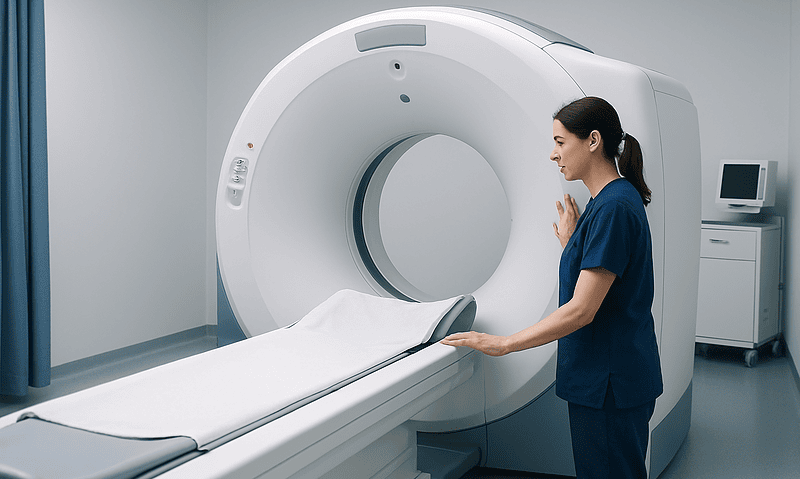
Here is the part that surprises many people: in pure HSP, the MRI is often normal or close to it. Magnetic resonance imaging uses a strong magnetic field and radio waves to build detailed pictures of soft tissue, as MedlinePlus explains, and it is excellent at showing structural damage. But HSP degeneration happens at the level of individual nerve fibers, below the resolution of a routine scan. Some people show mild thinning of the thoracic spinal cord, and that is about all a standard spine MRI may reveal.
So why order it at all? Because MRI is the primary tool for excluding conditions that mimic HSP and that may need entirely different management. Spinal cord compression from a herniated disc, a narrowed canal or a tumor can produce stiff, weak legs and brisk reflexes. Multiple sclerosis leaves characteristic inflammatory lesions in the brain and cord. Structural problems at the base of the skull, vascular malformations and certain infections all have imaging signatures. A clear scan in these respects is a meaningful result, not an empty one.
In complicated HSP, the brain MRI becomes more informative. A thin corpus callosum is a well-recognized finding in several recessive types, and it can be striking enough to shift the genetic strategy on its own. Changes in the white matter, the cerebellum or the deep gray structures each point toward particular gene groups.
Radiologists and neurologists read these scans with the clinical picture in mind. A report that says “no acute abnormality” is not the end of the diagnostic road in HSP; it is the clearing of one road so another can be followed. People sometimes leave an imaging appointment deflated because “nothing was found.” In this context, nothing found is frequently exactly what the team hoped to see.
Ruling out the conditions that look like HSP
Before anyone orders a gene panel, the team works through a list of acquired and treatable causes of leg spasticity. Missing one of these would be a far more serious error than delaying a genetic answer by a few weeks, and this stage is where the diagnostic process earns its reputation for being thorough rather than slow.
Blood tests are the workhorse. Vitamin B12 deficiency can damage the spinal cord’s posterior and lateral columns and produce stiffness, weakness and reduced vibration sense that closely resemble HSP; the NIH Office of Dietary Supplements describes these neurological consequences. Copper deficiency does something similar. Very long chain fatty acids may be measured in men and boys because a form of adrenoleukodystrophy affects the spinal cord in adulthood. Tests for syphilis, HIV and HTLV-1, a virus that causes a slowly progressive spastic paraparesis, may be included depending on history and region.
Nerve conduction studies and electromyography, which measure electrical signals in nerves and muscles, help when peripheral neuropathy is suspected alongside the spinal findings. A lumbar puncture, sampling the fluid around the spinal cord, is considered if inflammation or infection remains possible after imaging.
Two neurological diseases deserve particular care. Primary lateral sclerosis affects the same upper motor neurons as HSP but is not inherited and tends to involve the arms and speech over time. Motor neuron disease (ALS) usually combines upper motor neuron signs with muscle wasting and twitching, features that are not typical of HSP. The distinction rests on careful examination repeated over months and, increasingly, on genetic results.
Only when this list has been worked through does genetic testing move from “interesting” to “appropriate.” That sequencing is deliberate. A positive gene result can coexist with a vitamin deficiency, and nobody wants the second problem overlooked because the first one had a satisfying name.
HSP genetic testing: how it works and what it looks for
Genetic testing examines DNA, usually from a blood sample or saliva, for changes in the sequence of specific genes. MedlinePlus describes the broad categories of testing and what they can and cannot reveal. In HSP, the approach has shifted over the past decade from testing one gene at a time to testing many at once.
Three strategies are commonly used, and the choice depends on the clinical picture established in the earlier steps. Single-gene testing is reasonable when a close relative already has a confirmed gene change; the lab simply checks whether the same change is present. A multigene panel sequences a curated set of genes known to cause HSP and its close mimics, often several dozen at once. Exome or genome sequencing reads the protein-coding portion of all genes or the entire genome, and is increasingly used when panels return nothing or when the picture is unusual.
The lab is looking for several kinds of change. A single-letter substitution may alter the protein the gene builds. A small deletion or insertion may garble the instructions downstream. Larger deletions or duplications of whole sections of a gene are common in some HSP types and require a different detection method, so a thorough workup checks for both.
Inheritance pattern shapes interpretation. In autosomal dominant HSP, one altered copy of a gene is enough to cause symptoms, so each child of an affected parent has a 50 percent chance of inheriting it, a figure MedlinePlus gives for dominant conditions. In autosomal recessive forms, both copies must be altered, and parents are typically unaffected carriers. X-linked forms mostly affect males.
Pre-test genetic counseling is standard. It covers what the test can show, what it may leave unanswered, and how a result could affect relatives. Consent is required, and the decision to test always belongs to the person being tested, in discussion with the treating team.
Reading the genetic report: positive, uncertain, or negative
A genetic report rarely says simply “HSP: yes” or “HSP: no.” It classifies each variant found according to a tiered system, and understanding the tiers prevents a great deal of confusion.
A variant classified as pathogenic or likely pathogenic, in a gene whose known effects match the clinical picture, is the result that confirms the diagnosis. The gene name usually gives the HSP type its number, so a pathogenic SPAST change corresponds to SPG4. This is the moment when the walking assessment, the cleared MRI and the lab result finally lock together.
A variant of uncertain significance, or VUS, is a change whose effect on health is not yet known. It may be harmless; it may be the cause. Labs are cautious about upgrading a VUS, and a VUS alone does not confirm HSP. Testing affected and unaffected relatives can help: if the variant tracks with symptoms through the family, its significance rises. Labs also periodically reclassify variants as more data accumulate, so a report that reads “uncertain” today may be revised later. Asking how reclassification is communicated is a sensible question for the care team.
A negative result means no reportable variant was found in the genes tested. It does not rule out HSP. The clinical diagnosis stands on the examination and the exclusion of other causes; the genetic test simply failed to add a label. Reasons include genes not yet discovered, variants in regions the test did not read, or changes the method cannot detect. Many centers offer broader sequencing or re-analysis of existing data after an interval.
Incidental findings are also possible with exome or genome sequencing: variants unrelated to HSP that carry their own health implications. Whether you want to know about these is a choice made during counseling, before the sample is drawn, and it is entirely yours.
Who is usually offered testing, and who is usually asked to wait
Genetic testing is generally offered first to the person who has symptoms. That sounds obvious, but it has a practical logic: testing an affected person has the best chance of finding a clear answer, and once a family’s specific variant is known, testing relatives becomes simpler and far more interpretable.
Adults with a clinical picture consistent with HSP, after the rule-out workup, are the core group. Adults with a strong family history and early signs on examination are also candidates, even when symptoms are mild. Children with spasticity whose other causes (such as cerebral palsy from birth injury) have been excluded are often tested, particularly when the pattern is progressive rather than static. In children, a complicated picture with developmental differences frequently leads straight to broad sequencing because the differential diagnosis is wide.
Who is usually asked to wait? Healthy children of an affected parent, when the condition is an adult-onset form, are generally not tested until they can make the decision for themselves. Major genetics bodies advise against predictive testing in minors for conditions that will not affect them in childhood and for which no childhood intervention changes the course. The reasoning is about autonomy: a grown person may choose not to know, and that choice is lost if the test is done at six.
Unaffected adult relatives who want predictive testing are usually offered it only after counseling and only when the family variant is known, so that a negative result genuinely means something. Testing them against a panel without a known family variant produces results that are hard to interpret.
People in the middle of an acute medical crisis, or whose symptoms have not yet been fully evaluated, may be asked to complete the clinical workup first. The order protects the quality of the answer. In every case, the recommendation comes from the treating team, and the decision rests with the person being tested or, for a child, with the family in conversation with that team.
How the three pillars fit together: a side-by-side view
Each component of the HSP workup carries a different kind of evidence, and the diagnosis is strongest when they point the same way. The table below summarizes what each one contributes, what it typically shows in pure HSP, and what it cannot do on its own.
| Pillar | Core question it answers | Typical result in pure HSP | Main limitation |
|---|---|---|---|
| Clinical and walking assessment | Does the pattern fit upper motor neuron damage confined mainly to the legs? | Spastic gait, brisk leg reflexes, upgoing toes, mild loss of vibration sense | Cannot distinguish HSP from acquired causes with the same pattern |
| MRI of brain and spine | Is something else, such as compression or inflammation, causing this? | Normal or mild thinning of the spinal cord | Does not confirm HSP; its value is in what it excludes |
| Blood and electrical tests | Is there a treatable deficiency, infection or neuropathy? | Normal | A normal panel narrows the field but names nothing |
| Genetic testing | Can a specific inherited cause be identified? | Pathogenic variant in an HSP gene, most often SPAST in dominant families | Negative or uncertain results do not rule HSP out |
| Family history | Does the condition track through generations? | Often positive in dominant forms; frequently negative in recessive or de novo cases | Absence of history is common and proves nothing |
Read across a row and you see a tool; read down the column of limitations and you see why no single tool is trusted alone. A confirmed diagnosis is one where the clinical picture is characteristic, the alternatives have been excluded, and a pathogenic variant has been found. A clinical diagnosis of probable HSP is one where the first two conditions are met and the genetic result is negative or uncertain. Both are legitimate diagnoses, and both lead to the same management conversation with the treating team. The difference lies mainly in what can be offered to relatives and in eligibility for research studies that require a confirmed gene.
What the following weeks usually look like
The diagnostic process has a rhythm, and knowing it in advance takes some of the anxiety out of the waiting.
The first appointment produces the clinical impression and the order set: imaging, blood tests and sometimes nerve studies. MRI appointments are typically scheduled within days to a few weeks depending on the service, and most people have the scan and the bloodwork completed before the second visit. Results of routine blood tests usually return within days; specialized metabolic tests may take longer.
A second visit reviews those results. If an acquired cause has been found, the conversation changes direction entirely. If the rule-out workup is clear, genetic counseling is arranged, either at the same visit or as a separate appointment. Counseling itself is unhurried; expect a long conversation about inheritance, implications for relatives, insurance considerations where relevant, and the possibility of uncertain or incidental findings.
The sample is then drawn and sent. MedlinePlus notes that genetic test turnaround varies with the type of test, and in practice panels and exome sequencing are measured in weeks to a few months rather than days. This is the stretch people find hardest. Nothing visible is happening, yet the most decisive piece of information is being generated.
Results are returned at a dedicated appointment, usually with the neurologist or genetic counselor, and never by letter alone. Whatever the result, this visit covers next steps: referral to physical therapy and rehabilitation, bladder assessment if needed, discussion of symptom management options with the prescribing clinician, and a plan for follow-up intervals. If the result is uncertain or negative, the plan may include testing relatives or scheduling re-analysis.
Follow-up in HSP is typically spaced at intervals of months to a year, because the NHS and NINDS both describe progression as slow in most people. The baseline measurements from the first walking assessment become the reference point for every later visit.
What people often get wrong about how HSP is diagnosed
Several misunderstandings surface repeatedly in clinic, and each one can derail the process or cause unnecessary distress.
“A normal MRI means it is not neurological.” In pure HSP a normal MRI is expected. The scan’s job is to exclude other causes, and a clear result is consistent with, not against, the diagnosis.
“No family history, so it cannot be hereditary.” Recessive forms require two carrier parents who are themselves well. New variants arise without any family history. Mild cases in previous generations often went undiagnosed and were attributed to age or arthritis. A blank family tree is common in HSP.
“A negative gene test rules it out.” More than 80 genetic types are known according to NINDS, and panels do not cover every one of them. Technical limits mean some variants are missed. A clinical diagnosis stands on its own evidence when the genetic test is negative.
“A VUS is basically a positive.” Uncertain means uncertain. Treating a VUS as confirmation can lead relatives to be tested against a variant that later turns out to be harmless.
“Children should be tested so we know early.” For adult-onset forms, predictive testing in healthy children is generally not recommended, because it removes a decision that properly belongs to them as adults and offers no childhood benefit.
“HSP is a form of cerebral palsy, or of MS.” Cerebral palsy results from injury to the developing brain and does not progress; HSP is genetic and progressive, though slowly. Multiple sclerosis is inflammatory, visible on MRI and follows a different course. All three can produce spastic legs, which is exactly why the structured workup exists.
“The diagnosis changes life expectancy.” The NHS states that most people with HSP have a normal life expectancy. The diagnosis explains a pattern of symptoms and opens the door to targeted rehabilitation and symptom management; it does not predict the course for any individual.
Questions to ask your care team
A diagnostic workup goes better when the person at the center of it understands each step. These questions tend to produce useful answers; write them down before the visit, because the moment usually arrives faster than expected.
- Based on my examination, does my pattern look like pure or complicated HSP, and how does that shape which tests you are ordering?
- Which acquired causes are you ruling out with blood tests and imaging, and what would each one look like if it were found?
- If my MRI comes back normal, what does that mean for the diagnosis?
- Which genetic testing strategy are you recommending (single gene, panel, exome or genome) and why that one for me?
- What proportion of people with a picture like mine get a clear genetic answer, in your experience and in the published literature?
- If the result is uncertain or negative, what are the next steps, and does your lab re-analyze data over time?
- Do I want to be told about incidental findings unrelated to HSP, and how do I record that choice?
- How will the result affect my relatives, and who can talk to them if they want testing?
- What baseline measures are you recording today so that we can track change over the coming years?
- Who do I contact between appointments if something changes, and what kinds of change should prompt that call?
Notice that none of these questions asks for a prediction. Clinicians cannot say how quickly any one person’s HSP will progress, and a good team will say so plainly rather than offer reassurance it cannot support. Asking about process, evidence and follow-up gets honest, concrete answers; asking for a forecast gets a careful shrug. Both responses are useful information about how your team works.
When to call your doctor
HSP itself usually changes slowly, measured over years, which is why any rapid change deserves prompt attention: it may signal something other than HSP, including conditions that need urgent treatment. Contact your care team promptly, or seek emergency care, if any of the following occur.
- Sudden or rapid worsening of leg weakness over hours or days, rather than months
- New loss of bladder or bowel control, or inability to pass urine, especially if it appears suddenly
- Numbness spreading up the trunk, or a band-like tightness around the chest or abdomen
- New weakness, clumsiness or numbness in the arms or hands
- Difficulty swallowing, slurred speech or shortness of breath
- Severe back or neck pain accompanied by any change in leg function
- A fall resulting in a head injury, or falls that have become frequent enough to cause fear of walking
- Fever with worsening neurological symptoms
- New visual disturbance or double vision
Sudden spinal cord compression, inflammation and infection can all produce these signs, and they are time-sensitive. Spasticity that worsens abruptly without a clear cause can also indicate a hidden trigger such as a urinary infection, a pressure sore or constipation, each of which is manageable once identified.
For slower changes, such as needing a cane more often, new tripping or more frequent nighttime bathroom trips, a routine call to arrange an earlier follow-up is appropriate. These are the kinds of shifts the baseline walking assessment was designed to measure.
If you are a relative of someone with HSP and notice stiffness or tripping in yourself, the right first step is an appointment with your primary care clinician, who can examine you and refer to neurology if warranted. Self-diagnosis from symptom lists is unreliable in a condition with this many look-alikes. Every decision about testing, treatment and follow-up belongs with the treating team, who can weigh your individual picture in ways no article can.
Frequently asked questions
How is HSP diagnosed if there is no family history?
The same way as with a family history: clinical examination, imaging to rule out other causes, and genetic testing. Many people with HSP have no affected relatives because recessive forms require two unaffected carrier parents, new variants arise spontaneously, and earlier generations were often undiagnosed. A blank family tree lowers suspicion slightly but does not exclude the condition, and clinicians are trained not to rely on it.
What are the first hereditary spastic paraplegia symptoms a neurologist looks for?
The examiner looks for a stiff, slightly toe-dragging gait affecting both legs, exaggerated knee and ankle reflexes, an upgoing big toe when the sole is stroked, and mildly reduced vibration sense at the ankles. Bladder urgency is asked about. These findings are interpreted together and in the context of history; individually, each can occur in several other conditions, which is why they are assessed by a clinician rather than self-checked.
Is HSP genetic testing always needed to confirm the diagnosis?
Not always, but it is the only way to reach a confirmed molecular diagnosis. A clinical diagnosis of probable HSP can be made from examination plus exclusion of other causes. Genetic testing adds a specific label, clarifies inheritance for relatives, and may open research eligibility. The decision to test is made with the care team after counseling, and some people choose not to proceed.
What are typical spastic paraplegia MRI findings?
In pure HSP the MRI is frequently normal, sometimes showing mild thinning of the thoracic spinal cord. In complicated forms, brain MRI may show a thin corpus callosum, white matter changes or cerebellar changes depending on the genetic type. The scan’s main purpose is to exclude compression, inflammation such as multiple sclerosis, tumors and structural problems that can mimic HSP and may require different management.
What is the difference between pure vs complicated HSP?
Pure HSP affects essentially only the legs, with spasticity, weakness, brisk reflexes and often bladder urgency. Complicated HSP adds features such as cognitive impairment, seizures, peripheral neuropathy, coordination problems, retinal or hearing changes, or brain structural differences. The classification is made from the clinical examination and shapes how broad the rule-out workup and the genetic testing need to be.
How long does HSP genetic testing take to come back?
Turnaround depends on the type of test. MedlinePlus notes that genetic test timing varies, and in practice multigene panels and exome or genome sequencing are typically measured in weeks to a few months rather than days. Results are usually delivered at a dedicated appointment with a neurologist or genetic counselor so that the meaning of positive, uncertain or negative findings can be explained.
What does a variant of uncertain significance mean on my HSP report?
It means the lab found a genetic change whose effect on health is not yet known. It may be harmless or may be the cause, and on its own it does not confirm HSP. Testing affected and unaffected relatives can clarify whether the variant tracks with symptoms. Labs periodically reclassify variants as evidence grows, so ask your team how any future reclassification would be communicated to you.
Can children be tested for HSP before they have symptoms?
For adult-onset forms, predictive testing of healthy children is generally not recommended, because it offers no childhood benefit and removes a decision that belongs to them as adults. Children who already have progressive spasticity are a different situation and are commonly tested after other causes are excluded. Families discuss these choices with a genetic counselor and the treating team.
Which conditions are ruled out before HSP is diagnosed?
Clinicians exclude spinal cord compression, multiple sclerosis, vitamin B12 and copper deficiency, infections such as HTLV-1 and syphilis, adrenoleukodystrophy in males, primary lateral sclerosis and motor neuron disease, among others. MRI, blood tests, and sometimes nerve studies or lumbar puncture are used. Several of these are treatable, which is why this step comes before genetic testing rather than after.
Does a diagnosis of HSP mean my walking will get worse quickly?
No forecast can be made for an individual, but the NHS and NINDS both describe HSP as slowly progressive in most people, with change measured over years, and life expectancy is usually normal. Rapid worsening over days or weeks is not typical and should prompt a call to your care team, since it may indicate a separate problem that needs urgent assessment.
References
- Hereditary Spastic Paraplegia, National Institute of Neurological Disorders and Stroke (NIH)
- Hereditary spastic paraplegia, NHS
- Spastic paraplegia type 4, MedlinePlus Genetics
- What is genetic testing?, MedlinePlus Genetics
This article is for general information only and is not a substitute for professional medical advice. Please consult a qualified doctor about your individual situation.

More from the Blog

Supporting a Family Member Through Brain Tumor Treatment: Appointments, Rest and Recovery
Supporting someone with a brain tumor means helping with three practical things: getting to and understanding appointments, protecting rest and sleep during treatment, and…

Why Neurologists Ask for Videos of Abnormal Movements Before a Movement Disorder Evaluation
Neurologists ask for videos because many abnormal movements are intermittent, change with stress or medication timing, or quiet down in a clinic room, so…

What Does Autonomic Testing Involve? Tilt Table, Sweat and Heart Rate Tests Explained
Autonomic testing is a set of noninvasive measurements, usually completed in one visit of roughly one to two hours, that records how your heart…

Caring for Someone After a Traumatic Brain Injury: What Families Can Do in the First Weeks
In the first weeks after a traumatic brain injury, families help most by keeping the environment calm and predictable, protecting sleep, speaking slowly in…

When Is It Time to See a Neurologist for Migraine? How Frequency and Disability Guide It
Consider seeing a neurologist for migraine when attacks occur on about five or more days a month, when they regularly stop you from working…
What Does the Screening Injection Before Baclofen Pump Implantation Show?
A baclofen pump screening trial shows whether a person's spasticity loosens when a small test dose of baclofen is placed directly into the spinal…