

Cystoscopy, CT or MRI for Bladder Diseases: Which Test Is Ordered and What It Reveals?

Key Takeaways
- Cystoscopy is the only bladder test that can take a tissue sample, so a diagnosis of bladder cancer is confirmed by biopsy through the scope rather than by any scan.
- CT urography images the kidneys, ureters and lymph nodes in one pass of roughly 10–20 minutes, but can miss small or flat tumors on the bladder lining.
- MRI's distinct role is judging whether a known tumor has reached the bladder's muscle layer, a distinction that changes the treatment pathway.
- A flexible clinic cystoscopy typically takes about 5–15 minutes with numbing gel, and post-procedure burning and pink urine usually ease within 1–2 days.
- Visible blood in the urine, even a single painless episode that stops, is a standard reason for assessment that typically includes both cystoscopy and upper-tract imaging.
- Ultrasound and urine cytology are gentle first steps, but neither reliably excludes a bladder tumor, which is why they supplement rather than replace cystoscopy.
For most bladder problems, cystoscopy is the test that lets a doctor look directly at the bladder lining, while CT and MRI show what lies around and beneath it. Cystoscopy finds most surface tumors and inflammation; CT urography checks the kidneys and ureters and looks for spread; MRI helps judge how deep a known tumor goes. Which test is ordered depends on the symptom, the person's risk profile and the treating team's judgment.
The referral letter says three things in one line: cystoscopy, CT, possibly MRI. The man holding it has had blood in his urine twice, painless both times, and now he is trying to work out whether he is being over-tested or under-tested. His neighbor had a bladder camera and nothing else. A colleague went straight to a scan. Why does he need all of it?
That confusion is common, and it is reasonable. Bladder tests cystoscopy CT MRI are not three versions of the same thing. One is a direct look, one is a fast map of the whole urinary tract, and one is a slow, detailed study of tissue layers. Each answers a question the others cannot.
This explainer walks through what each test does, what it can miss, how uncomfortable it really is, and how a urology team decides the order. The aim is not to tell you which test you should have. It is to make sure that when your team explains its plan, you recognize the reasoning.
Why are there three different bladder tests: cystoscopy, CT and MRI?
The bladder is a muscular bag lined by a thin layer of cells called the urothelium. Almost every bladder disease that matters, from stones and chronic inflammation to cancer, starts in or under that lining. Yet the lining is a few cells thick and hidden inside a hollow organ that sits behind the pubic bone. No single test sees it well from every angle.
Cystoscopy solves the visibility problem by going inside. A thin tube with a camera passes through the urethra, the tube that carries urine out of the body, and the doctor watches the lining on a screen. Color, texture, small raised patches, a stone rolling in the base of the bladder: all of these are seen directly.
Imaging solves a different problem. A camera inside the bladder cannot see the kidneys or the ureters, the two narrow tubes that bring urine down from the kidneys. Blood in the urine can come from any of those places. CT, short for computed tomography, uses X-rays and a computer to build cross-sectional pictures of the whole urinary tract in a matter of minutes. MRI, magnetic resonance imaging, uses a magnetic field and radio waves instead of radiation and is particularly good at separating one type of soft tissue from another.
So the three tests are complementary rather than competing. The Mayo Clinic describes cystoscopy as the way to examine the lining directly and to take tissue samples, and describes CT and MRI as tools to look at the structures around the bladder and to check for spread. A urologist typically asks: where is the problem, how deep does it go, and is anything happening upstream? Each test answers one of those questions better than the others.
What actually happens during a cystoscopy?
Most first-time cystoscopies are done awake, in a clinic room, with a flexible scope about as wide as a drinking straw. You empty your bladder, lie on your back, and the area is cleaned. A local anesthetic gel, a numbing jelly, is placed into the urethra and given a minute or two to work. The scope is then passed slowly along the urethra into the bladder while sterile fluid flows through it to fill the bladder and smooth out its folds.
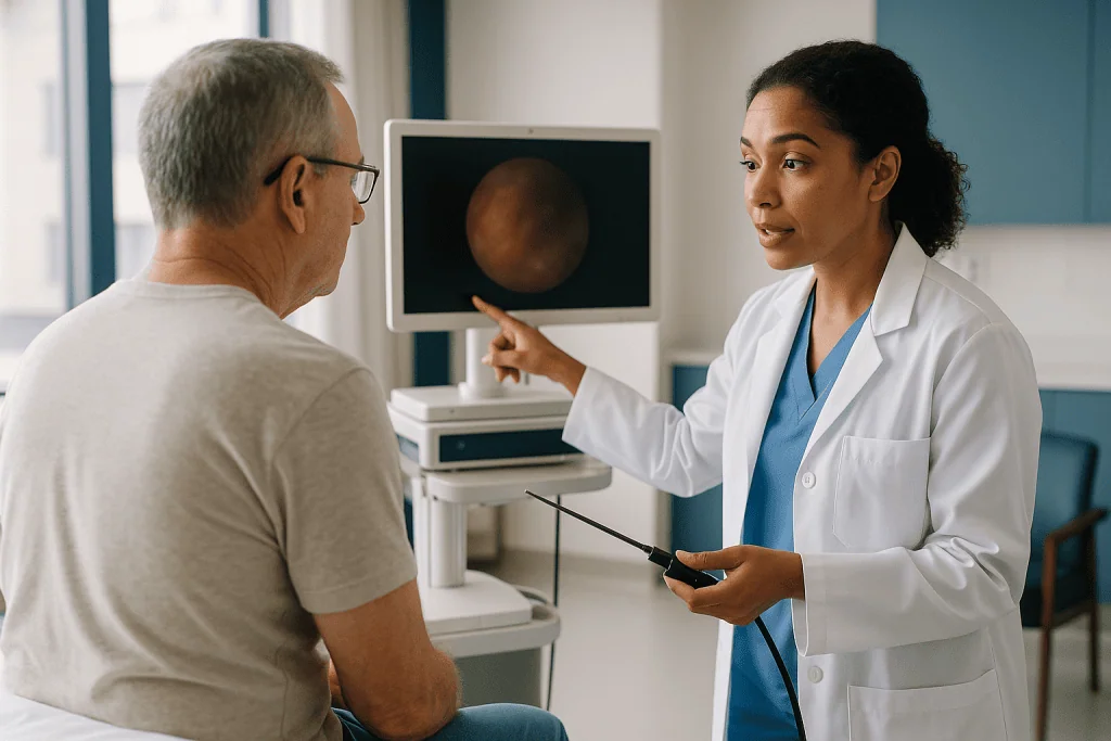
Filling is the odd part. The bladder stretches, and you feel a strong urge to urinate even though you cannot. The doctor rotates the scope to inspect every wall, the dome, the base and the two small openings where the ureters enter. Anything unusual can be photographed. If a sample is needed, small forceps pass through a channel in the scope, or the person is booked for a separate procedure under anesthesia.
According to the National Institute of Diabetes and Digestive and Kidney Diseases (NIDDK), a flexible cystoscopy in a clinic usually takes a few minutes, and the whole appointment is often under half an hour. Mayo Clinic gives a similar range of roughly 5–15 minutes for a simple outpatient examination.
A rigid cystoscopy is a different setting. The scope is straight and slightly wider, and it allows larger instruments to pass, so it is used when a tumor is going to be removed or a stone broken up. That is done under general or spinal anesthesia in an operating room, and the procedure that removes bladder tissue through the scope is called transurethral resection of bladder tumor, or TURBT. A rigid cystoscopy takes longer, and you go home the same day or after one night, depending on what was done.
Nothing is cut from outside. There are no incisions and no stitches, which is why cystoscopy is described as a procedure rather than an operation.
What does a CT scan of the bladder show, and can it see bladder cancer?
A CT scan can show bladder cancer, but with limits worth understanding. The bladder-specific version most people receive is called CT urography: a CT scan taken before and after an iodine-based contrast dye is injected into a vein. The dye passes through the kidneys into the urine, and when the bladder is full of contrast, a tumor stands out as a dark shape against the bright fluid, or as a thickened patch of wall.
What CT does well is scale and reach. In one session of roughly 10–20 minutes of room time, it images kidneys, ureters, bladder, lymph nodes, liver and bones. It picks up kidney stones, blockages, tumors in the ureter, and enlarged lymph nodes that suggest spread. The NHS describes CT urography as a standard test when blood in the urine needs a full explanation, precisely because it covers the whole urinary tract.
What CT does poorly is small and flat. Tumors a few millimeters across, or lesions that lie flat against the lining rather than growing into the bladder space, can be invisible on CT. A bladder that is not fully distended folds on itself, and folds can look like thickening. Inflammation from infection or a recent procedure can also mimic disease. This is why a normal CT does not, by itself, rule out a bladder tumor, and why guidelines still pair CT with cystoscopy when the concern is cancer.
Contrast dye brings its own considerations. It is processed by the kidneys, so people with reduced kidney function may need a modified protocol or a different test. Allergic reactions to iodinated contrast are uncommon but recognized, and anyone who has reacted before should tell the radiology team. CT also involves ionizing radiation, which is a legitimate reason to avoid repeating it unnecessarily, particularly in younger people who may need many years of follow-up.
What does an MRI of the bladder reveal that CT cannot?
MRI for bladder cancer answers a question that CT and cystoscopy struggle with: how deep? The bladder wall has layers. Innermost is the urothelium, beneath that a thin connective layer, then the muscle, then fat and neighboring organs. Whether a tumor has reached the muscle layer changes the entire treatment conversation, because non-muscle-invasive disease is usually managed through the scope, while muscle-invasive disease often calls for more extensive treatment.
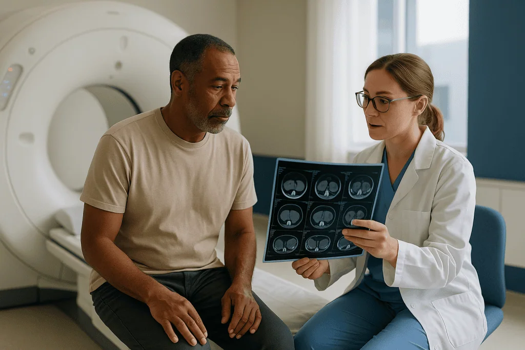
Cystoscopy shows the surface only. A tumor that looks small from inside may extend through the wall. CT shows the wall as a single gray band and cannot reliably separate its layers. MRI, using several image types in one examination, a method radiologists call multiparametric MRI, can often show the muscle layer as a distinct dark line and can show whether a tumor interrupts it.
The examination itself is longer and less comfortable than CT. You lie inside a tunnel-shaped magnet for roughly 30–45 minutes, hold still, and hear repetitive knocking sounds. A gadolinium-based contrast agent, a different dye from the one used in CT, is often injected part way through. There is no radiation, which is a genuine advantage for people who will be scanned repeatedly. Anyone with certain implanted devices, metal fragments, or severe claustrophobia needs to discuss this in advance, since some devices are not safe in a strong magnetic field.
MRI is not the first test for most people. It is slower, less available, and worse than CT at showing the kidneys and ureters in one sweep. Its role, as described by Mayo Clinic and in the radiology literature indexed on PubMed, is chiefly in staging a tumor that has already been found, and in guiding whether a second resection through the scope is needed. A standardized reporting scale for bladder MRI exists and is increasingly used, though it remains a support for the biopsy result rather than a replacement for it.
Cystoscopy vs CT scan: is cystoscopy more accurate?
For finding a tumor on the bladder lining, yes. For deciding whether that tumor has spread, no. The honest comparison depends entirely on what you are trying to find.
Cystoscopy is regarded across major urology guidelines as the reference test for detecting bladder tumors, the standard against which imaging is measured. It sees the surface directly at high magnification, and it allows the one thing no scan can do: taking a piece of tissue. A diagnosis of bladder cancer is made by a pathologist looking at that tissue under a microscope, not by a scan and not by the camera image alone. Mayo Clinic and the NHS both describe biopsy through the cystoscope as the step that confirms a diagnosis.
Cystoscopy has blind spots of its own. Flat, reddish lesions known as carcinoma in situ can look like inflammation. A very bloody bladder is hard to inspect. Some tumors sit in a fold or behind an enlarged prostate. Enhanced viewing techniques, such as blue-light cystoscopy after a dye is instilled into the bladder, have been developed to improve detection of flat lesions, and are used in some centers; their role remains a matter of ongoing study.
CT’s strengths lie exactly where cystoscopy is blind: above the bladder and outside its wall. A ureteral tumor, a kidney mass, or an enlarged pelvic lymph node will never be seen through a bladder scope. That is why a person with visible blood in the urine is commonly offered both, not one or the other.
A useful way to think about the cystoscopy vs CT scan question is to picture inspecting a house. Cystoscopy is walking through the rooms with a flashlight. CT is the aerial photograph of the whole property. You would not want only one of them if you suspected a problem you could not yet locate.
Who is usually offered which test, and who is usually asked to wait?
The strongest single trigger for a full workup is visible blood in the urine, known as gross hematuria, particularly when it is painless. The NHS advises that anyone who sees blood in their urine should be assessed, and most pathways for adults with visible hematuria include both cystoscopy and imaging of the upper urinary tract, because the risk of a significant finding is high enough to justify both. Age, smoking history and certain occupational chemical exposures push the concern higher, since these are the best-documented risk factors for urothelial cancer.
Blood that is found only on a dipstick or under a microscope, called microscopic hematuria, is handled more selectively. Younger adults with no risk factors and a plausible benign explanation are often offered an ultrasound and a repeat urine test first, with cystoscopy reserved for people whose risk is higher or whose blood persists. This is the group most often asked to wait, and the waiting is a deliberate choice to avoid an invasive test where the yield is low, not a sign of being dismissed.
Cystoscopy is also the routine test for people who have already had a bladder tumor. Because these tumors tend to recur on the lining, surveillance cystoscopy at intervals set by the treating team, based on the grade and stage of the original tumor, is standard practice described by Mayo Clinic and the NHS.
Other reasons a urologist orders cystoscopy include recurrent urinary infections without an obvious cause, difficulty emptying the bladder, suspected stones, persistent bladder pain, and follow-up after certain pelvic surgeries. CT is more likely when the concern is stones, blockage or spread. MRI is most often reserved for staging a tumor already confirmed on biopsy.
Timing can shift. A urinary infection is usually treated before cystoscopy, since instrumenting an infected bladder raises the chance of a more serious infection. Blood thinners, pregnancy and kidney function all alter which test comes first and how it is done. Those judgments belong to the treating team.
Bladder tests compared: cystoscopy vs CT vs MRI at a glance
Laying the three tests side by side makes the division of labor obvious. Each row below reflects general descriptions from Mayo Clinic, NIDDK and the NHS; individual departments vary in exactly how they run each test.
| Feature | Cystoscopy | CT urography | Bladder MRI |
|---|---|---|---|
| What it is | Camera passed through the urethra into the bladder | X-ray cross-sections after intravenous iodine contrast | Magnetic field and radio waves, often with gadolinium contrast |
| Best at | Seeing the lining directly; taking a biopsy | Kidneys, ureters, stones, lymph nodes, spread | Depth of tumor in the bladder wall |
| Weakest at | Anything above or outside the bladder | Small or flat lesions on the lining | Upper urinary tract; slow; limited availability |
| Typical time in room | About 5–15 minutes for a flexible clinic scope | About 10–20 minutes | About 30–45 minutes |
| Anesthesia | Numbing gel for flexible; general or spinal for rigid | None | None |
| Radiation | None | Yes | None |
| Main risks | Burning, minor bleeding, infection; rarely urethral injury | Contrast allergy; kidney strain; radiation exposure | Contrast reaction; unsafe with some implants; claustrophobia |
| Confirms cancer? | Yes, via biopsy | No, suggests only | No, suggests and stages |
Two points from the table deserve emphasis. First, only cystoscopy ends in a tissue sample, which is why a scan that looks worrying still leads back to the scope. Second, the tests carry different kinds of risk. Cystoscopy’s risks are local and short-lived; CT’s are about contrast and cumulative radiation; MRI’s are about the magnet and the time inside it. A team weighing which to order is balancing these against what each test can realistically find in your situation.
Bladder ultrasound vs cystoscopy: where do ultrasound and urine tests fit in?
Two quieter tests often come before any of the three headline ones. Ultrasound uses sound waves and a handheld probe on the lower abdomen. It has no radiation, no contrast and no discomfort beyond a full bladder, and it can show kidney size, blockage, stones, and larger bladder masses. NIDDK lists it among the first imaging tests for urinary symptoms for exactly those reasons.
Its limits are the flip side of its gentleness. Ultrasound sees the bladder from outside through the abdominal wall, so resolution of the lining is poor, small tumors are easily missed, and the ureters are largely invisible. A normal ultrasound is reassuring in a low-risk person but does not carry the weight of a normal cystoscopy in someone with visible blood.
Urine tests come in three forms. A dipstick and microscopy confirm that blood is really present and check for infection. Urine culture identifies bacteria. Urine cytology sends the cells shed into the urine to a pathologist who looks for abnormal ones. Cytology is good at catching high-grade tumor cells, which tend to shed readily, and poor at catching low-grade ones, so a negative result does not exclude disease. Mayo Clinic describes cytology as one of several tests used alongside cystoscopy rather than in place of it.
Newer urine-based molecular markers exist and are studied extensively in the PubMed literature. At present, major guidelines treat them as adjuncts and do not recommend replacing cystoscopy with them for diagnosis or surveillance. If a clinic offers one, it is reasonable to ask how the result will change the plan.
The practical sequence for many people, then, is urine tests and ultrasound first, cystoscopy and CT when risk or findings justify them, and MRI if a tumor has been confirmed and depth matters. The order is a funnel, widening or narrowing according to what each step reveals.
Does cystoscopy hurt? What is the most painful part?
Most people who have had a flexible cystoscopy describe two moments that stand out. The first is the scope passing the external sphincter, the ring of muscle you tighten to hold urine, which produces a brief pressure or sting lasting a few seconds. In men, the scope also passes through the prostate, and a second moment of pressure can occur there. The second notable sensation is the bladder filling with fluid, which brings a strong, sometimes urgent, need to urinate.
Neither is usually described as sharp pain. NIDDK and Mayo Clinic both characterize the experience as discomfort or a burning sensation rather than pain, and note that the numbing gel reduces it. Anxiety plays a measurable role: a tense pelvic floor makes the sphincter harder to pass. Slow breathing and deliberately relaxing the muscles you would use to stop urinating make a real difference, and an experienced operator will pace the scope to your breathing.
The burning generally returns after the procedure, when you first urinate. Mayo Clinic notes that this, along with a little blood and increased frequency, typically settles within 1–2 days. Drinking water helps dilute the urine and eases the sting.
People who have had a rigid cystoscopy under anesthesia feel nothing during the procedure itself. Afterward, the discomfort is greater than after a flexible scope, since tissue may have been removed, and a catheter may be left in briefly. Soreness on urinating and blood-tinged urine for several days are expected, and the treating team explains what is normal for the specific procedure done.
A minority find cystoscopy genuinely painful, particularly people with urethral narrowing, prior pelvic radiation, or chronic pelvic pain conditions. Saying so beforehand matters. Options include more anesthetic gel, a smaller scope, sedation, or performing the examination under anesthesia. Nobody should be made to endure a test in silence.
Is cystoscopy a serious procedure? Risks of each test in plain terms
Cystoscopy is invasive but not major. It is performed thousands of times a day in outpatient clinics, and NIDDK describes complications as uncommon. The main ones are worth knowing. A urinary tract infection can follow because a tube has passed through a channel that normally stays sterile; symptoms would be fever, chills and worsening burning in the days after. Bleeding is usually minor and brief. Rarely, the urethra can be scraped or narrowed by scarring, and very rarely the bladder wall can be perforated, which is more a risk of tissue removal than of simple inspection. Temporary difficulty urinating from swelling can occur and occasionally needs a short-term catheter.
Rigid cystoscopy under anesthesia carries the usual anesthetic considerations plus a higher chance of bleeding and infection, because it is typically combined with a resection or stone treatment. The team explains these against the specific procedure planned.
CT’s risks are silent. Iodinated contrast can trigger an allergic reaction ranging from hives to, rarely, a severe reaction; radiology departments screen for previous reactions and can pre-medicate or choose another test. Contrast passes through the kidneys, so people with reduced kidney function are assessed beforehand. Radiation exposure from a single CT is modest, but it accumulates, and this is why a young person on long-term surveillance may be steered toward MRI or ultrasound where possible.
MRI has no radiation, but the magnet is powerful enough to move or heat certain metal objects. Pacemakers, some cochlear implants, and older aneurysm clips are the classic concerns; many modern devices are MRI-conditional, meaning safe under specified settings. Gadolinium contrast is generally well tolerated; people with severe kidney disease are assessed individually. The confined space and noise are difficult for some, and open or wide-bore scanners, or mild sedation, are options.
None of these risks makes any of the tests dangerous for a typical patient. They make them tests to be ordered for a reason, which is what a careful urologist does.
Cystoscopy recovery time, preparation and what the following days look like
Preparation for a flexible cystoscopy is minimal. You can usually eat and drink normally, take regular medicines unless told otherwise, and drive yourself home. Many clinics ask for a urine sample first to check for infection. Tell the team about blood thinners, previous contrast reactions, and any difficulty passing urine, since these may change the plan. For a rigid cystoscopy under anesthesia, fasting instructions and a companion to take you home apply, as for any anesthetic.
The first day after a flexible cystoscopy is the main event. Expect burning when you urinate, a pink tinge to the urine, and the need to go more often. Mayo Clinic advises that these effects generally ease within 1–2 days and that drinking plenty of water helps. A warm cloth over the urethral opening or a warm bath can ease the sting. Most people return to work the same or the next day.
After a rigid cystoscopy with tissue removal, the timeline stretches. Blood in the urine can come and go for one to two weeks, often clearing and then reappearing around the time the internal scab separates. Strenuous activity, heavy lifting and sexual activity are typically paused for a period set by the surgical team. A catheter, if placed, is usually removed within a day or two. Pathology results from the biopsy take several days to a couple of weeks depending on the laboratory, and the follow-up appointment to discuss them is where the next steps are decided.
CT and MRI need no recovery. After contrast, drinking extra fluid over the following day helps the kidneys clear it. Results are read by a radiologist and sent to the ordering clinician, which usually means a wait of days rather than hours, and the report is interpreted alongside the cystoscopy findings rather than on its own.
If you are booked for more than one test, ask in what order they will happen and when the results will be brought together. Knowing the shape of the coming weeks reduces the anxiety of waiting for each piece.
What people often get wrong about bladder tests
Several misconceptions surface repeatedly in clinics and online, and each can lead to a poor decision.
A clear CT means the bladder is fine. It does not. CT misses small and flat lesions on the lining, and Mayo Clinic and the NHS describe cystoscopy, not CT, as the test that confirms or excludes a bladder tumor. A reassuring scan is welcome, but it does not cancel a recommended cystoscopy.
Cystoscopy is a surgery. A flexible cystoscopy is a clinic examination with no incisions, no anesthesia beyond numbing gel, and a return to normal activity the same day. Only the rigid version done under anesthesia resembles an operation, and even that is performed through the natural channel of the urethra.
Blood in the urine that stops on its own can be ignored. Hematuria from a bladder tumor is classically intermittent. The NHS advises assessment for any visible blood in the urine, whether or not it recurs. Its disappearance is not evidence that the cause has gone.
MRI is simply a better CT. The two answer different questions. MRI is superior for judging how deep a bladder tumor goes; CT is faster and better for the kidneys, ureters and a broad look for spread. Ordering MRI first for undiagnosed blood in the urine would leave the upper tract poorly examined.
Urine tests can replace the camera. Cytology and newer urine markers help, particularly for high-grade disease, but guidelines continue to treat them as supplements. A negative urine test in someone with visible blood does not close the question.
Only smokers get bladder cancer. Smoking is the largest modifiable risk factor, but a substantial share of people diagnosed have never smoked. Symptoms are what trigger testing, not lifestyle assumptions in either direction.
Correcting these ideas does not make the tests less uncomfortable or the wait less worrying. It does make the plan your team proposes easier to understand and harder to talk yourself out of.
Questions to ask your care team before bladder tests
Good questions do two things: they help you understand the plan, and they surface details about you that the team may not know. Take these to the appointment, in your own words.
- Which test are you ordering first, and what question is it meant to answer?
- If the first test is normal, will I still need the others? If it is abnormal, what happens next?
- Will the cystoscopy be flexible in the clinic or rigid under anesthesia, and why that choice for me?
- Is a biopsy likely during the cystoscopy, or would that be a separate visit?
- Will the CT use contrast dye? Do my kidney function and any past reactions affect that?
- Is MRI part of my pathway, and if so, at what stage and for what purpose?
- I take a blood thinner or have a heart device. Does that change how or when any of these tests are done?
- How long will results take, and who will explain them to me?
- If I found the last cystoscopy very painful, what options exist to make this one more comfortable?
- If I will need repeated checks, how often, and can radiation exposure be limited over time?
- What symptoms after the test should make me call the clinic rather than wait for follow-up?
It also helps to bring a short written history: when the blood or symptoms started, whether they were painful, any infections treated, smoking history, past pelvic surgery or radiation, and a current medication list. Clinicians make better decisions with better information, and a five-minute list can spare a repeat visit.
Finally, ask what the team is most concerned about and what it considers least likely. Hearing the range of possibilities, including the benign ones, gives the coming weeks a more realistic shape than a single word on a referral letter.
When to call your doctor
Most people get through bladder testing with nothing more than a day of burning and a pink tinge in the toilet bowl. A small number develop problems that need prompt attention, and knowing the difference matters more than any other piece of preparation.
After a cystoscopy, contact the clinic or urgent care the same day if you develop a fever above 100.4°F (38°C) or chills, since these can signal a urinary infection spreading beyond the bladder. Call if you cannot pass urine at all despite the urge, if the burning worsens after the second day instead of easing, or if the urine turns bright red, contains clots, or does not begin to clear within a couple of days after a flexible scope. Mayo Clinic and NIDDK both list fever, inability to urinate, and heavy or persistent bleeding as reasons to seek care.
After a rigid cystoscopy with tissue removal, some blood for a week or two is expected, but heavy bleeding, inability to urinate, worsening lower abdominal pain, or fever should be reported without delay. If a catheter was placed and stops draining, or the area around it becomes painful and swollen, call.
After a CT or MRI with contrast, seek emergency care for hives spreading rapidly, swelling of the lips or face, wheeze or difficulty breathing, or feeling faint. Milder reactions such as a small rash or nausea should still be reported to the radiology department so they are recorded for future scans.
Independently of any test, the NHS advises that anyone who sees blood in their urine should see a doctor, even if it happened once and stopped. New difficulty passing urine, pain in the flank with fever, or unexplained weight loss alongside urinary symptoms are also reasons for a prompt appointment rather than watchful waiting.
These are reasons to call, not signs of a diagnosis. The people who know your case are the ones to decide what they mean.
Frequently asked questions
What is the most painful part of a cystoscopy?
Most people find the scope passing the external sphincter, the muscle ring that holds urine in, the most uncomfortable moment, followed by the pressure of the bladder filling with fluid. Both last seconds to a couple of minutes and are usually described as stinging or urgency rather than sharp pain. Numbing gel, slow breathing and deliberately relaxing the pelvic floor reduce it, and anyone who finds it severe can ask about sedation or examination under anesthesia.
Can they see bladder cancer on a CT scan?
Often, yes, particularly larger tumors that grow into the bladder space or thicken the wall, which show up clearly when the bladder is filled with contrast dye. Small tumors and flat lesions on the lining are frequently missed, so a normal CT does not exclude bladder cancer. CT is valued mainly for what cystoscopy cannot see: the kidneys, ureters and lymph nodes. Confirming cancer still requires a biopsy taken through the cystoscope.
Is cystoscopy more accurate than a CT scan?
For detecting tumors on the bladder lining, cystoscopy is the reference test and is more accurate than CT, because it views the surface directly and allows a biopsy. For detecting disease in the kidneys and ureters or spread to lymph nodes, CT is far more useful, since the scope cannot see those areas. The two are complementary, which is why people with visible blood in the urine are usually offered both.
Is cystoscopy a serious procedure?
A flexible cystoscopy is a minor outpatient examination done with numbing gel, no incisions and a same-day return to normal activity. Complications such as infection or significant bleeding are uncommon. A rigid cystoscopy under anesthesia, used when tissue is removed or stones are treated, is more involved and carries higher risks of bleeding and infection, but it is still performed through the natural channel of the urethra without external cuts.
How long is cystoscopy recovery time?
After a flexible cystoscopy, burning on urination, slight blood and urinary frequency typically settle within 1–2 days, according to Mayo Clinic, and most people work the same or next day. After a rigid cystoscopy with tissue removal, blood in the urine can come and go for one to two weeks, and strenuous activity is paused for a period the surgical team sets. Biopsy results usually take several days to a couple of weeks.
When is MRI for bladder cancer used instead of CT?
MRI is usually ordered after a tumor has been found, to judge how deeply it extends into the bladder wall and whether it has reached the muscle layer, which CT shows poorly. It is also favored when radiation exposure should be limited or when iodinated CT contrast is unsuitable. MRI is rarely the first test for undiagnosed blood in the urine, because it is slower and less effective at surveying the kidneys and ureters.
Cystoscopy vs CT scan: do I need both if one is normal?
Frequently yes, because each test is blind where the other sees. A normal cystoscopy leaves the kidneys and ureters unexamined; a normal CT can miss small or flat bladder lesions. For visible blood in the urine, standard pathways pair cystoscopy with upper-tract imaging. For microscopic blood in a low-risk person, a team may reasonably start with ultrasound and urine tests alone. The decision depends on your risk profile and belongs to your treating clinician.
What is bladder ultrasound vs cystoscopy good for?
Ultrasound is a painless, radiation-free first look at kidney size, blockage, stones and larger bladder masses, which makes it a sensible opening test, especially for lower-risk people. Cystoscopy sees the bladder lining directly at high magnification and can take a biopsy, so it detects small and flat lesions ultrasound cannot. A normal ultrasound is reassuring but does not carry the same weight as a normal cystoscopy when bladder cancer is a concern.
Do I need anesthesia for a cystoscopy?
For a flexible cystoscopy, usually only a local anesthetic gel placed in the urethra, and you stay awake and can drive home afterward. A rigid cystoscopy, used when tissue is removed or larger instruments are needed, is done under general or spinal anesthesia in an operating room. People who have found a previous flexible scope very painful, or who have urethral narrowing or chronic pelvic pain, can ask about sedation or an examination under anesthesia.
Why does a urologist repeat cystoscopy after bladder cancer treatment?
Bladder tumors that are removed through the scope have a recognized tendency to recur elsewhere on the lining, so surveillance cystoscopy at intervals set by the treating team is standard practice described by Mayo Clinic and the NHS. The frequency depends on the grade and stage of the original tumor and how long you have been clear. Urine tests and imaging may be added, but they do not replace direct inspection of the lining.
References
- NIDDK (NIH): Cystoscopy and Ureteroscopy
- MedlinePlus Medical Encyclopedia: Cystoscopy
- NHS: Bladder cancer, Diagnosis
- Cleveland Clinic: Cystoscopy
This article is for general information only and is not a substitute for professional medical advice. Please consult a qualified doctor about your individual situation.

More from the Blog

How Do You Read a Urodynamic Report? Capacity, Pressure-Flow Curves and Leak Points Explained
A urodynamic report is read in order: the header and patient details, then the flow curve and the volume left after voiding, then the…

When Can You Lift, Drive and Exercise Again After Robotic Prostate Surgery?
Most people walk the day of robotic prostate surgery, drive again once the catheter is out and they are no longer taking opioid painkillers,…
How Peritoneal Dialysis Exchanges Work: Fill, Dwell and Drain Explained in Plain Words
A peritoneal dialysis exchange uses the lining of your own abdomen as a filter. Cleansing fluid flows through a soft catheter into the belly…

Reading a Bladder Disease Treatment Plan: Questions to Ask Your Urologist Before You Decide
Before agreeing to a bladder disease treatment plan, ask your urologist which specific condition is being treated, what test results support that diagnosis, why…

Is Tiredness After Hemodialysis Normal? Cramps, Low Blood Pressure and What to Report
Yes, feeling tired after hemodialysis is common and expected for many people, because a session removes fluid and waste quickly and shifts salts and…

Shockwave Therapy or Endoscopic Removal: Which Kidney Stone Treatment Fits Your Stone?
Shock wave lithotripsy breaks a kidney stone from outside the body using focused sound waves, while ureteroscopy passes a thin scope up the urinary…