

ESWL vs Ureteroscopy for Kidney Stones: Why Stone Size, Location and Hardness Decide
Key Takeaways
- Shock wave lithotripsy is generally limited to stones up to about 2 centimeters, according to the NHS, while larger stones are usually directed to keyhole surgery through the back.
- Stones in the lower pole of the kidney sit in a gravitational dead end, which is the most common reason a urologist steers a modest stone toward ureteroscopy instead of ESWL.
- The Hounsfield density reading on a non-contrast CT predicts how a stone will respond to shock waves; laser fragmentation is largely indifferent to hardness.
- ESWL removes nothing directly; it breaks the stone into fragments that pass in the urine over days to weeks, which is why follow-up imaging is needed to confirm clearance.
- Ureteral stents after ureteroscopy are typically temporary, but stent-related urgency and flank ache are the part of recovery patients most often say they were not warned about.
- Pregnancy, an uncorrected bleeding tendency, active urinary infection, and a blockage below the stone are standard reasons ESWL is not offered.
ESWL breaks kidney stones from outside the body with focused shock waves and relies on the fragments passing naturally; ureteroscopy passes a thin scope up the urinary tract to fragment and remove the stone directly. Neither is universally better. Urology guidelines steer the choice by stone size, position in the kidney or ureter, density on CT, and the patient's anatomy and health, so the treating team decides case by case.
The phone call usually comes a day or two after the emergency visit. The pain has eased, the CT report is in the portal, and a scheduler asks a question that sounds simple: would you prefer the shock wave appointment or the scope? Most people put the phone down and start searching eswl vs ureteroscopy, hoping one option is plainly safer, quicker, or more likely to work.
What they find is a tangle of forum posts about stents, videos of stones shattering like sugar cubes, and articles that quietly favor whichever machine the author owns. The honest answer is less dramatic. A urologist weighing the two treatments is not asking which is better in general. They are asking three narrower questions about the stone in front of them: how big it is, where it sits, and how hard it is.
This article walks through those three questions the way a clinician does, so the conversation with your care team feels like a discussion rather than a coin toss.
ESWL vs ureteroscopy: what actually happens in each procedure
Extracorporeal shock wave lithotripsy, or ESWL, is the only common stone treatment that never enters the body. You lie on a padded table, sometimes with light sedation or a general anesthetic, while a machine outside the skin sends focused pressure waves through a water-filled cushion toward the stone. Imaging, usually X-ray or ultrasound, keeps the target centered. Each pulse sounds like a firm tap on a tabletop. Thousands of them, delivered over a session that the Mayo Clinic describes as lasting roughly 45 to 60 minutes, cause the stone to crack into gravel that then travels down the ureter and out with urine over the following days or weeks.
Ureteroscopy takes the opposite approach: go to the stone rather than send energy toward it. Under general anesthesia, the urologist passes a ureteroscope, a very thin telescope about the width of a cooked spaghetti strand, through the urethra and bladder and up the ureter, the tube linking each kidney to the bladder. Rigid scopes reach stones lodged in the ureter; flexible ones bend into the kidney’s collecting system, the branching spaces where urine gathers. Once the stone is in view, a laser fiber threaded through the scope pulverizes it to dust or breaks it into pieces small enough to lift out with a wire basket. Nothing is left to pass on its own if the surgeon can help it.
The two procedures share a goal and almost nothing else. One trades precision for gentleness; the other trades a more invasive route for direct control. That difference explains nearly every clinical trade-off that follows, from how many sessions may be needed to why a stent is more often placed after the scope than after the shock waves.
Why stone size is the first thing urologists check
Size sets the floor and the ceiling. At the small end, stones under about 5 millimeters, roughly the width of a pencil eraser, frequently pass without any procedure at all, according to the National Institute of Diabetes and Digestive and Kidney Diseases. For those, the reasonable first step is often fluids, pain control, sometimes a medicine that relaxes the ureter, and a few weeks of patience. Both ESWL and ureteroscopy are held in reserve.
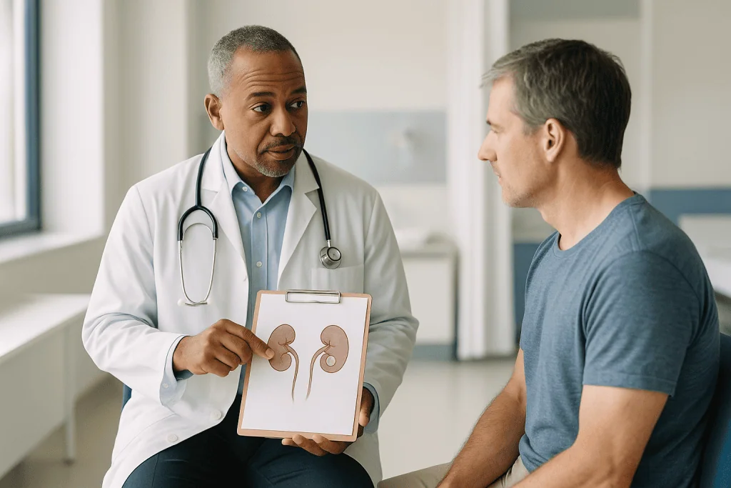
At the large end, the picture flips. The NHS notes that ESWL is generally reserved for stones up to about 2 centimeters. Beyond that, shock waves create too much gravel to pass comfortably, and the risk of fragments piling up in the ureter like a traffic jam, a complication called steinstrasse, German for stone street, climbs. Very large stones, particularly those filling the kidney’s collecting system, are usually directed toward percutaneous nephrolithotomy, a keyhole procedure through the back, rather than either option in this article.
Between those bookends lies the contested middle ground where the ESWL vs ureteroscopy question is genuinely open. A 7-millimeter stone and a 15-millimeter stone are both technically candidates for both approaches, but the odds shift as the number grows. Ureteroscopy clears larger stones in a single visit more reliably because the surgeon sees the pieces leave; ESWL may need a second or third session for the same stone, and each pass of fragments carries its own bout of discomfort.
Total stone burden matters as much as the biggest stone. Three 6-millimeter stones scattered across a kidney behave differently from one 18-millimeter stone. The scope can hunt them down individually; shock waves have to be re-aimed for each target. Ask your team not just how big the stone is, but how many there are.
Location: the lower pole problem and the ureter question
Think of the kidney as an upside-down funnel. Stones near the top or middle of the collecting system have gravity on their side once they shatter; fragments drift toward the outlet. Stones in the lower pole, the bottom cup of the kidney, sit in a gravitational cul-de-sac. Shock waves may crack them perfectly, yet the gravel has to climb uphill to reach the ureter. This is the single most common reason a urologist steers a patient with a modest-sized stone away from ESWL and toward a flexible scope, which can pluck fragments out of that pocket directly.
Anatomy within the lower pole matters too. A long, narrow tube leading from that cup to the main drainage area, or a steep angle between the two, makes spontaneous fragment clearance even less likely. Your radiology report may not spell this out, but your urologist will be looking at it.
Ureteral stones bring a different calculation. Stones in the upper ureter, near the kidney, often respond well to shock waves because the surrounding tissue transmits energy cleanly and the fragments have a short, downhill route. Stones in the lower ureter, close to the bladder, sit behind the bony pelvis, which can block or scatter shock waves; a rigid ureteroscope reaches them easily from below. The Mayo Clinic lists both approaches as standard for ureteral stones, and the Cleveland Clinic describes ureteroscopy as particularly suited to stones lodged in the ureter.
One further factor is how long the stone has been stuck. A stone wedged in one spot for weeks can become embedded in swollen ureteral tissue, which dampens shock wave energy and favors direct removal. A stone that arrived yesterday behaves like a loose marble. The same millimeter reading can therefore point to different treatments depending on the timeline.
Stone hardness: what the CT scan reveals before anyone touches you
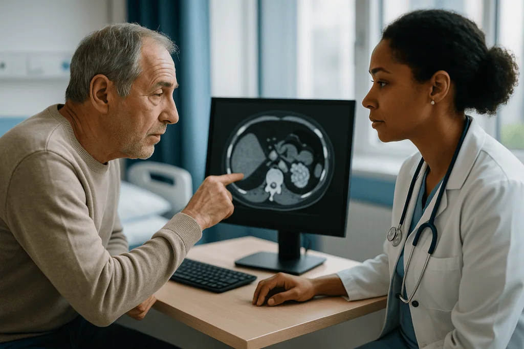
Not all stones crack alike. A non-contrast CT scan, the standard test for suspected kidney stones according to the Mayo Clinic, does more than locate the stone; it measures how strongly the stone blocks X-rays, expressed in Hounsfield units. A brighter, denser stone on the scan is usually a harder one, and hardness predicts how shock waves will behave. Softer stones fragment into fine sand. Very dense stones can shrug off thousands of pulses, leaving a few large chunks that are no easier to pass than the original.
Chemistry sits behind the density. Calcium oxalate monohydrate stones and brushite stones, both calcium-based, are notoriously tough. Cystine stones, which form in an inherited condition affecting how the kidneys handle an amino acid, are also poor shock wave targets. Uric acid stones tend to be softer and may even dissolve partly with medication that changes urine acidity, a decision that belongs to the treating team. Calcium oxalate dihydrate and struvite stones usually break more readily.
The catch is that a first-time stone former rarely knows their stone type in advance. The CT density reading is the proxy urologists lean on. When a report describes an unusually dense stone, many will counsel toward ureteroscopy, because a laser fiber does not care much how hard the stone is; it ablates the surface layer by layer regardless.
A second measurement from the same scan is skin-to-stone distance, the depth of tissue the shock wave must cross. Waves lose energy over distance and scatter through fat. For people with a higher body weight or a stone positioned deep in the back, that distance can exceed what the machine delivers effectively. This is a physics limitation, not a judgment, and it is one more reason a flexible scope, which travels inside the urinary tract and is indifferent to body habitus, may be recommended instead.
Shock wave lithotripsy vs laser: a side-by-side comparison
Laid out together, the differences fall into a pattern. The table below reflects guideline-level descriptions from the NHS, Mayo Clinic, and Cleveland Clinic rather than any single center’s results; individual experiences vary, and your team will adapt every row to your case.
| Factor | ESWL (shock waves) | Ureteroscopy (laser) |
|---|---|---|
| Route | Entirely outside the body | Scope through urethra, bladder, ureter |
| Anesthesia | Sedation or general, varies | Usually general |
| Typical stone size | Up to about 2 cm (NHS) | Small to moderate; larger with staged approach |
| Lower pole stones | Fragments may not clear | Direct retrieval possible |
| Very dense stones | May fragment poorly | Laser largely indifferent to hardness |
| Sessions | May need more than one | Often one, sometimes staged |
| Stent afterward | Less common | Common, usually temporary |
| Fragment passage | Expected over days to weeks | Minimal; pieces removed or dusted |
| Not usually offered when | Pregnancy, uncorrected bleeding disorder, active infection, blockage below stone | Untreated urinary infection; some strictures |
Read the rows as tendencies, not verdicts. A patient who dreads general anesthesia and has a soft, mid-kidney stone under a centimeter has a strong case for shock waves. Someone with a dense lower pole stone and a job that cannot accommodate repeat appointments and weeks of passing gravel may reasonably lean toward the scope. The technology is a tool; the fit is the decision, and it belongs to the person having the procedure and the team advising them.
Who is usually offered ESWL, and who is usually asked to wait
The classic ESWL candidate has a stone under about 2 centimeters, sitting in the upper or middle kidney or the upper ureter, of average or lower density on CT, in a body where the stone is not too far from the skin. Add a preference to avoid anything passing through the urethra, or a medical reason to keep anesthesia light, and the case strengthens. For many such patients it is a same-day procedure with a short recovery, which is why the NHS describes it as a common first-line option for suitable stones.
Certain situations take ESWL off the table entirely. Pregnancy is an absolute reason not to proceed, because shock waves cannot be safely aimed near a developing fetus. An uncorrected bleeding tendency, whether from a condition or from anticoagulant medicines, raises the risk of kidney bruising; whether and how to pause such medicines is strictly a decision for the prescribing clinician, never something to adjust on your own. An active, untreated urinary infection needs treatment first, since shattering an infected stone can release bacteria into the bloodstream. A blockage downstream of the stone, such as a narrowed ureter, means fragments would have nowhere to go.
Then there is the group asked simply to wait. If the stone is small, pain is controlled, kidney function is normal, and there is no infection, watchful waiting with fluids is a legitimate treatment plan rather than an absence of one. The NIDDK notes that many small stones pass on their own. Urologists typically set a review point a few weeks out and intervene sooner only if pain becomes unmanageable, infection develops, or imaging shows the stone has stopped moving. Waiting is not failure; it is often the least invasive path to the same destination.
Who is usually offered ureteroscopy instead
Ureteroscopy earns its place whenever direct visualization solves a problem shock waves cannot. The Cleveland Clinic lists stones lodged in the ureter, especially the lower ureter, as a prime indication, and adds stones that failed to break with ESWL, stones that need to be removed quickly, and stones in people whose anatomy or body size limits shock wave delivery.
Several patient groups tilt toward the scope almost by default. Pilots, commercial drivers, and others whose work prohibits any risk of sudden incapacitating pain generally need confirmation that no fragments remain, which only direct removal provides. People taking blood thinners that cannot be paused often do better with ureteroscopy, because the laser works within the urinary tract without bruising kidney tissue; this judgment, again, sits with the treating team and the clinician managing the anticoagulant. Those with a solitary kidney, or with reduced kidney function, may be offered the scope to avoid any period of fragment obstruction.
Hard stones and lower pole stones, covered earlier, round out the list. So does a patient who has already endured one or two shock wave sessions without clearance and would rather not gamble on a third.
The scope has its own limits. A ureter that is too narrow to admit the instrument may require a stent to be placed first, with the actual stone treatment scheduled a couple of weeks later once the tube has gently widened. Very large kidney stones may need staged ureteroscopy over more than one visit, or may be redirected to percutaneous surgery. Active urinary infection must be treated before either procedure. A frank conversation about how many sessions are realistic, and what happens if the first attempt cannot reach the stone, belongs in the consent discussion rather than the recovery room.
Can ESWL remove kidney stones completely, or does the work fall to you?
Strictly speaking, ESWL removes nothing. It converts one stone into many smaller ones and delegates removal to your ureter and bladder. This distinction, obvious once stated, drives most of the surprise people report afterward. The MedlinePlus lithotripsy overview is clear that fragments pass in the urine over the days and weeks following treatment, and that this passage can be uncomfortable.
What that feels like varies. Fine sand may go unnoticed apart from blood-tinged urine for a day or two. Larger fragments can reproduce a milder version of the original renal colic, the cramping flank pain that sent you to the emergency department, as each piece travels the ureter. Straining urine through a fine mesh, if your team asks for it, catches fragments for laboratory analysis, which identifies the stone type and guides prevention.
Some fragments never leave. Residual pieces too small to cause symptoms can sit in the lower pole indefinitely, and a proportion grow into future stones. Follow-up imaging, usually an X-ray or ultrasound a few weeks later, is how the team confirms whether the kidney is actually clear rather than merely quiet. Skipping that appointment because you feel fine is a common and avoidable mistake.
Ureteroscopy, by contrast, aims to leave the collecting system empty at the end of the operation, either by extracting fragments in a basket or by dusting the stone so finely that the powder washes out with irrigation fluid during the procedure. Surgeons still order follow-up imaging, because tiny remnants can hide behind folds of tissue, but the expectation is different: the patient is not the final step in the process. Whether that trade, more invasive surgery for less homework afterward, appeals to you is a legitimate part of the decision.
Which is the safest procedure to remove kidney stones? Risks side by side
Safety is the question people most want answered in a single word, and it is the one that least deserves it. Both procedures are considered low-risk, established treatments by every major reference; the Mayo Clinic, NHS, and Cleveland Clinic all describe them as routine. What differs is the shape of the risk, not simply its size.
ESWL’s risks cluster around the shock waves themselves and the fragments they create. Bruising of the kidney, sometimes visible as a small hematoma on imaging, is the most characteristic complication and usually resolves on its own; larger bleeds are uncommon but more likely in people with bleeding disorders or uncontrolled high blood pressure. Fragments can obstruct the ureter, causing pain and occasionally requiring a stent or a scope to clear the jam. Infection can follow if bacteria trapped in the stone are released. Skin bruising over the treatment area is common and harmless. Long-term concerns about ESWL raising blood pressure or affecting kidney function have been studied for decades without settled conclusions; the honest summary is that any effect appears small, and your team can discuss the evidence as it stands.
Ureteroscopy’s risks come from instruments inside a delicate tube. Minor injury to the ureter lining is common and heals without intervention. Perforation, a small tear, is uncommon and typically managed with a stent for a few weeks. Rarely, scarring can narrow the ureter later, which is one reason follow-up imaging is recommended. Infection after ureteroscopy can occasionally be serious, which is why untreated infections are cleared first. General anesthesia carries its own small, well-characterized risks.
Neither profile is worse across the board. For a specific person, though, one profile is usually more acceptable than the other, and that is the conversation worth having.
The stent question: why it matters more to patients than surgeons
Search forums on this topic and one word dominates: stent. A ureteral stent is a soft, hollow plastic tube, thinner than a drinking straw, that runs from the kidney to the bladder with a curl at each end to hold it in place. It keeps urine flowing past swelling or fragments while the ureter heals. The Cleveland Clinic describes stents as commonly placed after ureteroscopy and usually removed within days to a few weeks, depending on the surgeon’s judgment.
Surgeons tend to view the stent as a minor safeguard. Patients often experience it as the most memorable part of the whole episode. The tube can cause a persistent urge to urinate, a dull ache in the flank when the bladder empties, visible blood in the urine, and discomfort during activity. None of these are dangerous, but they are wearing, and they explain why many people ask whether the scope can be done without one.
Sometimes it can. Uncomplicated ureteroscopy for a small stone, with minimal ureteral trauma and no swelling, is increasingly performed stent-free in appropriately selected patients. When a stent is placed, it may be attached to a string that allows removal at home or in the clinic without a second scope, or it may require a brief office procedure. Ask which arrangement your surgeon plans and why.
ESWL needs a stent less often, but not never. A stent may be placed beforehand for larger stones to prevent steinstrasse, or afterward if fragments obstruct the ureter. Anyone choosing shock waves purely to avoid a stent should know that the guarantee does not exist in either direction.
Whatever the plan, a stent is temporary. Forgotten stents left in for many months can become encrusted with new stone and are difficult to remove, so keeping the removal appointment is not optional.
Ureteroscopy vs lithotripsy recovery: what the following days and weeks usually look like
After ESWL, most people go home the same day. The NHS and MedlinePlus both describe recovery as brief, with a return to ordinary activity often within a day or two once the sedation has worn off. The work of the next couple of weeks is passing fragments: expect pink or reddish urine initially, some cramping as pieces move, and possibly a request to strain your urine. Drinking enough fluid to keep urine pale is usually encouraged, unless your team has given different advice for a heart or kidney condition. A follow-up X-ray or ultrasound, typically a few weeks later, checks clearance and decides whether another session is needed.
After ureteroscopy, discharge is also usually the same day, though general anesthesia means someone should take you home. Burning with urination, urgency, and blood-tinged urine are common for several days, more so if a stent is in place. Many people return to desk work within a few days and to strenuous activity within a week or two; the Cleveland Clinic advises following your surgeon’s specific guidance. If a stent is present, its removal date is the real end of recovery for most patients, since the stent-related symptoms lift almost immediately once it is out.
Both paths converge on the same later milestone: stone analysis and prevention. If fragments were caught or retrieved, the laboratory identifies the type. Blood and urine tests may follow to look for underlying causes. The prevention plan that results, whether adjustments to fluid intake, diet, or in some cases a medicine, is where the long-term benefit lives. A procedure treats the stone you have. Only prevention addresses the one that might form next, and that discussion deserves as much attention as the choice between shock waves and scope.
What people often get wrong about ESWL and ureteroscopy
The first misconception is that ESWL is the gentle option and ureteroscopy the harsh one. Gentleness depends on outcome. A shock wave session that leaves fragments lodged in the ureter for three weeks, followed by an emergency stent, is not gentler than a single scope that ends with an empty kidney. The least invasive procedure on paper is not always the least disruptive in practice.
The second is that newer means better. Laser technology has advanced quickly, and flexible scopes now reach places that were once inaccessible. That progress is real, and it has shifted many stones from the shock wave list to the scope list. It has not made shock waves obsolete. For the right stone in the right place, ESWL remains a guideline-endorsed first choice, and choosing it is not settling for less.
Third, people assume the surgeon’s recommendation reflects preference or habit rather than evidence. Sometimes local expertise does shape the offer, and it is fair to ask about it. Far more often the recommendation follows the three factors this article has traced: size, location, density. If the reasoning is not explained, ask for it.
Fourth, many believe that a procedure marks the end of the stone story. The Johns Hopkins overview of kidney stones is blunt that recurrence is common in people who have formed one stone. Treatment clears today’s problem; prevention addresses tomorrow’s.
Finally, a stubborn myth holds that dissolving stones with home remedies, lemon juice, apple cider vinegar, or various herbal products, can substitute for either procedure. Adequate fluid and, for some, dietary changes have a place in prevention. There is no reliable evidence that any kitchen remedy breaks or dissolves an existing calcium stone, and delaying care for an obstructing stone while trying one can put the kidney at risk.
Questions to ask your care team before choosing
A good consultation about stone treatment should leave you able to explain the plan to a friend. These questions help get there, and none of them are impolite.
- How large is my stone, how many are there, and exactly where does each one sit?
- What did the CT show about the stone’s density, and does that change which option you would suggest?
- Is waiting a reasonable choice for me, and what would prompt you to intervene sooner?
- If you recommend ESWL, how likely is it that I would need more than one session, and how would we know?
- If you recommend ureteroscopy, do you expect to place a stent, for roughly how long, and how would it be removed?
- What type of anesthesia would I have, and what does that mean for the day of the procedure?
- Which complications are most relevant to my health history, and what would make you want to see me urgently afterward?
- Will you send any retrieved fragments for analysis, and when will we talk about preventing the next stone?
- Do any of my current medicines need attention before the procedure, and who will manage that?
- If the first attempt does not clear the stone, what is the next step?
Bring the list, a notepad, and if possible another person. Consultations move quickly, and the answers to the anesthesia and stent questions in particular tend to be the ones people wish they had written down. Any team accustomed to treating stones will have heard every one of these questions before and will welcome the chance to tailor the explanation to you.
When to call your doctor
Most recoveries after either procedure are uneventful, but a small number of complications need prompt attention, and the same red flags apply whether you are waiting for a stone to pass or recovering from treatment. The Mayo Clinic and NHS both advise urgent contact for the signs below.
Call your care team the same day, or use emergency services if symptoms are severe, for fever or chills, which can signal infection behind a blocked kidney and can progress quickly; pain that is severe, worsening, or not controlled by the medicines you were given; inability to pass urine, or passing only a trickle; nausea and vomiting that prevent you from keeping fluids down; heavy bleeding, meaning urine that looks like red wine rather than pink lemonade, or clots that make it hard to urinate; or new confusion, faintness, or a racing heartbeat.
Seek advice within a day or two, rather than waiting for a scheduled visit, if blood in the urine persists beyond the timeframe your team described, if stent discomfort is unmanageable, if you suspect the stent has slipped, or if you notice a fragment that seems stuck and pain that comes and goes in waves for more than a few hours.
Two situations deserve special mention. Fever combined with flank pain after any stone procedure is an emergency, not something to sleep on. And anyone with a single functioning kidney, diabetes, or a weakened immune system should have a lower threshold for calling about any of the above, because the margin for a blocked or infected kidney is smaller.
None of this is meant to alarm. It is meant to make the rare serious problem easy to recognize so it can be treated early, which is the outcome every team is working toward.
Frequently asked questions
Which is better for kidney stones, shock wave lithotripsy or laser removal?
Neither is better across all stones; the right choice depends on size, location, and density. Shock waves suit softer stones up to about 2 centimeters in the upper or middle kidney, and avoid any instrument entering the body. Laser ureteroscopy is favored for lower pole stones, dense stones, lower ureteral stones, and when complete clearance in one visit matters. Your urologist weighs these against your health and preferences.
What is the safest procedure to remove kidney stones?
Both ESWL and ureteroscopy are considered low-risk, well-established treatments, and neither is safer in every situation. ESWL carries a small risk of kidney bruising and fragment obstruction; ureteroscopy carries a small risk of ureteral injury and requires general anesthesia. For a particular person, one risk profile usually fits better than the other, which is why the decision is made individually with the treating team rather than by ranking the procedures.
Can ESWL be used to remove kidney stones?
ESWL breaks stones rather than removing them. Focused shock waves from outside the body fragment the stone into gravel, which then passes in the urine over the following days to weeks. Fragments too large or poorly positioned to pass may remain, so follow-up imaging is standard. Ureteroscopy, by contrast, retrieves or dusts the stone directly during the procedure, leaving little for the patient to pass afterward.
Is there an ESWL kidney stone size limit?
Yes, in practice. The NHS notes ESWL is generally used for stones up to about 2 centimeters. Above that, shock waves produce too much gravel to pass comfortably and the risk of fragments jamming the ureter rises, so larger stones are usually directed toward percutaneous nephrolithotomy or staged ureteroscopy. Very small stones, under about 5 millimeters, often pass on their own and may not need any procedure.
Lithotripsy or ureteroscopy: which is better for a lower pole stone?
Lower pole stones are the classic case where ureteroscopy is often preferred. Shock waves may break the stone, but fragments in the bottom cup of the kidney must travel uphill to reach the ureter and frequently stay put. A flexible ureteroscope can retrieve fragments directly from that pocket. For very small lower pole stones, watchful waiting or ESWL may still be reasonable, so the anatomy and size both matter.
How long does recovery take after ESWL compared with ureteroscopy?
Both are usually same-day procedures. After ESWL, MedlinePlus describes a return to normal activity within a day or two, followed by days to weeks of passing fragments. After ureteroscopy, most people resume light activity within a few days, with burning and urgency that often persist while a stent is in place. The stent, if used, is typically removed within days to a few weeks according to the Cleveland Clinic.
Why did my urologist recommend ureteroscopy when my stone is small?
Small size alone does not settle the choice. A small stone may still be very dense on CT, sit in the lower pole, lie deep beneath a long skin-to-stone distance, or be lodged in the lower ureter behind the pelvic bones, all of which reduce the odds of shock waves working. Medicines that thin the blood, a solitary kidney, or a job requiring guaranteed clearance can also favor the scope. Ask which factor applied to you.
Will I definitely need a stent after ureteroscopy?
Not always. Stents are common after ureteroscopy, but uncomplicated procedures for small stones with minimal ureteral irritation are increasingly done without one in suitable patients. When a stent is placed, it is temporary and may be attached to a string for simple removal. ESWL needs a stent less often but occasionally requires one before treatment of larger stones or afterward if fragments block the ureter.
Does ESWL damage the kidney or raise blood pressure long term?
Short-term kidney bruising after ESWL is recognized and usually resolves on its own. Whether repeated shock wave treatment raises blood pressure or reduces kidney function over the long term has been studied for decades without a settled answer; any effect appears small in the available evidence. Your urologist can discuss the current state of the research and how it applies to your kidney health and number of prior sessions.
What happens if the first ESWL session does not clear the stone?
Follow-up imaging a few weeks after treatment shows whether fragments remain. If the stone cracked but pieces are stuck, a second shock wave session or a ureteroscopy to retrieve them are the usual options. If the stone did not fragment at all, most urologists move to ureteroscopy rather than repeating ESWL, since a stone that resisted once is likely dense. Discuss the plan for this scenario before your first session.
References
This article is for general information only and is not a substitute for professional medical advice. Please consult a qualified doctor about your individual situation.

More from the Blog

Accompanying Someone to Urodynamic Testing: Privacy, Timing and the Journey Home
A companion can usually come to a urodynamic test, sit with the person in the waiting area and help with the journey home, but…

How Interstitial Cystitis Is Diagnosed: Ruling Out Other Causes, Bladder Diary and Cystoscopy
Interstitial cystitis is diagnosed by exclusion; no single test confirms it. A clinician takes a detailed history of bladder pain, pressure and urgency lasting…

How Long Does Epididymitis Take to Settle? A Week-by-Week View of Treatment and Recovery
Epididymitis usually begins to settle within about three days of starting an appropriate antibiotic, and most people feel largely better within two weeks, according…

Nerve-Sparing in Robotic Prostate Surgery: What the Robotic Approach Actually Changes
Nerve-sparing in robotic prostate surgery means removing the prostate while trying to preserve the neurovascular bundles that run along its sides and carry the…
Cystitis in Pregnancy: Why a Bladder Infection Is Treated Promptly and Monitored Closely
A bladder infection during pregnancy is treated promptly because hormonal and physical changes make it easier for bacteria to climb from the bladder to…

Questions to Ask Your Surgeon Before Robotic Prostate Surgery: Stage, Plan and Follow-Up
Before robotic prostatectomy, ask your surgeon to explain your stage, grade group and PSA in plain words; whether active surveillance or radiation are reasonable…