

Fracture Healing Stages Explained: From Swelling and Splint to Follow-Up X-Rays and Rehab
Key Takeaways
- Bone heals in a fixed sequence, hematoma, inflammation, soft callus, hard callus, remodeling, and no stage can be skipped, which is why early X-rays often look unchanged.
- Inflammation peaks around 24 to 48 hours after a break, so swelling that looks worse on day two than day one is expected, not a setback.
- Soft callus is cartilage and does not show on X-ray, so feeling better at two weeks while the film looks the same is normal.
- Most uncomplicated adult arm, wrist, and leg fractures unite in about six to eight weeks, but large weight-bearing bones and poorly supplied sites can take months.
- A splint is used before a cast because a rigid shell over a still-swelling limb can raise pressure enough to cause compartment syndrome.
- Smoking is the most consistently cited modifiable factor for slow healing and nonunion, while supplements beyond a documented deficiency have not been shown to speed healing.
Fracture healing stages usually follow a predictable sequence: a blood clot and inflammation form around the break in the first days, a soft cartilage callus bridges the gap over the following weeks, that callus hardens into woven bone, and remodeling then reshapes it for months. Most uncomplicated adult fractures take roughly six to eight weeks to knit, though age, location, and general health change the pace.
The first night is the strangest. You came home from the emergency department with a padded splint, a sling, a stack of discharge papers, and a hand that no longer looks like yours. The fingers are puffy. The ache has a pulse. Somebody said the words “we’ll see you in the fracture clinic in a week,” and now, at 2 a.m., you are staring at the ceiling wondering what exactly your body is doing under all that gauze.
Quite a lot, as it turns out. Bone is one of the few tissues in the adult body that heals without a scar. It does this by replaying a version of the process that built your skeleton in the first place, and it does it on a schedule that is remarkably consistent from one person to the next.
This article walks through the fracture healing stages in order, from that first swollen night to the day a physical therapist hands you a squeeze ball, with the evidence for each step and the honest gaps where the evidence runs thin.
What are the fracture healing stages? The five-step version and the three-step version
Search for this topic and you will find two competing counts. Some sources describe five stages of fracture healing; others describe three phases. Both are describing the same biology, sliced differently.
The five-stage version, common in patient education, runs like this: hematoma formation (a hematoma is a pool of clotted blood), inflammation, soft callus formation (a callus is the temporary repair tissue that bridges a break), hard callus formation, and remodeling. The three-phase version used in orthopedic reference texts such as the NIH Bookshelf overview simply groups the first two into an inflammatory phase, the middle two into a reparative phase, and keeps remodeling on its own.
What matters more than the count is the order, because the order never changes. Bone cannot skip to hard callus without first laying down soft callus, and it cannot remodel until a hard callus exists. That is why a clinician looking at a two-week X-ray is not expecting to see new bone yet, and why a six-week film that shows a fluffy cloud around the break is good news rather than a sign something went wrong.
One more distinction is worth defining early. Most casted fractures heal by secondary (indirect) healing, the callus route described above. Fractures fixed rigidly with a plate and screws may heal by primary (direct) healing, in which bone cells knit the edges together with little or no visible callus, according to the NIH Bookshelf overview. If your surgeon tells you not to expect much callus on your X-rays, that is often why.
Stage one: the blood clot and the swelling in the first days after a break
When a bone breaks, the small blood vessels running through it and its surrounding membrane, the periosteum, tear too. Blood leaks into the gap and clots within hours. That clot is not debris to be cleared away. It is the scaffold on which everything else is built.
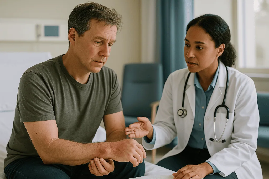
Inflammation follows almost immediately. Immune cells arrive, release signaling proteins, and recruit the stem cells that will eventually become cartilage and bone. The NIH Bookshelf fracture healing overview describes this inflammatory response peaking around 24 to 48 hours and largely settling within about a week. Clinically, this is the phase you feel most: heat, throbbing, and swelling that often looks worse on day two than on day one.
The swelling has a purpose, but it also has a cost, which is why emergency departments splint rather than cast at this stage. A splint is a rigid support that wraps only part of the way around the limb, leaving room to expand. A full circumferential cast applied over a limb that is still swelling can become dangerously tight (more on that in the splint section below).
Practical instructions at this stage are consistent across the NHS and Mayo Clinic: keep the limb raised above heart level when resting, keep the splint dry, and move the fingers or toes that are free. Ice wrapped in a cloth over the splint is commonly suggested for short periods; your discharge sheet will say how your team wants you to use it. Pain relief is a decision for the treating team, and the choice of medicine matters here for reasons covered later.
Stages two and three: how a soft callus turns into hard bone
Within days, the clot begins to organize into granulation tissue, a mixture of new blood vessels and cells. Over the next couple of weeks it becomes soft callus, a rubbery cuff of cartilage and fibrous tissue that wraps the broken ends like a sleeve. The NIH Bookshelf overview places soft callus formation at roughly two to three weeks after injury.
Soft callus is not strong. It cannot bear weight and it does not show on X-ray, because cartilage is invisible to X-rays. This is the phase in which many people feel noticeably better while their films still look unchanged, which can be confusing. The pain has dropped because the bone ends are no longer grinding, not because the bone is solid.
Hard callus is the next step. The cartilage undergoes a process called endochondral ossification (the same process by which a growing child’s cartilage growth plates turn into bone), and mineral is deposited into it. The result is woven bone, a disorganized but stiff material that finally bridges the gap. The NIH overview describes hard callus forming from around the third or fourth week and continuing for several months in adults.
This is the stage clinicians mean when they say a fracture has “united” or is showing “bridging callus.” On X-ray it appears as a hazy cloud that gradually becomes denser. Woven bone is bulkier than the original, which is why a healed break often feels like a lump under the skin for a long time. That lump is not a mistake; it is the body over-building for safety before it trims back.
The final fracture healing stage: remodeling, which you will never feel
Once woven bone bridges the break, the cast often comes off and life resumes. Biologically, though, the job is barely half done. Remodeling is the slow replacement of that bulky woven bone with organized lamellar bone, the layered, load-oriented tissue that makes up a normal skeleton.

Two cell types run this process in tandem. Osteoclasts dissolve bone that is not carrying load; osteoblasts lay new bone where stress runs. Because bone responds to mechanical force (an idea known as Wolff’s law), remodeling is guided by how you use the limb. Walking on a healed leg or gripping with a healed wrist is not just permitted, it is part of the repair.
How long does this take? The NIH Bookshelf overview describes remodeling continuing for months to years, and in children the bone may eventually look almost indistinguishable from the original. In adults, a faint line or slight thickening may remain permanently on X-ray without any consequence for function.
Remodeling matters for one very practical reason: the bone at the old fracture site is stronger than it looks on the day the cast is removed, but weaker than it will be six months later. Clinicians therefore often ask people to return gradually to high-impact sport or heavy lifting rather than all at once. Your team may put a rough timeline on this, and it will vary by bone and by what you do for a living. The absence of pain is not the same as full structural recovery, which is the single most useful idea to carry out of this section.
How long does a broken bone take to heal? Honest ranges, not promises
The most common figure quoted to patients is six to eight weeks. The NHS uses that range for a broken arm or wrist and for a broken leg; the Cleveland Clinic gives the same range for “most” fractures. It is a fair average, but averages hide a lot.
Three factors move the number most, according to those same sources. The first is age. Children heal faster than adults, partly because their periosteum is thicker and more biologically active; the NHS notes that children’s fractures typically mend more quickly. The second is the bone itself. Small bones with good blood supply, such as the fingers and collarbone, tend to sit at the shorter end; large weight-bearing bones such as the shin and thigh sit at the longer end and may take several months, as the NHS broken leg guidance describes. The third is blood supply at the specific site. A few areas, including the scaphoid in the wrist and the neck of the femur, are notorious for slow healing because their blood vessels enter from one direction and are easily disrupted.
Then come the modifiers in your control or your care team’s. Smoking is repeatedly linked to slower healing and higher rates of nonunion (a fracture that stops healing) in the NHS and Cleveland Clinic patient literature. Poorly controlled diabetes, low vitamin D, some long-term medicines, and infection at the fracture site can all lengthen the timeline.
Which is why a clinician who refuses to give you a firm date is not being evasive. They are describing a biological range and waiting for your X-rays to tell them where in it you sit.
Fracture healing stages at a glance: what happens, when, and what you notice
The table below pulls the sequence together. Timings come from the NIH Bookshelf fracture healing overview and the NHS patient guidance; they describe typical adult, uncomplicated fractures and will differ for children, for large weight-bearing bones, and for anyone with the risk factors covered above.
| Stage | Typical timing (adult) | What is happening | What you usually notice |
|---|---|---|---|
| Hematoma | Hours | Blood clots in and around the gap, forming a scaffold | Sharp pain, rapid swelling, bruising begins |
| Inflammation | Peaks 24–48 hours, settles over about a week | Immune cells clear damage and recruit repair cells | Heat, throbbing, swelling often worst on day two |
| Soft callus | About weeks 2–3 | Cartilage and fibrous tissue cuff the bone ends | Pain eases; X-ray still looks unchanged |
| Hard callus | From about weeks 3–4, continuing for months | Cartilage mineralizes into woven bone; fracture “unites” | Firm lump at the site; X-ray shows cloudy new bone |
| Remodeling | Months to years | Woven bone replaced by organized, load-oriented bone | Nothing; lump slowly shrinks, strength keeps rising |
Two things stand out when the stages sit side by side. The first is how much of the visible drama, the swelling and bruising and pain, is concentrated in the opening week, before any bone has formed at all. The second is how long the invisible tail is. The date the cast comes off is not the finish line; it marks the moment the bone is strong enough for ordinary use while it keeps rebuilding in the background.
Why a splint first and a cast later: what swelling does inside a rigid shell
People are often puzzled to leave the emergency department in a splint and be asked to come back days later for a cast. It can feel like a job half done. It is deliberate.
A limb keeps swelling for roughly the first two to three days after a fracture, in step with the inflammatory stage described above. A cast is a closed cylinder. If the tissue inside expands and the shell does not, pressure rises. At a certain point that pressure squeezes small blood vessels shut, and muscle and nerve begin to starve. This is compartment syndrome, a surgical emergency, and the Mayo Clinic and NHS both list it among the serious complications of fractures and tight casts.
A splint sidesteps the problem by leaving one side open, held with elastic bandage that can stretch. Once the swelling has peaked and begun to recede, a cast can be fitted snugly enough to hold the bone still without risking that pressure trap. Some clinics apply a full cast at the first visit and then split it lengthwise for the same reason.
What you can do in the meantime, per NHS advice, is keep the limb elevated, ideally above heart level, and move the free joints often. Both reduce swelling by helping fluid drain. Keep the splint dry and resist the temptation to loosen or re-wrap it yourself.
Once the cast is on, it will loosen slightly over the following weeks as swelling resolves and muscle shrinks from disuse. A little looseness is expected. A cast that has become so loose the limb shifts inside it is a reason to call, because a fracture that moves cannot hold its alignment.
How can I tell if a broken bone is healing? Signs a fracture is healing that you can and cannot trust
The honest answer is that you cannot fully tell from the outside, and neither can your clinician without an X-ray. But there are patterns that track with normal progress, and knowing them saves a great deal of worry.
Pain that steadily eases week by week is the most reliable everyday signal. It reflects the soft callus stabilizing the bone ends so they no longer shift with every movement. Swelling and bruising that fade over the first two weeks are the second. The Cleveland Clinic and Mayo Clinic both describe this gradual settling as the expected course.
Some signs feel reassuring but mean little. A firm lump at the fracture site is hard callus and does confirm bone formation, but its size says nothing about strength. Being able to wiggle the fingers or toes comfortably is good news about nerves and circulation, not about the fracture itself. Itching under the cast is skin, not bone.
Then there are things that should prompt a call rather than patience: pain that plateaus and then climbs, a sensation of the bone shifting or grinding, or new deformity. These can suggest the cast has loosened, the alignment has slipped, or healing has stalled.
Ultimately, the fracture clinic decides whether healing is on track by combining three things: your account of pain and function, an examination for tenderness directly over the break, and a follow-up X-ray looking for bridging callus. None of the three alone is sufficient. That is why your appointments are scheduled as they are, and why skipping one to save a trip is rarely a good trade.
Does a broken bone hurt more as it heals? Separating healing pain from warning pain
A healing fracture should, broadly, hurt less over time, not more. The sharpest pain belongs to the first days, when the bone ends are mobile and the tissue is inflamed. As soft callus forms and stiffens the site, most people describe the pain shifting from a constant throb to an ache that flares with movement and settles at rest.
That said, there are ordinary reasons for pain to spike within an overall downward trend. Swelling that builds at the end of a day on your feet can make a cast feel tight and the limb throb; elevation usually settles it overnight. Muscle cramps are common inside a cast as unused muscle tightens. The first days out of a cast are often uncomfortable, because joints that have been still for weeks are stiff and the skin is tender. Early physical therapy produces soreness of its own, the kind that eases as the session’s effects wear off.
What healing does not do is produce steadily increasing pain, pain that wakes you and will not settle with elevation, or pain out of proportion to what has happened. The Mayo Clinic’s fracture guidance flags escalating pain, numbness, and pale or bluish digits as reasons to seek care promptly, because they can signal pressure on nerves and vessels rather than healing.
Pain relief itself deserves a word. Anti-inflammatory painkillers are widely used after fractures, but some laboratory and observational research has raised the question of whether they slow bone healing by damping the inflammatory stage that starts the whole process. The evidence in humans is mixed and mostly short-term. It is a reasonable topic to raise with the prescribing clinician, who will weigh it against your pain and other health conditions.
How to stimulate bone growth after a fracture: what the evidence supports and what it doesn't
Almost everyone with a fracture wants to know what they can do to hurry it along. The frustrating truth is that the biggest levers are about not slowing healing rather than accelerating it.
Nutrition comes first. Bone is roughly two-thirds mineral by weight, and building callus demands calcium, which in turn depends on vitamin D for absorption, as the NIH Office of Dietary Supplements explains in its fact sheets on both nutrients. Protein supplies the collagen framework. A varied diet that meets ordinary requirements covers this for most people; whether someone with a documented deficiency benefits from supplementation is a question for their clinician, informed by a blood test rather than a guess.
Smoking is the single most consistently cited modifiable factor. Nicotine narrows blood vessels and reduces oxygen delivery to the fracture site; the NHS and Cleveland Clinic both list smoking among the reasons a bone may heal slowly or fail to unite. Cutting back or stopping during healing is an area where the evidence and common sense point the same way.
Controlled loading helps. Once your team clears it, using the limb within the limits they set signals bone cells to build where stress runs. This is why partial weight-bearing is prescribed rather than complete rest for many leg fractures.
Where the evidence thins out is in the devices and supplements marketed to “speed up” healing. Low-intensity pulsed ultrasound and electrical bone stimulators have been studied for slow-healing fractures with inconsistent results; they are used selectively by some surgeons for specific situations, not as routine boosters. Collagen powders, herbal mixes, and mineral blends have not been shown in good trials to shorten healing time in people without a deficiency. Spend the energy on sleep, food, and not smoking.
Who heals in a cast and who is usually asked to wait, or offered surgery
Not every fracture gets the same pathway, and the decision is made on the shape of the break rather than on how much it hurts.
A cast or splint alone is usually the plan when the broken ends are close together and well aligned, or can be gently pulled back into alignment (a maneuver called reduction), and when the bone is likely to stay put once immobilized. Many wrist, forearm, ankle, and finger fractures fall into this group, per NHS guidance on broken arms, wrists, and ankles.
Surgery to hold the bone with plates, screws, pins, or a rod is more likely when the fragments are widely displaced, when the break runs into a joint surface (where even a millimeter of step can lead to arthritis), when the fracture is unstable and keeps slipping, when the skin is broken over it (an open fracture, which carries infection risk), or when a bone with poor blood supply needs the extra stability. The Mayo Clinic broken wrist and broken ankle pages set out these considerations in plain terms.
Then there is the group asked to wait. Sometimes a fracture clinic will re-X-ray at one or two weeks before committing to a plan, because early swelling can hide how stable the position really is. Sometimes surgery is postponed until swelling subsides so the skin can close safely afterward. And at the far end, a fracture that has not shown expected callus by the time it should, delayed union, or that has clearly stopped healing, nonunion, may lead to a second conversation about surgery, bone graft, or the stimulators mentioned earlier.
Every one of these forks belongs to the treating team, who are weighing X-ray geometry, your bone quality, your other health conditions, and what you need the limb to do.
What the weeks after the cast comes off usually look like: follow-up X-rays and rehab
Cast removal is anticlimactic in a way that catches people out. The saw is loud but does not cut skin. The limb underneath is pale, thin, flaky, and often surprisingly weak. The joint may not straighten fully. None of this is a complication; it is what several weeks of stillness does to muscle and skin.
The follow-up X-ray taken at this visit is looking for bridging callus across the fracture line, the cloudy new bone described earlier. If it is present and the site is no longer tender to firm pressure, the team will usually declare the fracture clinically united and shift the focus to function. If callus is thin, another period of protection or a further X-ray may be advised. Both outcomes are normal parts of the pathway.
Rehabilitation, whether guided by a physical therapist or by a home exercise sheet, has three broad phases. The first restores range of motion: gentle, frequent movements of the stiff joint, often in warm water, which also soothes the skin. The second rebuilds strength, starting with light resistance and progressing as the bone remodels. The third returns you to the specific demands of your life, whether that is typing, lifting a toddler, or landing from a jump.
The NHS broken arm and wrist guidance notes that it can take several months for a limb to regain full strength and movement after the cast is removed, longer than the healing period itself. Expect stiffness in the mornings, aching after activity, and gradual improvement week over week rather than day over day. Setbacks are usually a sign of doing too much too soon, and the fix is to step back a level, not to stop.
What people often get wrong about the fracture healing stages
Fractures attract folklore. Some of it is harmless; some of it slows recovery.
“If I can move it, it isn’t broken.” Many fractures, especially those that are cracked but not displaced, allow near-normal movement. The NHS explicitly advises against using movement as a test. Persistent pain, swelling, and tenderness over bone after an injury deserve assessment regardless.
“A healed bone is stronger than before.” The hard callus is temporarily bulkier than the original bone, which is where this idea comes from. Bulk is not strength. Once remodeling finishes, the bone returns to roughly its prior strength, not above it.
“When the cast comes off, I’m done.” The visible healing stage ends at cast removal; remodeling and rehabilitation continue for months, and the limb is weaker at that moment than it will be later.
“Complete rest is best.” Bone builds in response to load. Beyond the first stage, controlled use within your team’s limits helps rather than harms. Total inactivity also raises clot risk in the legs.
“Calcium tablets will speed it up.” Adequate calcium and vitamin D are necessary, but the NIH Office of Dietary Supplements fact sheets describe them as supporting normal bone health, not as accelerators. Extra beyond need has no demonstrated benefit for healing speed.
“Kids and adults heal the same way.” Children heal faster, remodel more completely, and can even correct some degree of angulation as they grow, which is why pediatric fracture care sometimes accepts positions that would be operated on in an adult.
“Pain means something is wrong.” Pain that eases over time is healing. Pain that escalates is a warning. The direction of travel is what matters.
Questions to ask your care team at each fracture clinic visit
Fracture clinic appointments are brisk, and the questions that occur to you at midnight rarely survive to the consulting room. Writing them down helps. These are the ones that tend to matter most, organized by the stage you are in.
At the first visit or after the emergency department:
- Is this fracture in a good position, or might it need to be moved or fixed?
- Will you re-X-ray it to check it has stayed in place, and when?
- What signs of a too-tight splint or cast should make me call today rather than wait?
- Are there pain relief options you would prefer I avoid given how bones heal?
Once the cast is on:
- Which joints should I keep moving, and how often?
- Can I put any weight through this limb, and if so how much?
- Is there anything about my health or medicines that might slow this down?
- How will you decide when the cast can come off?
At cast removal and after:
- Does the X-ray show bridging callus, and is the fracture clinically united?
- What movements are safe now, and which should I hold back on?
- Do I need a referral to physical therapy, or a home program?
- When would you expect me to return to work, driving, or sport, and what would change that?
- Is there anything on this X-ray I should know about for the long term?
A final question worth asking at any stage is simply: what would make you worried about this fracture? Clinicians answer that one readily, and it tells you exactly which changes to watch for between appointments.
When to call your doctor: red-flag signs during any fracture healing stage
Most fractures heal quietly. A small number develop problems that are far easier to fix early than late, and the signs are consistent enough to list. Drawn from NHS, Mayo Clinic, and Cleveland Clinic guidance on fractures and cast care, the following warrant a same-day call to your fracture clinic, or urgent care if the clinic is closed.
- Pain that keeps increasing despite elevation and the pain relief you have been given, or pain that feels out of proportion to the injury.
- Numbness, tingling, or pins and needles in the fingers or toes beyond the cast.
- Fingers or toes that turn pale, blue, or cold, or that you cannot move.
- Severe swelling below the cast, or a cast that has become so tight it feels like a tourniquet.
- A cast that has become loose enough for the limb to shift inside it, or that has cracked, softened, or gotten soaked through.
- A foul smell, discharge, or wetness from under the cast, or fever with a hot, red area of skin at the cast edge, which can indicate infection or a pressure sore.
- Burning or rubbing pain at one spot under the cast that does not ease.
- A sensation that the bone has moved or a new visible bend in the limb.
Two situations are emergencies rather than phone calls. The first is the combination of severe, escalating pain, tightness, and numbness in a casted limb, which can indicate compartment syndrome. The second, particularly with leg fractures or after surgery, is calf pain, warmth, or swelling in the other leg or above the cast, or sudden breathlessness or chest pain, which can signal a blood clot. Both need to be seen immediately.
No article can replace the judgment of the team that has seen your X-rays. If you are unsure whether something counts, call. Fracture clinics would far rather reassure you than find a problem late.
Frequently asked questions
What are the 5 stages of fracture healing?
The five stages usually listed are hematoma formation, inflammation, soft callus formation, hard callus formation, and remodeling. Orthopedic texts often compress these into three phases: inflammatory, reparative, and remodeling. The order is fixed. A blood clot forms first, inflammation recruits repair cells, cartilage bridges the gap, that cartilage mineralizes into woven bone, and finally the bulky new bone is reshaped over months.
How long does a broken bone take to heal in an adult?
Roughly six to eight weeks for many common fractures of the arm, wrist, ankle, and leg, according to the NHS and Cleveland Clinic. Large weight-bearing bones such as the thigh and shin can take several months. Age, blood supply at the site, smoking, diabetes, and infection all shift the timeline, so your team will use follow-up X-rays rather than the calendar to judge progress.
How can I tell if a broken bone is healing?
You cannot confirm it without an X-ray, but pain that eases steadily week by week and swelling that fades over the first two weeks are the everyday signs that track with normal healing. A firm lump at the site is hard callus. Pain that plateaus and then climbs, a grinding sensation, or new deformity should prompt a call rather than patience.
Does a broken bone hurt more as it heals?
No. Pain should trend downward as soft callus stabilizes the bone ends. Temporary flares are common at the end of an active day, with muscle cramps inside a cast, or in the first days after the cast comes off when stiff joints start moving. Steadily increasing pain, or pain with numbness and pale digits, is a warning sign, not a healing sign.
How can I stimulate bone growth after a fracture?
The best-supported steps are not smoking, eating a diet that meets ordinary calcium, vitamin D, and protein needs, and using the limb within the limits your team sets, because bone builds in response to load. Devices such as ultrasound or electrical stimulators are used selectively for slow-healing fractures, and over-the-counter supplements have not been shown to shorten healing in people without a deficiency.
Why did the emergency department give me a splint instead of a cast?
Because the limb keeps swelling for two to three days after a fracture. A full cast is a closed cylinder, and swelling inside it can raise pressure enough to compress blood vessels and nerves, a condition called compartment syndrome. A splint leaves room to expand. Once swelling has peaked, a snug cast can be applied safely at a follow-up visit.
What do follow-up X-rays after a fracture actually show?
Early films, at one to two weeks, mainly check that the bone has stayed in position. Later films look for bridging callus, a cloudy band of new bone crossing the fracture line, which appears from roughly the third or fourth week onward. Cartilage soft callus is invisible on X-ray, so an unchanged early film does not mean nothing is happening.
Are the signs a fracture is healing different in children?
The stages are the same, but children move through them faster and remodel more completely, according to NHS guidance. Their thicker periosteum is more biologically active, and growing bone can correct a degree of angulation over time. This is why pediatric fracture care sometimes accepts positions that would be surgically fixed in an adult, and why children’s casts often come off sooner.
Can painkillers slow down fracture healing?
Some laboratory and observational research suggests anti-inflammatory painkillers might blunt the inflammatory stage that starts bone repair, but human evidence is mixed and mostly short-term. It has not changed routine practice for most people. It is a reasonable question to raise with the clinician prescribing your pain relief, who will weigh it against your pain, kidney function, and other conditions.
What is a nonunion, and how common is it?
A nonunion is a fracture that has stopped healing and will not unite without further treatment; a delayed union is one healing more slowly than expected. Risk is higher with smoking, poor blood supply at the site, infection, open fractures, and inadequate stabilization. Reliable overall rates vary by bone and are not given here; your team will tell you if your fracture is at particular risk.
References
- Fracture Healing Overview – StatPearls, NCBI Bookshelf (NIH)
- Broken arm or wrist – NHS
- Broken leg – NHS
- Bone Fractures – Cleveland Clinic
This article is for general information only and is not a substitute for professional medical advice. Please consult a qualified doctor about your individual situation.

More from the Blog

How Arthritis Wears a Joint Down: When Damage Reaches the Point Where Replacement Is Discussed
Arthritis wears a joint down by thinning the cartilage that cushions the bone ends, roughening the bone beneath, and inflaming the joint lining, until…
How X-Rays and CT Scans Guide Trauma Surgery Decisions After a Serious Injury
After a serious injury, X-rays and CT scans give trauma surgeons a map of what is broken, bleeding or displaced before anyone decides to…

Preparing for Hand Surgery: Nerve Tests, Imaging and the Medicines Your Team Reviews
To prepare for hand surgery, most people complete nerve tests such as a nerve conduction study or EMG, imaging such as X-ray, ultrasound or…

Hip Flexor Pain: Tight, Strained or Something Else, and the Exercises That Help
Hip flexor pain is soreness at the front of the hip or groin where the iliopsoas and rectus femoris muscles lift the thigh. Most…
Does the Robot Operate on Its Own? Robotic Knee Replacement Myths, Corrected
No. In robotic knee replacement, the surgeon performs the operation and the robotic arm acts as a precision tool. The surgeon plans the implant…

How a Pavlik Harness Keeps Your Baby’s Hips in Place So the Socket Can Deepen
A Pavlik harness works by holding a baby's hips bent up and gently spread apart, so the ball at the top of each thigh…