

How a Dental Anatomy Evaluation Guides Your Plan: Tooth Charts, Bite Checks and Imaging
Key Takeaways
- A full adult set has 32 teeth and a child's set has 20, and children have no premolars at all, which is why a child's chart looks different from an adult's.
- Enamel is the hardest substance in the body but contains no nerves, so decay is painless until it reaches the softer dentin beneath.
- The numbers read out during a check are pocket depths in millimeters; the trend between visits matters more than any single reading.
- Gingivitis is reversible with cleaning and home care, while periodontitis involves bone loss that can be halted and managed but does not regrow on its own.
- Dental X-ray frequency is set by individual risk rather than a fixed schedule, and a run of clear images is a reason to space them further apart.
- NHS guidance places check-up intervals anywhere from three months to two years depending on what the evaluation finds, so a longer gap can be a sign of good health.
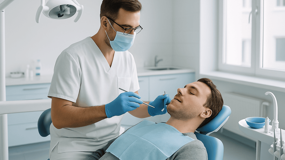
A dental anatomy evaluation is a structured, tooth-by-tooth assessment in which a dentist records each tooth on a numbered chart, checks how your upper and lower teeth meet, measures the gums around every tooth, and uses X-rays when the exam alone cannot answer a question. The findings are combined into a written record that guides which problems need treatment, which can be monitored, and how often you should return.
You are lying back in the chair, mouth open, and the hygienist is reciting what sounds like a lottery draw. “Three, two, three. Two, three, four.” Then the dentist joins in: “Fourteen, occlusal, watch.” Nobody is talking to you, exactly, yet everything being said is about you. It is a strange feeling, being described in a code you were never taught.
That code is the working language of a dental anatomy evaluation, and it is far more logical than it sounds. Every number is a location or a measurement. Every word is a surface, a finding or a decision about whether to act. Once you can follow it, a routine exam stops being something done to you and becomes something you can take part in.
This explainer walks through the four instruments of that evaluation: the tooth chart, the bite check, the gum measurements and the imaging. It also covers what the results usually lead to, what people commonly misunderstand, and which symptoms should never wait for the next scheduled visit.
What does a dental evaluation consist of?
Strip away the jargon and a dental evaluation is a structured inventory. The dentist wants to know what is in your mouth, what condition each part is in, how the parts move against one another, and what lies beneath the surface that a mirror and light cannot reach. Those four questions map neatly onto the four tools in this article’s title.
A complete visit usually begins before anyone looks at a tooth. Your medical history matters because conditions such as diabetes, pregnancy and heart valve disease, along with several classes of medicine, change how gums heal and how treatment should be timed. Next comes the head and neck: lymph nodes, the jaw joint, lips, tongue, palate and the floor of the mouth. Mayo Clinic lists this soft-tissue check, including a look for signs of oral cancer, as a standard part of a routine exam rather than an optional extra.
Only then does the tooth-by-tooth work start. Each tooth is examined for decay, cracks, wear, existing fillings and crowns, and any looseness. Gums are measured with a fine probe. The bite is assessed with the jaw closed and while it moves. Radiographs, the clinical word for X-ray images, are taken when the history and exam suggest they will change a decision, not out of habit.
The output is a record rather than a verdict. Everything is charted so the next visit can be compared against it, and that comparison is where much of the diagnostic value hides. A gum measurement that stays the same year after year is reassuring; one that keeps growing is a story worth hearing. The visit closes with a conversation about what was found, what it might mean, and which findings can simply be watched for now.
Can you give a detailed anatomy of a tooth? Here it is in plain language
Picture a tooth as a hard shell wrapped around a living core, anchored in bone by a sling of fibers. That single image explains most of what a dentist is looking for.
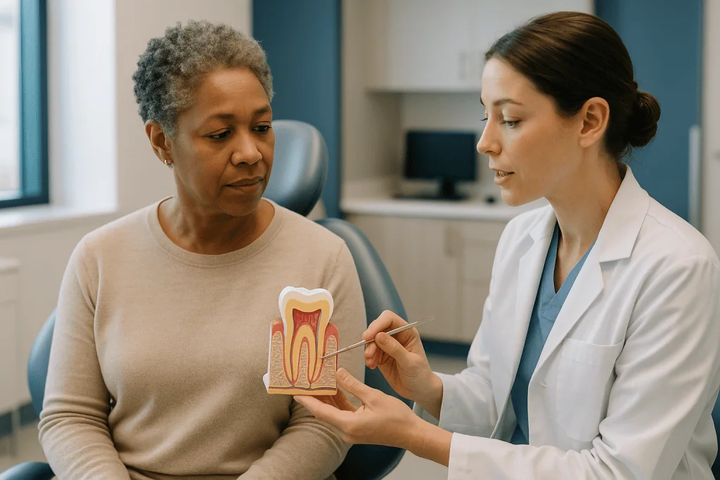
The crown is the part you can see above the gum. The root is the hidden portion, and molars often have two or three of them. Where crown meets root, at the gum line, is the neck of the tooth, a spot where decay and wear like to start because the protective coating thins there.
The crown is coated in enamel, which MedlinePlus describes as the hardest substance in the human body. Enamel has no nerve supply and no living cells, so it cannot feel and cannot repair itself once a cavity has formed. Beneath it sits dentin, a softer, slightly porous layer that makes up most of the tooth and does transmit sensation. This is why a cavity that was silent in enamel suddenly stings with cold once it reaches dentin.
At the center is the pulp: blood vessels and nerve fibers that keep the tooth alive and register pain. The pulp runs down the root inside the root canal and exits through a tiny opening at the tip, the apex, where it connects to the body’s wider blood and nerve supply.
Roots are covered not by enamel but by cementum, a thin, bone-like layer. Attached to it is the periodontal ligament, the fibrous sling that suspends the tooth in its bony socket and cushions it with every bite. The word periodontal simply means “around the tooth.”
Each layer answers a clinical question. Decay confined to enamel may be monitored. Decay into dentin usually calls for a filling. Involvement of the pulp changes the plan entirely. Loss of the ligament and surrounding bone is the definition of gum disease. The evaluation is, in effect, a layer-by-layer check of this structure.
Types of teeth and what each one does
A full adult set contains 32 teeth, according to Cleveland Clinic, and children have 20. The difference is not just number. Children have no premolars, and their molars are eventually replaced rather than added to.
Adults have four types, and each is shaped for a job. The eight incisors at the front are thin, chisel-edged blades for cutting. The four canines at the corners are pointed for tearing and carry the longest roots in the mouth, which is why they tend to be the last teeth standing when others are lost. Behind them, eight premolars have two cusps, the raised points on a chewing surface, and act as a bridge between cutting and grinding. The twelve molars at the back, including the four third molars most people call wisdom teeth, are broad and grooved for crushing.
Timing matters in an evaluation because it tells the dentist what should be present. Cleveland Clinic notes that primary teeth begin to appear at around six months of age, that permanent teeth start arriving at about six years, and that wisdom teeth usually come through in the late teens or early twenties, if they come through at all. A child of eight with a molar erupting behind the baby teeth is on schedule; an adult with a wisdom tooth that never appeared may have one lying sideways under the gum, which only imaging can confirm.
Shape also predicts trouble spots. Molar grooves trap plaque and are where most childhood decay starts. Incisors, being thin, chip on hard foods and during falls. Premolars sit where the bite forces concentrate and are frequent sites of cracks. Canines, exposed at the corner of the smile, often show gum recession first. When a dentist calls out a tooth and a surface, they are usually thinking about which of these risks applies to that particular shape.
How the tooth numbering chart maps your mouth
The numbers you hear are addresses. In the United States most practices use a system in which each adult tooth carries a number from 1 to 32. Counting begins at the upper right third molar, sweeps across the upper arch to the upper left third molar at 16, drops to the lower left third molar at 17, and returns along the lower arch to finish at the lower right third molar, 32. Primary teeth are lettered A through T along the same route. Dentists trained elsewhere may use a two-digit international code instead, so if your records travel, a number can look different while referring to the same tooth.

Every tooth also has named surfaces. Occlusal is the chewing surface of a back tooth; incisal is the biting edge of a front one. Mesial faces the midline of the mouth and distal faces away from it. Buccal or facial faces the cheek or lip, and lingual faces the tongue. “Fourteen, occlusal” therefore pinpoints the chewing surface of the upper left first molar.
On the chart itself, findings are drawn in color or code. Existing fillings, crowns, implants and missing teeth are marked one way; new decay, cracks and areas to monitor another. A good chart is a map that a stranger could read and understand what has been done and what is being watched.
Why should you care about this? Three reasons. Records that use precise locations can be compared over years, which is how slow changes get caught. Referrals to a specialist make sense only if everyone agrees which tooth is meant. And you can ask, quite reasonably, to see the chart and have the marks explained. Most dentists are glad to do so; it is far easier to discuss tooth 30 while pointing at a diagram than while pointing into a mouth.
What actually happens during a dental anatomy evaluation, step by step
The order varies between practices, but the sequence below is typical for a comprehensive visit, as described by Mayo Clinic and others.
First, the conversation. You will be asked about medical conditions, medicines, allergies, previous dental treatment and anything that hurts or worries you. Mention pregnancy, bleeding tendencies and any planned medical procedures; each can change what is done today.
Second, the outside view. The dentist feels the lymph nodes under the jaw and along the neck, watches the jaw open and close, and may press on the joint just in front of each ear. Clicking, deviation or tenderness is noted.
Third, the soft tissues. Lips, cheeks, tongue (including underneath), palate and throat are inspected. This is the oral cancer screen; it takes a minute and most people barely notice it.
Fourth, the teeth. Using a mirror and a fine explorer, each tooth is checked in turn while findings are called out to be charted. You may feel light pressure or hear a scratching sound; a sticky spot on enamel is one sign of decay.
Fifth, the gums. A slim, blunt probe marked in millimeters is slipped gently between tooth and gum at several points around each tooth to measure the depth of the pocket, the small groove where gum meets tooth. Bleeding, recession and looseness are recorded.
Sixth, the bite. You will be asked to close, to slide the jaw side to side and forward, and sometimes to bite on thin colored paper that leaves marks where the teeth touch.
Seventh, imaging, if indicated. Small sensors are held inside the mouth for close-up views, or a machine circles the head for a wider image.
Finally, the talk. Findings are explained, a preliminary plan is sketched, and you decide together what happens next. Nothing beyond the exam itself should be started that day without your understanding and agreement.
The dental bite assessment: how a dentist reads your occlusion
Occlusion is the way the upper and lower teeth meet when the mouth closes and how they glide against each other during chewing. Malocclusion is any departure from a comfortable, even fit. It is one of the most under-appreciated parts of an evaluation, because teeth that are individually healthy can still be damaged by the way they collide.
The dentist watches you bite down and looks at several relationships. How far the upper front teeth overlap the lower ones vertically and horizontally. Whether any back teeth on one side sit inside the opposing arch, a crossbite. Whether the front teeth fail to touch at all when the back teeth are together, an open bite. Whether teeth are crowded, rotated or spaced. Then the jaw moves: sliding left, right and forward reveals which teeth guide the movement and whether any tooth takes an early, heavy hit.
Wear tells the longer story. Flat, shiny patches on cusps, called wear facets, suggest grinding or clenching, often at night. Small notches at the gum line, chipped edges on front teeth and cracked fillings all raise the same question. The jaw joint and chewing muscles are checked for tenderness at the same time.
Why does this matter for planning? A filling or crown placed onto a heavy contact will fail sooner. A tooth with a hairline crack under a wear facet may be better protected by a covering than a patch. Grinding may prompt a discussion about a night guard, a custom plastic shield worn during sleep.
One honest caveat: the link between bite problems and symptoms such as headaches or jaw pain is real for some people but far from universal, and the research on correcting bites to relieve pain is mixed. A careful dentist will say what the bite shows and what it may or may not explain, rather than promising that adjusting it will make discomfort disappear.
Gum measurements: why those numbers matter as much as the teeth
The string of numbers read out during a check is periodontal charting, and it is arguably the most predictive part of the whole visit. Teeth rarely fall out because of decay; they are lost when the bone and ligament holding them give way.
Each number is the depth, in millimeters, of the pocket between gum and tooth at one spot. Healthy gums hug the tooth closely, so the probe stops quickly. When the gum is inflamed and the attachment has begun to detach, the probe slides deeper. Alongside depth, the chart records whether the spot bled when touched, how far the gum has receded from its original position, whether the tooth moves under gentle pressure, and whether the probe can pass between the roots of a molar.
These findings separate two conditions. Gingivitis is inflammation of the gum alone: redness, puffiness and bleeding on brushing. NIH’s dental research institute describes it as reversible with professional cleaning and better daily care. Periodontitis is the stage at which the supporting bone has begun to be lost. That bone does not grow back on its own, so the aim becomes stopping further loss and keeping the teeth stable.
Imaging completes the picture. Pocket depth tells the dentist how far the attachment has receded from the crown; X-rays show the actual height of bone between the teeth. A deep pocket with bone still intact is treated differently from a deep pocket with bone gone.
The practical consequence for your plan is sequencing. When active gum disease is present, most clinicians stabilize it before placing new fillings, crowns or implants, because restorations built on an inflamed foundation tend to have a shorter life. If your charting numbers are higher than last time, expect that conversation to come first, and expect the interval between visits to shorten.
Types of dental X-rays and what each one shows
Roughly half of each tooth, and all of its supporting bone, is invisible to direct inspection. Cleveland Clinic explains that dental X-rays fill that gap by revealing decay between teeth and under fillings, bone levels, infections at root tips, impacted teeth and problems inside the jaw.
| Image type | What it captures | Typical reason it is taken |
|---|---|---|
| Bitewing | Crowns of upper and lower back teeth in one view, plus the bone crest between them | Finding decay between teeth and checking early bone loss |
| Periapical | One or two whole teeth from crown to root tip | Assessing a painful tooth, root infection, cracks or a failed filling |
| Panoramic | Both jaws, all teeth, sinuses and jaw joints in a single wide image | Overview for new patients, wisdom teeth, jaw growth, planning extractions |
| Cone-beam CT | A three-dimensional scan of a selected region | Implant planning, complex root anatomy, locating impacted teeth or hidden fractures |
Cone-beam CT, often shortened to CBCT, is a rotating scanner that builds a 3D model of the jaw; it gives more information than a flat image but uses more radiation, so it is reserved for questions that flat images cannot answer.
How often should imaging happen? There is no fixed answer. Cleveland Clinic and Mayo Clinic both describe frequency as a judgment based on individual risk: history of decay, gum disease, age, symptoms and what previous images already show. Someone with a decade of clean bitewings needs them less often than someone with new cavities each year. Children, whose tissues are more sensitive to radiation, are imaged on a risk basis too.
Modern digital sensors use a low dose, and protective aprons or collars are commonly offered. Always tell the team if you are or might be pregnant; imaging can be done when genuinely needed, but non-urgent images are often deferred. Reasonable questions are “What will this image tell us that the exam could not?” and “When were my last ones?” If earlier images exist elsewhere, ask whether they can be transferred rather than repeated.
How a dental anatomy evaluation turns findings into a plan
A chart full of marks is not a treatment plan. The plan emerges from sorting those marks into priorities, and the sorting follows a fairly consistent logic across guideline-based practice.
Pain and infection come first. A tooth with a swollen root tip or a pulp that is dying is dealt with before anything elective, because infection can spread and because pain undermines everything else.
Disease control comes second. Active gum disease is treated and re-measured. Decay that has reached dentin is removed and the tooth restored. The goal at this stage is to stop things getting worse before investing in anything larger.
Restoration comes third: replacing lost teeth, rebuilding worn or cracked ones, correcting a bite that is damaging restorations. Purely cosmetic changes come last, once the mouth is stable.
Not every finding leads to treatment. Early enamel changes that have not broken the surface, small pockets that do not bleed, and old fillings with slightly rough edges are often best watched. “Watch” is a legitimate clinical decision, not an oversight, and it is why the chart notes the finding: so it can be checked next time rather than forgotten.
The evaluation also sets your return interval. The NHS, drawing on NICE guidance, describes check-up spacing that ranges from every three months for people at higher risk to as long as two years for adults with consistently healthy findings. Where you fall on that range is a direct product of what the chart, the probe and the images showed.
Two things should be true of any plan you are offered. It should be explained in terms of your own findings, ideally with the chart or images in front of you. And it should include the alternatives, including the option of doing nothing for now and what that would likely mean. The choice remains yours, made with your treating team.
Who a full evaluation is usually for, and who is usually asked to wait
A comprehensive dental anatomy evaluation is the default starting point for anyone new to a practice, anyone who has not been examined for several years, and anyone whose situation has changed: a new medical diagnosis, a new medicine that dries the mouth, pregnancy, or a plan for braces, implants or major reconstruction. Some medical teams also request a dental assessment before treatments that affect healing or immunity, such as radiotherapy to the head and neck or certain bone-strengthening medicines, because dental infection is easier to address beforehand than during.
Children are examined too, but the format adapts. The NHS advises that children be seen from when their first teeth appear, and the early visits are as much about familiarity and habit as about findings. Charting, probing and imaging are scaled to age and cooperation.
Some people are asked to postpone the full version, not because it is unsuitable but because something else comes first. Severe pain or facial swelling calls for a focused emergency exam of the problem area; the complete survey follows once the acute issue is settled. Someone who has recently had a heart attack, stroke or major surgery is usually asked to wait until their physician confirms it is safe to proceed with anything beyond a basic check. Patients on blood thinners are not excluded, but the team will want details before any procedure that could bleed.
Pregnancy does not delay the examination itself. Cleaning and assessment are considered safe throughout, and gum changes are common enough during pregnancy that a check is often encouraged. What tends to be deferred is non-urgent imaging and elective treatment, which are commonly scheduled around the pregnancy rather than through it.
People with strong anxiety, a sensitive gag reflex or limited stamina can ask for the evaluation to be split across shorter visits. Thoroughness matters more than speed, and a good team will say so.
What the following days and weeks usually look like
The evaluation itself has almost no recovery. Gums that were probed may feel slightly tender for the rest of the day, and if a cleaning was done at the same visit, a brief spell of cold sensitivity is common as the surfaces adjust. Bleeding on brushing that was already present often improves noticeably within days of a professional cleaning combined with better home care, which is one of the quickest and most encouraging changes in dentistry.
In the days that follow, expect a written or printed plan if one was not handed over on the day. It should list findings by tooth number, proposed treatment in sequence, items to monitor, and any referrals. Referrals to specialists such as a periodontist (gum specialist), endodontist (root canal specialist) or orthodontist (alignment specialist) are arranged in this window.
Over the following weeks, treatment appointments happen in the order the plan set out: urgent work, then disease control, then restoration. If gum treatment was performed, a re-measurement visit is usually scheduled some weeks later to see whether pockets have shallowed and bleeding has stopped; that second set of numbers, compared against the first, decides whether the gums are stable enough to move on.
Then the cycle resets. Your recall interval, anywhere from three months to two years according to NHS guidance, is set by risk and revised at each visit. Imaging is repeated only when the interval and your history justify it.
Between visits, your part is straightforward and evidence-backed: NHS guidance is to brush twice a day for about two minutes with a fluoride toothpaste, clean between the teeth daily, and limit how often sugar passes through the mouth. Frequency of sugar exposure matters more than total amount, because each exposure gives plaque bacteria a fresh chance to produce acid. If anything on the chart was marked “watch,” these habits are what tip a watched spot toward stable rather than toward a filling.
What people often get wrong about dental evaluations
“If nothing hurts, nothing is wrong.” Enamel has no nerves, gum disease is usually painless until late, and decay between teeth can grow for years unseen. Pain is a late signal, not an early one, which is the entire argument for evaluation before symptoms.
“Follow the 3-3-3 rule.” This popular phrase, brushing three times a day, for three minutes, within three minutes of eating, appears in no major guideline. NHS advice is twice daily for about two minutes with fluoride toothpaste, and it specifically cautions against brushing straight after acidic food or drink, because softened enamel is more easily worn. More brushing is not better brushing; technique and fluoride matter more.
“X-rays every visit are standard.” They are not. Frequency should reflect your risk, as Cleveland Clinic and Mayo Clinic both describe, and a run of clear images is a reason to space them out.
“X-rays are too dangerous to have at all.” Also untrue. Dental imaging is low dose, and the risk of missing a spreading infection or bone loss is real. The sensible position sits between the two extremes: image when it will change a decision.
“Bleeding gums are normal.” Bleeding is inflammation. It is common, but it is a finding, not a baseline.
“Baby teeth don’t matter because they fall out.” They hold space for permanent teeth, guide jaw growth, and an infected baby tooth can damage the developing tooth beneath it.
“Wisdom teeth always need to come out.” Guidance in the UK and elsewhere favors removing them when they are causing or clearly likely to cause problems, not by default.
“A chart full of numbers means bad news.” Every tooth gets numbers whether healthy or not. The chart is a map, not a bill.
“Enamel can grow back.” Early mineral loss can be halted and partly re-hardened with fluoride, but a cavity, once the surface has broken, does not close.
Questions to ask your care team
The most useful questions are the ones that turn a list of findings into a shared understanding. Consider bringing a few of these.
- Can you show me the chart and point to what you found on each tooth you mentioned?
- Which findings need treatment soon, which can wait, and which are you simply watching?
- What would happen if I did nothing about this particular tooth for the next year?
- What did the X-rays show that the exam alone could not? When were my last images, and can earlier ones be transferred?
- What are my gum measurements compared with last time, and is the trend stable?
- Does my bite explain any of the wear, cracks or sensitivity you noted?
- What are the alternatives for each proposed treatment, including a smaller or more conservative option?
- What is the sequence, and why that order?
- How often should I return, and what would change that interval?
- Is there anything in my medical history or medicines that affects the plan?
People also ask how to tell whether an evaluation was thorough. Rather than judging a clinician, look at the process. A complete assessment includes a medical history, a soft-tissue and neck check, tooth-by-tooth charting, gum measurements, a look at the bite, and imaging justified by need. Findings should be explained in language you understand, with the option of watching as well as treating, and you should never feel rushed into deciding in the chair. If large amounts of treatment are proposed without images, measurements or a clear explanation of why each item is needed, it is reasonable to ask for those details or to seek a second opinion before proceeding. Seeking a second view is a normal part of healthcare, and a confident team will not be offended by it.
When to call your doctor
Most of what a dental evaluation finds can wait for a scheduled appointment. A small number of situations should not, and some belong in an emergency department rather than a dental chair.
Seek urgent medical care, by emergency services if necessary, for swelling of the face, jaw or neck that is spreading or makes it hard to swallow, breathe or open the mouth; for a high fever alongside severe tooth or jaw pain; or for bleeding from the mouth that does not slow with firm pressure. These can signal an infection moving beyond the tooth, and they are treated as medical emergencies.
Contact your dentist the same day for a tooth knocked out or pushed out of position by injury, for a broken tooth exposing a pink or bleeding center, for throbbing pain that wakes you at night or is not eased by the pain relief your clinician has advised, for a lump or abscess on the gum, or for numbness of the lip or chin after a blow to the jaw.
Book a prompt appointment, without waiting for your next routine visit, for a mouth ulcer, white or red patch, or lump that has not healed within three weeks, which the NHS advises should always be checked; for a tooth that has become loose without injury; for gums that bleed heavily or have begun to pull away noticeably; or for a filling or crown that has fallen out and left a sharp or sensitive edge.
Tell your medical doctor as well as your dentist if you have a heart valve condition, a recent joint replacement, a bleeding disorder or a weakened immune system and develop any dental infection, because coordination between the two teams may be needed. In every case, the treating clinicians decide what happens next; your job is simply to make the call early rather than late.
Frequently asked questions
What does a dental evaluation consist of?
A dental evaluation consists of a medical history review, a check of the head, neck and soft tissues of the mouth, a tooth-by-tooth examination recorded on a numbered chart, gum measurements around each tooth, an assessment of how the teeth meet, and X-rays when they are needed to answer a question the exam cannot. It ends with a discussion of findings, options and how soon you should return.
What is the 3-3-3 rule for dental care, and is it evidence-based?
The 3-3-3 rule is a popular saying that recommends brushing three times a day, for three minutes, within three minutes of eating. It is not part of any major clinical guideline. NHS advice is to brush twice daily for about two minutes with fluoride toothpaste and to avoid brushing immediately after acidic food or drink, when enamel is temporarily softened. Consistency and fluoride matter more than frequency.
Can you provide a detailed anatomy of a tooth?
A tooth has a visible crown and one or more hidden roots. The crown is coated in enamel, the hardest tissue in the body, over a softer sensitive layer called dentin. At the center lies the pulp, containing nerves and blood vessels, which runs down each root through a canal to the tip. Roots are covered in cementum and held in the jawbone by the periodontal ligament, a fibrous sling that cushions each bite.
What are red flags that a dental evaluation was not thorough?
Signs worth noticing include no questions about your medical history, no charting of individual teeth, no gum measurements, no check of the soft tissues or bite, imaging that is not explained, and a large treatment plan presented without images or a clear reason for each item. Feeling pressured to decide in the chair is another. Asking for the chart, the images and the alternatives, or seeking a second opinion, is a normal response.
How often should I have a dental anatomy evaluation?
It depends on your risk. The NHS, following NICE guidance, describes check-up intervals ranging from every three months for people with active gum disease or frequent decay to as long as two years for adults whose findings have been consistently healthy. Your dentist sets and revises the interval at each visit based on your chart, gum measurements and imaging history rather than applying a single fixed schedule.
What types of dental X-rays are there and are they safe?
The common types are bitewings, which show the crowns of upper and lower back teeth together; periapicals, which show a whole tooth to the root tip; panoramic images of both jaws; and cone-beam CT, a 3D scan for complex questions. Cleveland Clinic describes dental X-rays as low dose, with frequency based on individual risk. Tell your team if you are or might be pregnant so non-urgent imaging can be timed appropriately.
How does a tooth numbering chart work?
In the United States, adult teeth are usually numbered 1 to 32, starting at the upper right third molar, sweeping across the upper arch to 16, then continuing along the lower arch from the lower left third molar at 17 back to the lower right at 32. Baby teeth are lettered A through T. Each tooth also has named surfaces such as occlusal (chewing) and buccal (cheek side), so a finding can be located precisely.
What does a dental bite assessment check?
A bite assessment looks at how the upper and lower teeth meet when closed and how they slide during chewing. The dentist checks overlap of the front teeth, crossbites, open bites, crowding, wear facets that suggest grinding, and tenderness in the jaw joint and muscles. Colored paper may be used to mark heavy contacts. The findings influence how fillings and crowns are shaped and whether a protective night guard is discussed.
What do the numbers the hygienist calls out during my check mean?
Those numbers are gum pocket depths, measured in millimeters at several points around each tooth with a fine probe. Shallow, non-bleeding readings indicate healthy attachment; deeper readings that bleed suggest inflammation or loss of the tissue and bone holding the tooth. The most useful information comes from comparing readings with earlier visits, which is why they are recorded every time even when everything is healthy.
Do I need a full dental evaluation if nothing hurts?
Yes, because pain is a late sign in dentistry. Enamel has no nerves, gum disease is usually painless until advanced, and decay between teeth can grow unseen for years. A dental anatomy evaluation exists precisely to find these changes while they are small enough to watch or treat simply. How often you need one is then set by what the evaluation finds rather than by symptoms.
References
- MedlinePlus: Tooth anatomy
- Cleveland Clinic: Teeth: Anatomy, Types, Function & Care
- Cleveland Clinic: Dental X-Rays
- NIH National Institute of Dental and Craniofacial Research: Periodontal (Gum) Disease
This article is for general information only and is not a substitute for professional medical advice. Please consult a qualified doctor about your individual situation.

More from the Blog

Tooth Decay Myths: Sugar-Free Means Safe, Brushing Harder Helps and Other Errors
Most tooth decay myths fall apart under evidence. Sugar-free does not mean safe, because acidic drinks and starchy snacks still feed decay or erode…

Chipped or Loose Porcelain Veneer: Repair, Rebonding or Replacement Explained
A chipped or loose porcelain veneer can often be managed without replacing it. Small edge chips may be smoothed or patched with tooth-colored composite…
What Does a Dental Spa Visit Include? Cleaning, Polishing and Gum Care in a Calmer Setting
A dental spa visit usually includes the same core clinical steps as a standard hygiene appointment: an examination of the teeth and gums, removal…

Anesthesia for Impacted Tooth Removal: Local, Sedation or General and What You Feel
Impacted tooth removal is usually done with one of three anesthesia approaches: a local anesthetic that numbs the jaw while you stay awake, sedation…

Smile Makeover Timeline: How Whitening, Bonding and Veneers Are Sequenced Visit by Visit
A smile makeover timeline usually spans several weeks to a few months and follows a fixed order: a full dental examination and any needed…

What an Oral and Maxillofacial Surgeon Treats: Impacted Teeth, Cysts, Trauma and Jaw Alignment
An oral and maxillofacial surgeon treats conditions of the mouth, jaws and face that go beyond routine dentistry: impacted or difficult wisdom teeth, cysts…