

How Arthroscopy Works: The Camera, the Portals and What Surgeons Can Fix Through Keyholes
Key Takeaways
- The arthroscope is a rigid, pencil-width tube carrying a light and camera, and the joint is filled with sterile saline so the surgeon can see cartilage, ligaments and lining magnified on a screen.
- Two or three separate portals are needed so the camera and instruments can approach the target from different angles, a skill surgeons call triangulation.
- The NHS puts most arthroscopies at 30 minutes to 2 hours with same-day discharge, and describes serious complications as occurring in fewer than 1 in 100 cases.
- Portals heal in days, but repaired tendons, ligaments and labral tissue heal on a timetable measured in months regardless of incision size.
- Trials and major guidelines do not support arthroscopic washout or debridement for degenerative knee osteoarthritis unless the knee is truly locking.
- Calf swelling, sudden breathlessness, fever with a hot joint, or worsening pain after day three are red flags that need a same-day call rather than a wait for follow-up.
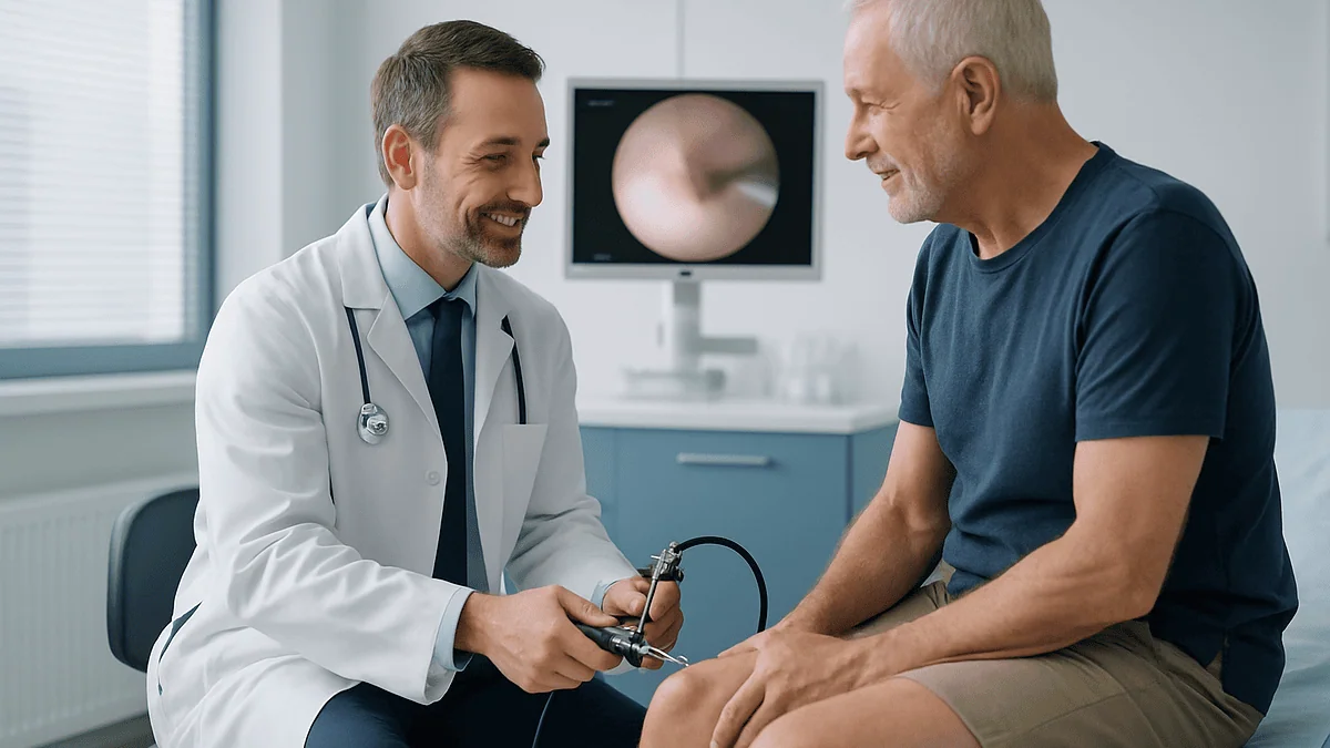
Arthroscopy works by passing a pencil-width tube with a light and a video camera into a joint through an incision a few millimetres long, then filling the joint with sterile fluid so the surgeon can see the cartilage, ligaments and lining on a screen. Through one or two further keyhole openings called portals, slender instruments trim, repair or remove damaged tissue without a long cut.
The consent form says “arthroscopic partial meniscectomy,” and the man holding it is mostly thinking about his knee locking on the stairs last Tuesday. His surgeon drew three small dots on a printout and said, “That is the whole operation.” Three dots. It seems too small to fix anything.
That gap between how minor the incisions look and how real the surgery is sits at the center of most questions about how arthroscopy works. People want to know what the camera actually shows, why the surgeon needs more than one hole, and how anything useful gets done through openings the size of a buttonhole.
This explainer walks through the equipment, the choreography inside the joint, the conditions that suit a keyhole approach and the ones that do not, and what the days afterward usually feel like. Where the evidence is firm, it says so. Where it is thinner than the brochures suggest, it says that too.
How arthroscopy works, in plain language
An arthroscopy is an operation to look inside a joint, and often to treat what is found, using a narrow tube with a camera on the end. The word comes from Greek roots meaning “joint” and “to look.” The tube itself is the arthroscope: a rigid rod roughly the width of a pencil that carries a light source and relays a magnified picture to a monitor in the operating room, according to the Mayo Clinic.
The basic sequence is the same in every joint. Once you are numb or asleep, the surgeon makes an incision a few millimetres long and slides in the arthroscope. Sterile fluid is pumped through the scope, gently expanding the joint so there is room to see. A second small opening, and sometimes a third, admits instruments: graspers, scissors, shavers, tiny sutures on curved needles. The surgeon watches the screen, not the joint, while working.
The NHS puts the whole procedure at about 30 minutes to 2 hours depending on what needs doing, with most people going home the same day. The openings are closed with a stitch or a small adhesive strip, and the joint is wrapped.
The clinical logic is straightforward. A traditional open operation must cut through skin, fat and sometimes muscle to expose the joint surface. Arthroscopy borrows a principle from plumbing: if you can put a camera and a tool down separate small channels, you do not need to open the whole pipe. The trade-off is that everything must be done in a fluid-filled space the size of a plum, working from a two-dimensional image, which is why surgeons train specifically for it and why some problems still call for an open approach.
What the arthroscope camera actually sees
The picture on the monitor is not a grainy endoscopy image. Modern arthroscopes deliver a bright, magnified, high-definition view of surfaces that no scan can show as clearly: the glassy white articular cartilage that caps the bone ends, the crescent-shaped meniscus in the knee, the ring of cartilage called the labrum that deepens the shoulder and hip sockets, and the synovial lining that produces joint fluid.

Magnification is the point. A frayed edge of meniscus, a flap of cartilage lifting off the bone, or a partial tear in a ligament can be missed on an MRI yet be obvious at arthroscopy. The Johns Hopkins patient guide notes that a surgeon may probe tissue with a small hooked instrument to test whether it is firm or soft, something imaging cannot do. This is why arthroscopy was historically used for diagnosis as well as treatment, although better scanning means purely diagnostic arthroscopy is now uncommon.
The camera has limits worth knowing. It shows the inside surface of the joint, not the bone beneath the cartilage or the tendons outside the capsule, unless the surgeon deliberately moves into those spaces. The view is two-dimensional, so depth is judged from experience and from the way instruments interact with tissue. And the picture depends on clear fluid; bleeding clouds the field and can slow the operation.
Most arthroscopes have an angled lens, commonly 30 degrees, so rotating the scope in place sweeps the view around a corner without moving the entry point. Surgeons describe it as looking around a room through a letterbox while turning a periscope. Learning to interpret that rotating, magnified, fluid-filled image is a large part of arthroscopic training.
Why portals, and where the keyholes go
A portal is simply the small opening through which the arthroscope or an instrument enters the joint. Its position is not arbitrary. Each joint has a small number of established portal sites chosen to pass between nerves, blood vessels and tendons rather than through them, and to give the best angle on the structures most often treated.
In the knee, the two standard portals sit either side of the patellar tendon just below the kneecap; one takes the camera, the other the working instruments, and they can be swapped during the operation. The shoulder typically uses a portal at the back for the camera and one or two at the front or side for instruments. The hip, a deep ball-and-socket joint, needs gentle traction on the leg to open a gap for the scope, and portals are placed on the outer side of the hip.
Surgeons usually make portals through a short cannula, a hollow sleeve that protects the skin and soft tissue as instruments pass in and out many times. The Cleveland Clinic describes the incisions as roughly the size of a buttonhole. Each one is usually closed with a single stitch or adhesive strip and leaves a dot-like scar.
Why not one hole? Because a camera and an instrument entering side by side through the same opening would be fighting for the same line of sight, like trying to thread a needle while your other hand blocks the view. Separate portals let the surgeon triangulate: the scope looks from one angle, the tool approaches from another, and the two meet at the target. Triangulation is the core motor skill of arthroscopy and the reason two or three keyholes are almost always needed.
Fluid, light and the instruments that fit through a keyhole
Three supporting systems make the camera useful. The first is fluid. Sterile saline is pumped into the joint under controlled pressure, which pushes the joint surfaces apart, washes away debris and blood, and gives the light something clear to travel through. Some of that fluid seeps into the surrounding tissue during surgery, which is why the joint often looks puffy for a day or two afterward; the MedlinePlus knee arthroscopy entry lists swelling as an expected early feature rather than a complication.
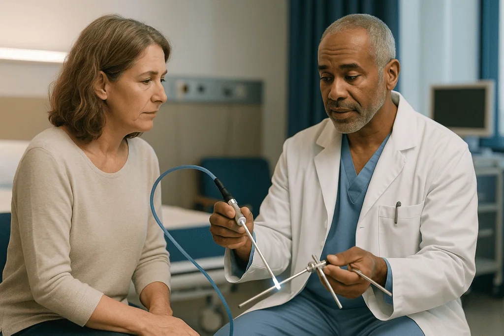
The second is light. A fiber-optic cable runs alongside the lens and floods the joint with cold light so that the camera can pick up detail.
The third is the instrument set, all built to pass through a portal a few millimetres wide:
- A probe, the hooked feeler used to test tissue firmness and lift flaps.
- Graspers and punches that bite out small pieces of torn meniscus or loose cartilage.
- A motorized shaver, a rotating blade inside a suction tube, that trims frayed tissue and removes inflamed lining.
- Radiofrequency wands that smooth surfaces and seal small bleeding vessels.
- Suture anchors, tiny screws or plugs with thread attached, that let a surgeon stitch a torn labrum or rotator cuff back to bone.
Repairing rather than removing tissue is technically harder through a keyhole, because knots must be tied with long instruments in a fluid-filled space. Instrument makers have developed knotless anchors and pre-loaded suture passers to shorten that step. None of this changes the biology of healing; a stitched tendon still needs weeks to reattach, which is why recovery from a repair is longer than from a simple trim even though the incisions look identical.
What is arthroscopy used for? Joints and problems that suit keyholes
Arthroscopy is best suited to mechanical problems on the inside surfaces of a joint: something torn, loose, frayed or catching. The knee and shoulder account for most procedures, but the technique is established in the hip, ankle, wrist and elbow as well. The Mayo Clinic and NHS list the common indications, summarized here.
| Joint | Problems commonly treated arthroscopically | Typical goal |
|---|---|---|
| Knee | Torn meniscus, anterior cruciate ligament (ACL) tears, loose cartilage fragments, kneecap tracking problems | Trim or repair the meniscus, reconstruct the ligament, remove loose bodies |
| Shoulder | Rotator cuff tears, torn labrum after dislocation, impingement, frozen shoulder that has not settled | Reattach tendon or labrum, shave bone spur, release tight capsule |
| Hip | Labral tears, femoroacetabular impingement (bone shapes that pinch), loose fragments | Repair labrum, reshape bone edge |
| Ankle | Cartilage lesions, impinging scar tissue or bone spurs, unstable ligaments | Remove spurs, treat cartilage defect, tighten ligament |
| Wrist and elbow | Ligament tears, cartilage damage, loose bodies, some stiffness | Trim, repair or remove |
The pattern across the table is consistent. Arthroscopy treats discrete structural faults. It is not a treatment for diffuse wear of the whole joint surface, and it cannot regrow cartilage that has worn away. That distinction matters most in the knee, where the temptation to “clean out” an arthritic joint is strong and the evidence, discussed later, does not support it.
A meniscus, for definition, is one of two C-shaped cartilage cushions between the thigh bone and shin bone. A labrum is the rim of cartilage around a socket joint. Both are common arthroscopic targets because they tear in patterns a camera can see and an instrument can reach.
Knee arthroscopy procedure, step by step
The knee is the most common arthroscopy site and a useful walk-through. MedlinePlus and the Mayo Clinic describe the sequence in similar terms.
Before the operation, you will have fasted as instructed and met the anesthesia team. Knee arthroscopy can be done under general anesthesia (fully asleep), a spinal block (numb from the waist down), or a regional block with sedation. The choice depends on your health, the planned procedure and your preference, and it is a decision for you and the anesthesiologist together.
In the operating room the leg is cleaned and a tourniquet may be placed high on the thigh to reduce bleeding. The surgeon marks the portal sites, makes the first small incision and inserts the scope, then starts the fluid flow. A systematic tour follows: the space above the kneecap, both compartments of the joint, the ligaments in the center, and the back of the knee where cartilage flaps like to hide.
Once the problem is confirmed, the second portal is made and the appropriate instrument introduced. A frayed meniscus edge might be trimmed back to a smooth rim with a punch and shaver. A tear near the meniscus’s blood supply might instead be stitched, because tissue with circulation can heal. Loose fragments are grasped and removed. Photographs are usually taken for your record.
At the end the fluid is drained, the portals are closed, and a bulky dressing goes on. In the recovery area, staff check sensation and circulation, and a physical therapist may show you how to walk with or without crutches before discharge. The NHS notes that most people go home the same day. The whole visit often takes longer than the surgery itself, because the preparation and recovery phases bracket a procedure that may last well under an hour.
Who arthroscopy is usually for, and who is usually asked to wait
The strongest case for arthroscopy is a clear mechanical fault, confirmed on examination and usually on imaging, that is causing symptoms not settling with time and non-surgical care. A young athlete whose knee locks because a meniscal flap is catching, or someone whose shoulder keeps dislocating because the labrum is torn, fits that description well. So does a person with a loose fragment of bone or cartilage floating in the joint.
Many people are appropriately asked to wait, and the reasons are evidence-based rather than a matter of rationing. Guideline bodies in several countries advise against arthroscopic “washout” or debridement for knee osteoarthritis unless there is a true mechanical block, because trials comparing keyhole surgery with physical therapy or with sham surgery for degenerative knee pain have not shown a meaningful benefit. Someone in their sixties with a worn knee and a degenerative meniscal tear on MRI is therefore usually offered a structured exercise program first, and often does as well without an operation.
Timing also matters for people whose joint is still acutely inflamed, whose skin is broken or infected near the joint, or who have not yet completed a reasonable trial of rehabilitation. Surgeons frequently ask for a period of physical therapy before deciding, partly because it helps in its own right and partly because it clarifies which symptoms are truly mechanical.
General health shapes the decision too. Uncontrolled diabetes, active infection elsewhere, and clotting disorders raise the risk of complications and may need to be addressed first. None of these are absolute bars; they are factors the treating team weighs against the expected benefit. The decision to operate, and when, sits with you and that team, informed by your symptoms, your imaging and what the evidence says about people in your situation.
Arthroscopy vs open surgery: what the comparison really shows
The obvious advantages of arthroscopy are visible on the skin. Smaller incisions mean less disturbance to muscle and capsule, and the Mayo Clinic and NHS both list the practical results: usually same-day discharge, less early pain, smaller scars, and a lower wound-infection risk than a long open cut. Many people can bear weight on an arthroscopically treated knee within a day or two, whereas an open joint operation of the same era would have meant a hospital stay.
| Feature | Arthroscopy | Open surgery |
|---|---|---|
| Incision | Two or three openings a few millimetres long | One incision several centimetres long |
| View | Magnified, two-dimensional camera image | Direct, three-dimensional view |
| Tissue disturbed | Minimal; muscle usually not cut | Skin, capsule and sometimes muscle divided |
| Hospital stay | Usually same day | Often overnight or longer |
| Best suited to | Discrete tears, loose bodies, labral and cuff repairs | Joint replacement, complex fractures, large reconstructions |
The honest caveat is that the healing inside the joint is the same whichever route the surgeon takes. A rotator cuff tendon stitched arthroscopically still needs weeks of protected movement while it reattaches to bone. The keyhole approach reduces the collateral damage of getting there; it does not speed up biology.
Some operations remain open by necessity. Joint replacement requires removing and re-shaping bone ends and seating an implant, which cannot be done through a portal. Complex fractures and large ligament reconstructions often need direct access. And in a minority of arthroscopic cases the surgeon finds something that cannot be safely addressed through the scope and converts to a small open incision during the same operation, which is why consent forms usually mention that possibility. Neither approach is superior in every situation; the right one is the one that fits the problem.
Is arthroscopic surgery very painful?
Most people find arthroscopy less painful than they feared, but “minimally invasive” is not “pain-free,” and expectations set too low can make the first days feel like something has gone wrong. The Mayo Clinic describes pain and swelling that ease over several days as part of the normal course.
Where the pain comes from is worth understanding. The portals themselves are small and usually sting only briefly. The deeper ache comes from the joint: the tissue that was trimmed or stitched, the lining irritated by instruments and fluid, and the pressure of swelling in a confined space. Procedures that involve bone, such as shaving a spur or anchoring a tendon, tend to hurt more than a simple trim, and shoulder and hip arthroscopy are often reported as more uncomfortable than knee arthroscopy because those joints are surrounded by large muscles that go into spasm.
Pain control is planned before you leave. Local anesthetic is often injected around the portals or into the joint at the end of surgery, and a nerve block may provide several hours of numbness, so the first evening can feel deceptively easy and the following morning noticeably worse as the block wears off. Your team will discuss oral medication options by class, typically starting with non-opioid pain relievers, and the dose and duration are decisions for the prescribing clinician. Ice, elevation and a compression wrap do real work in the first days by limiting swelling.
Pain that steadily improves over the first week is expected. Pain that steadily worsens after the first two or three days, especially with heat, redness or fever, is not, and belongs in the red-flag section below rather than in the category of normal recovery.
Arthroscopic surgery recovery time: what the first days and weeks look like
Recovery has two clocks. The wound clock is short; the tissue clock depends entirely on what was done inside the joint. Confusing the two is the most common source of frustration after arthroscopy.
In the first two or three days the joint is swollen and stiff, the dressing is bulky, and the goal is simple: protect the portals, control swelling and start gentle movement as instructed. MedlinePlus advises keeping the leg elevated and using ice to reduce swelling after knee arthroscopy. Crutches may be needed for balance even when full weight bearing is allowed. Portal stitches, if used, typically come out at a follow-up visit around one to two weeks.
By the end of the first week most people with a straightforward trim are walking without aids and returning to desk-based work; the NHS suggests about a week off for a sedentary job and longer for manual work. Driving waits until you can control the pedals confidently and are no longer taking medication that impairs alertness, which is a judgment your team should confirm.
From the second week onward the tissue clock takes over. A meniscal trim may feel near normal within a few weeks. A meniscal or labral repair, a rotator cuff reattachment or an ACL reconstruction follows a protected rehabilitation program measured in months, because healing tissue must be shielded while it regains strength. The NHS describes full recovery as taking anywhere from weeks to months depending on the procedure.
Physical therapy is not optional decoration. Restoring range of motion early prevents scar tissue from stiffening the joint, and progressive strengthening restores the muscle protection the joint relies on. The most consistent predictor of a good result within any given procedure is completing the rehabilitation program, not the size of the scars.
What not to do after an arthroscopy
Most post-operative rules exist to protect two things: the healing tissue inside the joint and the portals on the outside. The specific list depends on your procedure, and your surgeon’s written instructions take precedence over anything general. The following are the errors that surgical teams see most often.
- Do not soak the wounds. Showering is usually permitted once the dressing is changed, but baths, pools and hot tubs are typically off limits until the portals have sealed, because standing water carries infection risk into a fresh opening.
- Do not test the joint early. A repaired meniscus or labrum that feels fine at two weeks is held together by sutures, not by healed tissue. Loading it before the tissue clock allows can undo the repair.
- Do not skip the exercises because it hurts a little. Gentle prescribed movement prevents stiffness; complete rest breeds it. The distinction is between discomfort that eases as you move and sharp pain that stops you.
- Do not drive too soon. Both the Mayo Clinic and NHS advise waiting until your team confirms you can react safely and you are off sedating medication.
- Do not sit still for hours. Immobility after lower-limb surgery raises the risk of a blood clot in the deep veins of the leg (deep vein thrombosis). Regular ankle pumps, short walks and any clot-prevention measures your team prescribed matter, particularly in the first two weeks.
- Do not remove or change the dressing early unless told to, and do not apply creams to the portals.
- Do not decide alone that an imaging finding is fixed. If your team said other joint changes were left untreated, plan for them rather than expecting the operation to have addressed everything.
A short paragraph on seeking care belongs here as well. Calf pain or swelling, sudden breathlessness, spreading redness, discharge from a portal, or fever are reasons to contact your team the same day rather than wait for the scheduled follow-up.
What are the downsides of arthroscopy? Risks explained honestly
Arthroscopy is among the safest orthopedic operations, and the NHS states that serious complications occur in fewer than 1 in 100 cases. That reassuring number still deserves unpacking, because “rare” is not “never,” and the less serious downsides are more common than the headline suggests.
The expected downsides are swelling, bruising and stiffness in the first weeks. A few people develop a persistent joint effusion, fluid that keeps collecting, which may need to be drawn off. Numbness in a patch of skin near a portal, from a small sensory nerve branch disturbed on the way in, is not unusual and often improves over months, though it can be permanent.
The uncommon but serious risks listed by the Mayo Clinic and NHS include infection inside the joint, which requires urgent treatment; deep vein thrombosis and, rarely, a clot travelling to the lung; damage to nerves or blood vessels near the portals; and instrument breakage inside the joint. Anesthesia carries its own small risks, discussed separately by the anesthesia team. Continued or worsening symptoms are a downside too: if the pain was coming from wear rather than the tear that was treated, the operation may not help, which is why patient selection is discussed so carefully earlier in this article.
Two downsides are less obvious. First, removing meniscal tissue reduces the knee’s shock absorption, and larger resections are associated in long-term studies with faster development of arthritis in that compartment; surgeons therefore preserve as much as they safely can. Second, a keyhole procedure is still surgery, with recovery time, rehabilitation effort and time off work, and those costs in time and energy are real even when the incisions are tiny.
Alternatives always exist, from continued physical therapy and activity modification to injections or, for advanced arthritis, joint replacement. Weighing them against arthroscopy for your particular problem is the conversation to have with your treating team.
What people often get wrong about how arthroscopy works
“It’s just a look inside, not real surgery.” Almost all modern arthroscopy is therapeutic, not diagnostic; tissue is cut, removed or repaired. The anesthesia, the tourniquet and the rehabilitation are the same category of event as any operation. Treating it as trivial leads people to skip exercises and return to sport early.
“Small scars mean fast healing inside.” The portals heal in days. A stitched tendon or ligament graft heals on its own timetable, measured in months, regardless of how it was reached. The size of the incision tells you nothing about the size of the repair.
“An arthroscopic clean-out helps an arthritic knee.” This is the myth with the strongest evidence against it. Trials comparing arthroscopic debridement or lavage with physical therapy, and with placebo surgery, have not shown meaningful benefit for degenerative knee pain, and major guidelines advise against it unless the knee is truly locking. Wear on a joint surface cannot be shaved back to health.
“The camera sees everything, so a normal arthroscopy means nothing is wrong.” The scope sees joint surfaces. Pain from tendons outside the capsule, from bone beneath the cartilage, from referred sources such as the hip or spine, or from nerve sensitization is invisible to it.
“The surgeon can fix whatever they find.” Some findings, such as widespread cartilage loss, cannot be repaired through a scope or by any current technique. Surgeons often document these and leave them, and a good consultation explains this possibility beforehand.
“Once it’s fixed, it’s fixed for good.” A repaired meniscus can re-tear; a stabilized shoulder can dislocate again with the same mechanism of injury. Arthroscopy treats a problem; it does not make a joint injury-proof.
Each of these misunderstandings shares a root: mistaking the size of the door for the size of the room. The camera and portals are the access route, not the treatment itself.
Questions to ask your care team before arthroscopy
A ten-minute consultation moves fast, and the questions that matter most are easy to forget once the consent form appears. Taking a written list is not overcautious; surgeons generally welcome it. These are the ones that most reliably change how people experience the operation and its aftermath.
- What exactly do you expect to find, and what do you plan to do about it? Ask whether the plan is to remove tissue, repair it or both, because recovery differs sharply.
- What will you do if you find something different from the scan? Ask about the chance of converting to an open procedure and how that decision is made while you are asleep.
- How much of my pain is likely to come from the problem you are treating, and how much from wear you cannot treat? This is the single most useful question for a knee with mixed findings.
- What happens if I choose not to have surgery now, and what would make you advise waiting or reconsidering?
- What type of anesthesia do you recommend for me, and why?
- When can I put weight on the joint, drive, return to my specific job, and return to my specific sport? Ask for ranges and what each depends on.
- Will I have a brace or sling, and for how long?
- How is pain managed in the first days, and who do I contact if it is not controlled?
- What is my personal risk of a blood clot, and do I need any preventive measures?
- When and where will physical therapy start, and how many sessions are typical for this procedure?
- What signs mean I should call you, and what number do I use out of hours?
- Can I have the operative photographs and report for my records?
Writing down the answers, or bringing someone to listen, turns the consultation into a plan you can actually follow. The final decision about whether and when to proceed rests with you and the treating team.
When to call your doctor after arthroscopy
Expected recovery is a slow, steady improvement: swelling that peaks in the first two or three days and then eases, pain that fades week by week, portals that dry and seal. The signs below break that pattern and warrant a same-day call to your surgical team or, where indicated, emergency care. The Mayo Clinic, NHS and MedlinePlus list these as reasons to seek prompt attention.
- Fever or chills, particularly with a joint that feels hot, increasingly swollen or increasingly painful. Infection inside a joint is uncommon but needs urgent treatment.
- Redness spreading from a portal, thick or foul-smelling discharge, or a wound that opens.
- Pain that is worsening rather than easing after the first few days, or pain not controlled by the plan you were given.
- Calf pain, calf tenderness or new swelling in the lower leg, especially on one side. These can signal a deep vein thrombosis.
- Sudden shortness of breath, chest pain or coughing up blood. These may indicate a clot in the lung and are an emergency; call emergency services rather than the clinic.
- Numbness, tingling or weakness in the limb below the joint that is new or worsening, or a foot or hand that turns pale, cold or dusky.
- Bleeding that soaks through the dressing and does not stop with firm pressure.
- A joint that locks, gives way or will not move in a way it did before the operation and that does not settle.
- Persistent nausea, vomiting or inability to pass urine in the first day, which can relate to anesthesia.
None of these signs confirms a complication on its own, and most calls end with reassurance. The reason to make them promptly is that the few problems arthroscopy can cause are far easier to manage when caught early. If you are unsure whether something is normal, the safer choice is always to ask the team that operated. They know what was done inside the joint and what your recovery should look like.
Frequently asked questions
How long does it take to heal after an arthroscopy?
The skin portals usually seal within one to two weeks, but the tissue inside heals on its own schedule. The NHS suggests about a week off for desk work, longer for manual jobs, and full recovery from weeks to months depending on the procedure. A simple meniscal trim recovers far faster than a ligament reconstruction or tendon repair, which are protected for months. Your surgeon can give ranges specific to what was done.
What are the downsides of arthroscopy?
Expected downsides are swelling, stiffness and bruising for a few weeks, plus time off work and a rehabilitation commitment. Uncommon but serious risks listed by the NHS and Mayo Clinic include joint infection, blood clots, nerve or vessel injury and anesthesia complications; the NHS estimates serious complications at fewer than 1 in 100 cases. If pain was coming from wear rather than a treatable tear, symptoms may not improve.
Is arthroscopic surgery very painful?
It is usually less painful than open surgery but not pain-free. The deeper ache comes from the treated tissue and swelling inside the joint rather than the small incisions. Shoulder and hip arthroscopy, and procedures involving bone or repairs, tend to hurt more than a simple knee trim. Local anesthetic or a nerve block often makes the first evening easy, with more discomfort the next day. Pain should ease steadily over the first week.
What not to do after an arthroscopy?
Avoid soaking the wounds in baths or pools until the portals seal, driving until your team confirms it is safe, and loading a repaired structure before the tissue has healed. Do not skip prescribed exercises, and do not sit immobile for long periods, because stillness raises the risk of a blood clot. Follow your surgeon’s written instructions, which override general advice.
What is arthroscopy used for most often?
Most commonly, arthroscopy treats torn menisci and ligament injuries in the knee and rotator cuff or labral tears in the shoulder. It is also used in the hip for labral tears and impingement, in the ankle for cartilage damage and bone spurs, and in the wrist and elbow for ligament and cartilage problems. It suits discrete mechanical faults rather than diffuse arthritis.
How does the knee arthroscopy procedure differ from shoulder arthroscopy?
The principle is identical: camera in one portal, instruments through another, fluid to open the space. The knee uses two portals just below the kneecap and is often done under spinal or general anesthesia. The shoulder typically uses a rear camera portal and front or side working portals, is more often done with the patient partly upright, and generally involves more muscle irritation and a longer sling period.
Arthroscopy vs open surgery: which is safer?
Neither is universally safer; each fits different problems. Arthroscopy disturbs less tissue, so early pain, wound infection risk and hospital stay are usually lower. Open surgery gives a direct three-dimensional view and is necessary for joint replacement, complex fractures and some large reconstructions. Occasionally a surgeon converts from keyhole to open during the operation if the problem cannot be safely reached through the scope.
Can arthroscopy help knee osteoarthritis?
Evidence does not support it for most people. Randomized trials comparing arthroscopic debridement or lavage with physical therapy, and with sham surgery, have not shown meaningful benefit for degenerative knee pain, and major guidelines advise against it unless the knee has a true mechanical lock from a loose fragment or displaced tear. Structured exercise is the usual first recommendation.
Will I be awake during arthroscopy?
It depends on the anesthesia chosen. Options include general anesthesia, where you are fully asleep; a spinal block that numbs the lower body; or a regional nerve block with sedation. Some people under spinal anesthesia choose to watch the screen. The anesthesia team weighs your health, the planned procedure and your preference, and the decision is made together with you before surgery.
What happens if the surgeon finds something unexpected during arthroscopy?
Surgeons plan for this. The consent conversation usually covers the likely findings and what would be done for each, including whether a repair might become a removal or whether a small open incision could be needed. Findings that cannot be treated through the scope, such as widespread cartilage wear, are usually photographed and documented so your team can discuss them with you afterward.
This article is for general information only and is not a substitute for professional medical advice. Please consult a qualified doctor about your individual situation.

More from the Blog

Soft Tissue Injury Healing Stages: Why Sprains, Strains and Bruises Heal at Different Rates
Soft tissue injury healing time depends on what was damaged and how well it is supplied with blood. Bruises usually fade within about two…

What Happens Years After Meniscus Surgery? Cartilage Wear and Realistic Expectations
Years after meniscus surgery, the knee's future depends mainly on how much meniscus tissue was removed and how healthy the cartilage was beforehand. Partial…

Does a Sprain or Strain Need an MRI? When Imaging Adds to a Soft Tissue Injury Exam
Most sprains and strains do not need an MRI. Clinicians usually diagnose them by asking how the injury happened and examining the joint or…

Does Bursitis Come Back? Why Flares Recur and What Long-Term Results Usually Look Like
Yes, bursitis can come back. Most flares settle within a few weeks with rest, ice and reduced pressure on the area, but recurrence is…
Does a Torn ACL Always Need Surgery? Who Copes Without Reconstruction and Who Does Not
Not always. Many people with a torn ACL, particularly those who do not play pivoting sports, regain a stable, working knee through structured rehabilitation…

Is Swelling After Ankle Arthroscopy Normal? What Helps and When to Call Your Team
Some swelling after ankle arthroscopy is expected and usually peaks in the first few days before easing over several weeks, because the joint is…