

How Cryotherapy Freezes Skin Lesions: Liquid Nitrogen, Freeze Time and What You Feel
Key Takeaways
- Liquid nitrogen boils at about minus 196 degrees Celsius, far colder than the roughly minus 50 to minus 60 degree refrigerant in home wart sprays, which is why clinic freezes reach thicker lesions.
- Cryotherapy destroys lesions through three mechanisms: ice crystals rupturing cell membranes, clotting of small blood vessels during the thaw, and an inflammatory response that clears dead tissue and can alert the immune system to wart virus.
- A slow thaw and a repeated freeze-thaw cycle kill tissue more completely than a single long freeze, so clinicians let the frozen zone warm on its own and reserve double cycles for tougher lesions.
- Melanocytes, the pigment cells, die at warmer temperatures than surrounding skin cells, which is why permanent lightening is the most common lasting side effect and why shorter freezes are favored on the face and on darker skin.
- The NHS notes that cryotherapy for warts often needs repeating at one to two week intervals because freezing kills infected cells but does not remove the virus from surrounding skin.
- An intact blister is a sterile natural dressing; leaving it in place, keeping the site clean and lightly moisturized, and avoiding sun on the new pink skin do more for healing than any product.
Cryotherapy treats skin lesions by applying liquid nitrogen, a liquid colder than minus 190 degrees Celsius, to a wart, actinic keratosis or similar growth for a matter of seconds. Water inside the cells freezes, membranes rupture and tiny blood vessels clot, so the damaged tissue dies, blisters and peels away over one to three weeks while healthy skin grows underneath. Most people feel a sharp cold sting that fades within minutes.
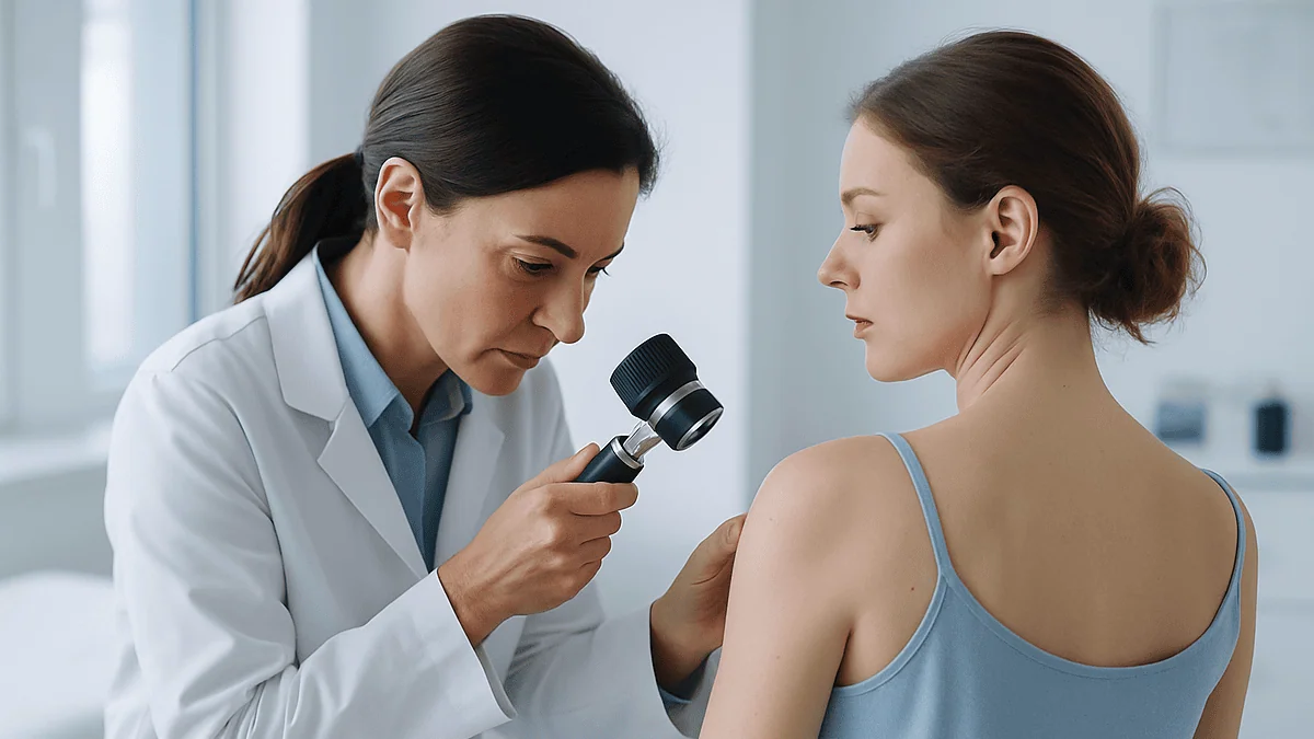
The spot on her left temple had been there for a year. Flat, a little scaly, the color of weak tea. She had covered it with concealer, scratched it off twice, and finally mentioned it at a routine appointment. The clinician looked, said the word “actinic keratosis,” picked up what looked like a small metal thermos with a nozzle, and asked whether she wanted it frozen today.
That is how most people meet cryotherapy: not as a planned procedure, but as a ten-second decision in an exam room. The canister hisses, the skin goes white, something stings, and then the clinician says the spot will blister and fall off. Few people get a proper explanation of what just happened to their cells.
This article is that explanation. It walks through cryotherapy for skin lesions, how it works at the level of ice crystals and capillaries, what freeze time means, why the sting feels the way it does, and what the following two weeks usually look like. It also corrects a few persistent myths, including the one about putting ice on the blister.
What is cryotherapy for skin lesions and how does it work?
Cryotherapy is the deliberate destruction of unwanted tissue with extreme cold. In dermatology the cold almost always comes from liquid nitrogen, which boils at about minus 196 degrees Celsius (minus 321 Fahrenheit) and is stored in insulated flasks until the moment it is sprayed or dabbed onto skin.
Cryotherapy for skin lesions works by exploiting a simple weakness shared by every living cell: it is mostly water. When skin is cooled fast enough, water inside and between the cells turns to ice. Those crystals are sharp. They puncture cell membranes, pull water out of the cells and disrupt the proteins that keep a cell functioning. A keratinocyte, the type of cell that makes up most of the outer skin and most warts and sun spots, cannot survive that kind of insult.
A second mechanism kicks in as the tissue thaws. The tiny blood vessels in the frozen zone are injured, platelets gather and small clots form. Blood flow to the lesion stops, so any cells that survived the ice are starved afterward. Finally the body mounts an inflammatory response, sending immune cells to clear the dead tissue. That response is thought to contribute to the treatment of viral warts, because it brings the immune system face to face with wart virus proteins it had been ignoring.
The result is a controlled, shallow injury. Over the next days the dead lesion separates from the living tissue beneath it, often as a blister, then a crust, and the skin underneath repairs itself. Mayo Clinic describes this sequence for actinic keratoses: the area blisters or peels, then heals, and new skin appears within a few weeks.
The craft lies in freezing deeply enough to destroy the lesion but not so deeply that the scar, pigment change or nerve irritation outlasts the problem it was meant to solve.
Why liquid nitrogen, and how the cold is delivered
Clinicians have used many cryogens, the word for any substance that produces freezing. Carbon dioxide snow, nitrous oxide and refrigerant sprays sold for home wart treatment all work on the same principle. Liquid nitrogen dominates in clinics because it is colder than the alternatives, cheap to store and inert, meaning it does not react with skin or leave a chemical residue. A thicker or deeper lesion needs the tissue itself to reach a destructive temperature, and only a very cold source can push the freeze front far enough, fast enough.

Speed matters. Slow cooling lets cells adapt by shifting water outward, so ice forms mainly between cells and some survive. Rapid cooling forms ice inside the cells, which is far more lethal. The faster the freeze, the more complete the kill within the target zone.
There are three common ways to apply it. The spray technique uses a hand-held flask with a fine nozzle; the clinician holds it a centimeter or two from the lesion and sprays in short bursts until a white halo of frozen skin extends a millimeter or so beyond the visible edge. The contact probe technique chills a metal tip and presses it onto the lesion, giving a more sharply defined freeze that is useful near the eye or on small, raised growths. The oldest technique dips a cotton-tipped applicator in liquid nitrogen and touches it to the skin; it is gentler, freezes less deeply and is often chosen for children or thin facial skin.
Cleveland Clinic notes that whichever method is used, application typically lasts only seconds. The white frozen appearance that spreads outward is the clinician’s main visual guide; it shows how far the ice front has traveled across the surface, which correlates roughly with how deep it has gone.
Freeze time, thaw time and the freeze-thaw cycle
Three numbers shape what happens to a lesion: how long the freeze lasts, how long the tissue takes to thaw, and how many times the cycle is repeated. None of these is fixed, and a clinician adjusts all three to the lesion in front of them.
Freeze time is counted from the moment the skin turns fully white, not from the first puff of spray. A flat, thin actinic keratosis on the cheek might need only a brief freeze. A thick plantar wart on the sole, protected by a cap of hardened skin, may need considerably longer or may be pared down with a blade first so the cold can reach the living tissue. Cleveland Clinic describes the application as lasting a few seconds in most cases, with the clinician judging duration by the lesion’s thickness and location.
Thaw time is the quieter half of the story. As ice melts, water rushes back into cells that have been dehydrated and weakened, and many of them burst. A slow thaw is more destructive than a fast one, which is why clinicians let the frozen zone warm on its own rather than rubbing it or applying heat.
Repeating the cycle, freezing again once the first freeze has fully thawed, destroys tissue more completely than a single long freeze of the same total duration. Clinicians reserve the double freeze-thaw for tougher lesions, since each cycle also increases the risk of blistering, pigment change and scarring.
Many lesions need more than one visit. The NHS notes that cryotherapy for warts may need to be repeated at intervals of one to two weeks, because the virus sits in skin layers that a single freeze may not fully reach. This is normal practice, not a sign that the first treatment failed.
What you actually feel during cryotherapy for skin lesions
People expect cold. What they usually describe is a burn. The nerve endings that detect painful cold fire intensely in the first few seconds, so the sensation resembles a lit match held close to the skin or a sharp sting that quickly becomes an ache. It peaks during the thaw, when blood returns to the area, and most people say the worst of it has settled within a few minutes.
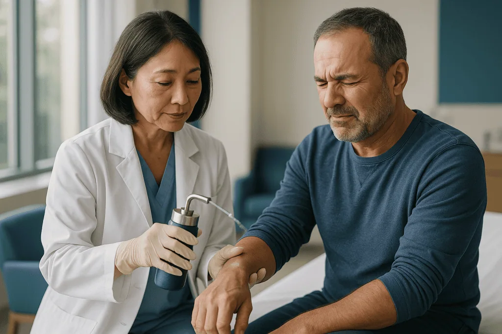
Location changes everything. Fingertips, the sides of the nose, the lips and the area around the eye are richly supplied with nerves and feel the freeze more sharply. The back and upper arms are forgiving. The soles of the feet sit somewhere in between, though thick plantar warts often need a longer freeze, which lengthens the discomfort.
Children and adults with low pain tolerance can be offered a topical numbing cream applied beforehand, or a small injection of local anesthetic for larger lesions. Many clinicians skip anesthetic for small, single spots because the injection itself can sting as much as the freeze. That is a conversation to have before the canister comes out, not after.
In the hour after treatment the spot throbs mildly and swells. Over the next twelve to twenty-four hours the ache fades and is replaced by a feeling of tightness as fluid gathers under the dead skin. A freeze near the eye can produce startling puffiness of the lid the next morning, which is harmless and settles on its own; it is a matter of gravity and loose tissue, not a complication.
If pain is increasing rather than easing after the first day, or if it comes with spreading redness or warmth, that pattern is not typical and is covered in the section on when to call your doctor.
Which skin lesions are treated with freezing, and who is asked to wait
Cryotherapy suits lesions that are superficial, well defined and benign or precancerous. The most common targets are viral warts on the hands and feet, actinic keratoses (rough, scaly patches caused by years of sun exposure that carry a small long-term risk of turning into squamous cell skin cancer), seborrheic keratoses (harmless, waxy, stuck-on brown growths that appear with age), skin tags and small benign growths such as molluscum contagiosum, a viral skin infection common in children.
Some thin, low-risk skin cancers are occasionally frozen, but that is a specialist decision made with a biopsy result in hand, and surgical removal remains the usual approach when a lesion needs to be fully examined under a microscope.
Who is usually asked to wait, or offered something else?
- Anyone whose lesion has not been confidently identified. A pigmented spot that could be a melanoma should never be frozen, because freezing destroys the tissue a pathologist would need to examine.
- People with darker skin tones, for whom the risk of permanent lightening of the treated area is higher, may be offered a shorter freeze or a different method, particularly on the face.
- People with poor circulation, uncontrolled diabetes or neuropathy in the feet, where a blister on the sole can become a slow-healing ulcer.
- People with cold-triggered conditions such as cold urticaria, cryoglobulinemia or Raynaud phenomenon, in whom cold exposure can provoke a systemic or vascular reaction.
- Lesions over the lower legs in older adults, where healing is slow and the risk of ulceration is higher.
None of these is an absolute bar. Each is a reason for the treating clinician to weigh freezing against alternatives and to tell the patient why they chose one path over the other.
Cryotherapy for actinic keratosis on the face: what the evidence shows
Actinic keratoses sit at the center of the cryotherapy story because they are so common and because the face is where most of them appear. They are not cancer. They are a marker of sun damage and a small fraction of them, over years, progress to squamous cell carcinoma. Clinicians treat them to remove that possibility and because they are often itchy, rough and cosmetically bothersome.
Both Mayo Clinic and the NHS list cryotherapy among the standard treatments for individual actinic keratoses. It works well for a handful of discrete, well-separated spots: each is frozen for a few seconds, blisters or flakes, and heals. The NHS describes it as a straightforward outpatient treatment that can be repeated if a patch returns.
The question people search for is whether it is “effective” for precancerous areas on the face. The honest answer is that freezing reliably clears many individual lesions, but it does nothing for the surrounding sun-damaged skin, where new keratoses are already forming invisibly. Dermatologists call this field change. When a patient has dozens of spots across the forehead and scalp, freezing each one becomes impractical and clinicians often turn to field treatments: prescription creams that provoke an immune or chemical reaction across the whole area, or photodynamic therapy, in which a light-sensitizing cream is applied and the region is then illuminated to destroy abnormal cells.
On the face, cosmetic outcome matters. A freeze that is slightly too long can leave a pale, shiny patch that is more noticeable than the original keratosis, particularly on olive or brown skin. Experienced clinicians tend toward shorter freezes on the face, accepting that a second visit may be needed, rather than risking permanent lightening. That trade-off is worth discussing before the spray starts.
Common lesions compared: freeze approach, healing and what to expect
The table below summarizes how cryotherapy is typically approached for the lesions most often frozen in a skin clinic. It reflects general patterns described by the NHS, Mayo Clinic and Cleveland Clinic; the clinician treating you will adjust everything to the size, depth and location of your particular lesion.
| Lesion | Typical freeze approach | Sessions commonly needed | Typical healing pattern | Points to know |
|---|---|---|---|---|
| Common wart (hands) | Spray or probe; may be pared first | Often several, 1–2 weeks apart (NHS) | Blister, crust, heals in about 1–3 weeks | Virus may persist; recurrence is common |
| Plantar wart (sole) | Longer freeze after paring thick skin | Often several | Slower; walking delays healing | Can be painful to walk on for a few days |
| Actinic keratosis | Brief spray, single cycle | Usually one, repeated if it returns | Scaling or small blister, heals within weeks (Mayo Clinic) | Does not treat surrounding sun damage |
| Seborrheic keratosis | Spray until lesion whitens | Usually one | Lifts off as a crust | Pale patch possible on darker skin |
| Skin tag | Probe or forceps-held freeze | One | Darkens and drops off | Often considered cosmetic |
| Molluscum (children) | Light cotton-tip freeze | Sometimes several | Small blister, quick healing | Often left to resolve on its own |
Two threads run through the table. First, thicker lesions and lesions on weight-bearing or poorly perfused skin need longer freezes, heal more slowly and are more likely to need repeat visits. Second, the lesions most likely to come back are the viral ones, because freezing destroys infected cells but does not eliminate the virus from the surrounding skin.
Healing times here are ranges drawn from the cited sources, not promises. Individual healing depends on age, circulation, location and how the treated area is looked after.
The first 48 hours: redness, swelling and the cryotherapy blister, what to do
Within minutes of the freeze the treated spot flushes red and swells slightly as blood rushes back into thawing tissue. Over the next several hours a blister often forms. This is not a complication; it is the plan. The freeze has killed the top layer of skin and loosened its attachment to the layer beneath, and fluid collects in the gap. Blisters are most dramatic on thin skin, on the hands and around the eyes. Some lesions, particularly flat actinic keratoses, never blister at all and simply turn dark and flaky.
The blister may be clear or blood-tinged. A dark purple blister looks alarming but reflects bleeding from the small vessels that the freeze was meant to injure, and it heals the same way as a clear one.
What should you do with it? Cleveland Clinic and the NHS give consistent advice: leave it alone. An intact blister is a sterile dressing made by your own body. It protects the raw skin beneath, keeps bacteria out and lets the new layer form undisturbed. Popping it exposes the wound, hurts more and raises the chance of infection and scarring.
If a blister sits where it will inevitably be rubbed or burst, such as a fingertip or the sole of the foot, a clinician may advise draining it with a sterilized needle while leaving the roof in place, then covering it with a simple dressing. That is a judgment call for the care team, not a default.
Keep the area clean with gentle washing, pat dry and apply a plain protective ointment if the clinic recommended one. Avoid picking at the edge. In this first phase the most useful thing a patient can do is nothing.
How long does cryotherapy take to heal? Days and weeks after treatment
Healing follows a predictable arc, though the speed varies with the thickness of the lesion and where it sits on the body.
In the first two to four days the blister flattens, or the frozen lesion darkens, and the area may weep slightly. The ache fades. By the end of the first week a crust or scab has usually formed over the site. Underneath it, new skin cells are migrating in from the edges and from hair follicles, which act as reservoirs of regenerating cells.
Somewhere between one and three weeks the crust lifts off, taking the dead lesion with it, and reveals pink new skin. Cleveland Clinic gives this range for most small lesions on the face and upper body. Mayo Clinic describes a similar timeline for actinic keratoses, with new skin visible within a few weeks of the blister or peeling phase.
The lower legs are the exception. Circulation there is slower, gravity pools fluid in the tissue and a frozen spot on the shin of an older adult can take considerably longer, occasionally forming a shallow ulcer that needs wound care. That is why many clinicians prefer other methods below the knee.
The pink patch left behind is not the final result. It fades toward normal skin color over the following weeks to months. Some sites end up slightly lighter than the surrounding skin, especially after a longer freeze or on darker skin, because the pigment-producing cells, called melanocytes, are more sensitive to cold than the surrounding keratinocytes. That lightening can be permanent.
For viral warts, judge the outcome at the follow-up visit rather than at the moment the crust falls off. Warts that look gone can return within weeks if infected cells remain at the margin, which is why repeat freezes are often spaced one to two weeks apart.
Liquid nitrogen wart removal aftercare: showering, dressings and healing faster
The most common aftercare question is the simplest: can I shower after liquid nitrogen treatment? Yes. The treated area can get wet from the day of treatment. Warm water and a mild cleanser do no harm; what matters is to avoid scrubbing the site, soaking it for long periods, or directing a strong jet of water at a fresh blister. Pat the area dry rather than rubbing.
Dressings are optional for most lesions. A spot on the face or upper arm can usually be left open to the air. A blister on a finger, the sole or anywhere that clothing rubs is more comfortable under a light adhesive dressing changed daily. Plain petroleum-based ointment under the dressing keeps the healing surface from drying out and sticking to the fabric. Antibiotic ointments are not routinely needed on a clean, intact wound and can cause skin irritation in some people.
People also ask how to make cryotherapy heal faster. The honest answer is that wound healing has a biological pace that cannot be hurried much, but it can easily be slowed. The things that help are modest:
- Leave blisters and crusts in place until they separate on their own.
- Keep the site clean and lightly moisturized rather than letting it dry and crack.
- Protect the new pink skin from sun with clothing or sunscreen, since fresh skin pigments unevenly.
- Do not smoke; nicotine narrows blood vessels and delays healing.
- Keep blood sugar well controlled if you have diabetes.
For plantar warts, reducing pressure with a cushioned insole or a doughnut-shaped pad makes walking more comfortable and protects the blister. For hand warts, gloves for wet work keep the site clean.
Resume normal activity, including exercise and swimming, once any blister has flattened and the site is no longer raw. The treating clinician will tell you if your particular lesion needs anything different.
Risks and side effects of freezing skin lesions, in plain language
Cryotherapy is a minor procedure, but minor is not the same as risk-free, and a patient deserves a clear picture of what can go wrong.
The expected effects, which are part of the treatment rather than complications, are pain during and shortly after the freeze, redness, swelling and blistering. These resolve over days.
The common side effects that persist longer:
- Hypopigmentation, meaning permanent lightening of the treated spot. It is the most frequent lasting side effect and is more visible on darker skin, because melanocytes die at warmer temperatures than the surrounding skin cells.
- Hyperpigmentation, meaning darkening, which usually fades over months but can linger.
- A small depressed or shiny scar, more likely after deep or repeated freezes.
- Loss of hair in the treated area if hair follicles were frozen.
Less common problems include infection of the blister (signaled by increasing pain, spreading redness, warmth and pus), bleeding from a blood blister, and nerve injury. The last is specific to a few sites where sensory nerves run just beneath the skin, such as the sides of the fingers, the back of the hand and the area in front of the ear; freezing deeply there can cause numbness or tingling that may take months to recover. Clinicians are cautious in those zones for exactly this reason.
Rare but important: a lesion that was misidentified. Freezing a skin cancer that was assumed to be a benign keratosis removes the chance to diagnose it and can let it grow back deeper. This is why any spot that is bleeding, changing, irregular in color or failing to heal should be assessed, and often biopsied, before anyone picks up a canister.
Treatment failure, meaning the lesion persists or returns, is not a side effect but it is a realistic outcome, especially for warts. The NHS is explicit that wart treatments, including freezing, do not always work and often need repeating.
Alternatives to cryotherapy: when another method makes more sense
Freezing is one tool among several, and the right choice depends on what the lesion is, where it sits and what the patient values most.
For warts, the first-line alternative is a topical treatment that softens and peels the thickened skin over weeks. The NHS lists this alongside cryotherapy as a standard option and notes that both require persistence. Watchful waiting is also legitimate: many warts in children and young adults clear on their own within months to a couple of years as the immune system catches up. Other clinic-based options include scraping and cautery under local anesthetic, laser treatment and, for stubborn warts, immunotherapy approaches that provoke an immune response at the site.
For actinic keratoses, individual spots can be frozen, but widespread sun damage is often better managed with field treatments. Mayo Clinic describes prescription creams that trigger inflammation or a chemical reaction across the damaged area, and photodynamic therapy using a light-sensitizing agent followed by illumination. These treat the visible spots and the invisible early ones at once, at the cost of a longer, more uncomfortable reaction across a larger area.
For seborrheic keratoses and skin tags, the alternatives are curettage (scraping the lesion off with a looped blade), shave removal, snip excision for tags, and electrocautery, which uses heat rather than cold. Shave and curettage have the advantage of producing tissue that can be sent for examination if there is any doubt about the diagnosis.
For anything suspicious, surgical excision with a margin of normal skin remains the standard, because the whole lesion goes to the laboratory.
The decision is not freezing versus everything else. It is a weighing of certainty of diagnosis, depth of lesion, cosmetic sensitivity of the site, skin tone, convenience and the patient’s tolerance for repeat visits, and it belongs with the treating team.
What people often get wrong about cryotherapy for skin lesions
Several ideas about freezing circulate widely enough to be worth correcting.
“Home freezing kits are the same as the clinic.” They are not. Over-the-counter sprays use a refrigerant mixture that reaches roughly minus 50 to minus 60 degrees Celsius, far warmer than liquid nitrogen. They can treat small, thin warts on hands and feet, which is what the NHS says they are for, but they cannot freeze deeply enough for thick lesions and should never be used on the face, genitals or any spot that has not been identified by a clinician.
“Put ice on it afterward to reduce swelling.” The treated skin has already been frozen once. More cold adds injury, not relief. Swelling is part of the process and settles on its own.
“Pop the blister so it heals faster.” The opposite is true. The blister roof is a natural dressing; removing it slows healing and invites infection.
“One freeze and the wart is gone for good.” Freezing destroys infected cells but does not remove the virus from surrounding skin. Warts commonly need several sessions and can return even after apparent clearance.
“If it comes back, the clinician did it wrong.” Recurrence after cryotherapy reflects the biology of the lesion more than the technique, particularly for warts and for actinic keratoses in heavily sun-damaged skin.
“Cryotherapy treats skin cancer.” It is occasionally used for selected very superficial, biopsy-confirmed lesions by specialists, but it is not a general skin cancer treatment and should never be applied to an unidentified pigmented spot.
“The white patch left behind will fade.” Pinkness fades. True hypopigmentation, loss of pigment cells, is often permanent, which is why a cautious freeze on the face is worth a second visit.
Questions to ask your care team before the canister comes out
A ten-second procedure still deserves a two-minute conversation. These questions tend to surface the information that matters most.
- What do you think this lesion is, and how confident are you? If there is any doubt, would a biopsy or shave removal be better than freezing it?
- Why cryotherapy rather than a cream, curettage or simply watching it?
- How many sessions do you expect this lesion to need, and how far apart?
- Given my skin tone and the location, how likely is a permanent pale patch, and would a shorter freeze with a possible repeat visit be a reasonable trade?
- Should I have numbing cream or a local anesthetic, especially for a child or a sensitive site?
- What should the blister look like, and what should I do if it bursts on its own?
- Can I shower, swim and exercise, and from when?
- Do I need a dressing, and if so, what kind and for how long?
- What signs would mean the site is infected or healing badly, and who do I contact if that happens?
- If the lesion returns, what is the next step?
- Are there other spots on my skin you noticed today that I should keep an eye on?
Write the answers down or ask for them in writing. Aftercare instructions given in the moment, with a stinging spot and a hand on the door, are easily forgotten.
A clinician who welcomes these questions is doing their job well. The answers will not change the physics of liquid nitrogen, but they will change how prepared you are for the fortnight that follows.
When to call your doctor after cryotherapy
Most treated spots follow the arc described above without any need for a return visit before the scheduled follow-up. A few patterns fall outside that arc and should prompt a call to the clinic or, out of hours, to a general medical service.
Call promptly if you notice:
- Pain that is increasing after the first 24 to 48 hours instead of easing, or pain that is out of proportion to the size of the treated area.
- Redness spreading outward from the site, especially with warmth, swelling or red streaks tracking up the limb.
- Thick yellow or green discharge, a foul smell, or a crust that keeps re-forming over pus.
- Fever, chills or feeling generally unwell in the days after treatment.
- Bleeding that does not stop with ten minutes of firm pressure.
- A blister larger than expected, or one that keeps refilling after it has been drained by the clinic.
- Numbness, tingling or weakness in the fingers or hand after a freeze near a nerve, which the team should document even if it is likely to recover.
- A wound that has not crusted over within about two weeks, or that is enlarging, particularly on the lower leg.
- Any treated lesion that grows back quickly, bleeds, changes color or develops an irregular edge; this needs assessment rather than another freeze.
Seek urgent care the same day if spreading redness is accompanied by fever or rapid worsening, since skin infections can progress quickly in people with diabetes, poor circulation or a weakened immune system.
Finally, keep the follow-up appointment even if the spot looks healed. Whether a wart has truly cleared, or an actinic keratosis has gone and its neighbors need attention, is a judgment for the clinician who saw it before the freeze. Every decision about repeat treatment, switching methods or taking a biopsy sits with that team.
Frequently asked questions
How does cryotherapy for skin lesions actually destroy a wart or sun spot?
It freezes the water inside the lesion’s cells so quickly that ice crystals form within them, rupturing membranes and denaturing proteins. As the tissue thaws, small blood vessels clot and cut off blood supply, and the body’s inflammatory response then clears the dead tissue. The lesion separates from healthy skin beneath as a blister or crust and is shed, usually within one to three weeks.
How long does cryotherapy take to heal?
Most small treated spots on the face, hands and upper body crust and heal within about one to three weeks, according to Cleveland Clinic and Mayo Clinic. Thick plantar warts and lesions on the lower legs take longer because of pressure and slower circulation. The pink patch left behind continues to fade toward normal color over weeks to months, though some sites remain permanently lighter.
Can I shower after liquid nitrogen treatment?
Yes, the treated area can get wet from the day of treatment. Use warm water and a mild cleanser, avoid scrubbing or directing a strong spray at a fresh blister, and pat the site dry rather than rubbing. Long soaks are best avoided while the skin is raw. Swimming and exercise can usually resume once any blister has flattened, unless your clinician advises otherwise.
What should I do about a cryotherapy blister?
Leave it intact if at all possible. The blister roof protects the new skin forming underneath, keeps bacteria out and reduces scarring. If it is in a spot that will inevitably burst, such as a fingertip or the sole, your clinician may advise draining it with a sterile needle while leaving the roof in place and covering it. Watch for spreading redness, increasing pain or pus, which suggest infection.
How can I make cryotherapy heal faster?
Healing cannot be hurried much, but it can be protected. Leave blisters and crusts alone until they separate, keep the site clean and lightly moisturized with a plain ointment, protect new pink skin from sun, avoid smoking, and keep blood sugar controlled if you have diabetes. For plantar warts, cushioning the area reduces pressure. Picking, popping and over-cleaning are the main things that slow healing.
Is cryotherapy effective for precancerous areas on the face?
For a few discrete actinic keratoses, freezing is a standard treatment listed by Mayo Clinic and the NHS, and it clears many individual lesions. It does not treat the surrounding sun-damaged skin where new spots are forming, so people with widespread damage are often offered field treatments such as prescription creams or photodynamic therapy. On the face, clinicians usually favor shorter freezes to limit permanent pale patches.
What is the aftercare after liquid nitrogen wart removal?
Keep the site clean, apply a plain protective ointment if advised, and use a light dressing only where the blister will be rubbed, such as fingers or soles. Do not pick at the crust. Expect a possible repeat freeze at one to two week intervals, as the NHS notes, since warts often need several sessions. Call the clinic if pain increases, redness spreads or the wound has not crusted within about two weeks.
Can I get cryotherapy on the NHS?
It depends on the lesion and the local area. The NHS treats actinic keratoses and other lesions with a medical reason for removal, and cryotherapy for warts is offered in some areas though not all, since many warts clear on their own. Removal of purely cosmetic lesions such as skin tags is generally not available on the NHS. Your general practitioner can explain what applies locally.
Does cryotherapy hurt, and is anesthetic used?
It typically feels like a sharp cold burn or sting that peaks during the thaw and fades within a few minutes, with a mild ache for a few hours afterward. Sensitive sites such as fingertips, lips and around the eyes hurt more. Numbing cream or a small local anesthetic injection can be offered, especially for children or larger lesions, though many single small spots are frozen without it.
Will cryotherapy leave a scar or a white mark?
A small scar is possible, especially after deep or repeated freezes. More common is a permanently lighter patch, because pigment-producing cells are more sensitive to cold than surrounding skin cells; this is more noticeable on darker skin tones. Temporary darkening can also occur and usually fades over months. Discuss your skin tone and the lesion’s location with your clinician before treatment so the freeze can be tailored.
References
- Cleveland Clinic: Cryotherapy
- NHS: Actinic keratoses (solar keratoses)
- NHS: Warts and verrucas
- NHS: Skin tags
This article is for general information only and is not a substitute for professional medical advice. Please consult a qualified doctor about your individual situation.

More from the Blog
Before Scar Treatment: Sun, Tanning and the Products to Pause in the Weeks Before
Preparing for scar treatment usually means protecting the area from tanning and unprotected sun for several weeks beforehand, pausing self-tanner, waxing and strong exfoliating…
Surgical Tattoo Removal vs Laser: When Cutting the Tattoo Out Makes More Sense
Surgical tattoo removal cuts the inked skin out in one or a few operations and always leaves a scar, while laser removal breaks pigment…

Laser Resurfacing Side Effects: Expected Crusting vs Pigment Change or Infection Signs
Expected laser resurfacing side effects include redness, swelling, oozing, crusting, itching and peeling, most intense in the first days and settling over one to…
Who Is a Candidate for Laser Resurfacing? Skin Tone, Recent Tanning and Healing History
Typical laser resurfacing candidates have skin concerns such as fine lines, sun damage, uneven texture or certain scars, no active tan, no recent isotretinoin…
Microneedling vs Chemical Peels for Texture and Scars: Which Approach Fits Which Skin
Microneedling and chemical peels both improve skin texture and shallow scarring, but through different routes. Microneedling creates controlled micro-injuries that prompt new collagen and…
Which Collagen Treatment Fits Your Skin Concern? How Dermatologists Match Method to Goal
The right collagen treatment depends on the specific concern, not on the popularity of the method. Fine lines and texture usually point toward topical…