

How Ear Reconstruction Works: Rib Cartilage Framework, Staged Surgery and Skin Coverage

Key Takeaways
- Rib cartilage ear reconstruction carves a framework from two to four of the patient's own costal cartilages and buries it under the skin, where suction drains pull the skin into every groove to create definition.
- The operation is staged because a buried framework needs months for blood vessels to grow into the overlying skin before that skin can be lifted again to elevate the ear.
- Johns Hopkins describes rib cartilage reconstruction as usually starting around school age, often six years or older, because younger ribs do not hold enough cartilage for a full-size ear.
- The chest donor site, not the ear, is the main source of pain after surgery, and deep breathing is encouraged early because shallow breathing raises the risk of chest infection.
- Rebuilding the outer ear does not restore hearing; the CDC notes that anotia and microtia affect about 1 in 3,800 US babies, and hearing is addressed separately, often with a bone-conduction device.
- Alternatives include a porous polyethylene framework, a removable prosthesis, or no reconstruction, and published comparisons describe trade-offs rather than one clearly superior option.
Rib cartilage ear reconstruction rebuilds a missing or underdeveloped outer ear using the patient's own rib cartilage. A surgeon removes cartilage from several ribs, carves and assembles it into an ear-shaped framework, and places it under the skin on the side of the head. Later operations lift the ear away from the scalp and cover the back with tissue and skin grafts. It is usually done in stages over months, in school-age children or adults.
The question usually arrives at a kitchen table. A parent turns a laptop toward a partner and says, quietly, so the child in the next room does not hear: they take it from the ribs. Or it arrives in a mirror, decades later, when an adult who has always combed hair over one side finally types a phrase into a search bar late at night.
Rib cartilage ear reconstruction is one of those operations that sounds almost medieval when you first read about it and turns out, on closer inspection, to be an exercise in patience and carpentry. Cartilage is borrowed from the chest, carved into the ridges and hollows of an ear, and tucked under skin that has to be coaxed into wrapping around every curve. Nothing about it is quick.
What follows is an honest walk through how that works, why it is done in stages, what the chest and the new ear feel like along the way, and where the real uncertainties lie.
What is rib cartilage ear reconstruction, and why is the ear so hard to rebuild?
Rib cartilage ear reconstruction, sometimes called autologous costal cartilage reconstruction, is surgery that builds a new outer ear from the patient’s own rib cartilage. Autologous simply means the tissue comes from your own body rather than a donor or a manufactured material. Costal means rib.
The outer ear, the auricle, looks like a simple flap of skin. It is not. Underneath sits a single sheet of elastic cartilage folded into a helix (the outer rim), an antihelix (the inner ridge), a concha (the bowl), and a tragus (the small bump in front of the ear canal). Each fold has a specific depth, and the whole structure projects from the head at an angle. Cover any one of those landmarks incorrectly and the eye notices immediately, even if it cannot say why.
Two things make this harder than most reconstruction. First, ear cartilage has almost no equivalent elsewhere in the body; rib cartilage is stiffer and thicker but is the largest supply a person carries that can be carved into shape. Second, the skin over a normal ear is thin, almost like tracing paper, and it clings to every contour. Skin on the side of the head in someone born without an ear is thicker and flatter, so surgeons must persuade it to drape into the new grooves.
The overall logic is straightforward even if the execution is not: harvest enough rib cartilage, sculpt an ear-shaped scaffold, bury it under the skin so the skin shrink-wraps to it, and then, months later, lift the whole thing off the head so it stands out like the other ear. Every subsequent section of this article is a variation on that four-step idea.
Who is rib cartilage ear reconstruction usually for, and who is usually asked to wait?
Most people who have this operation were born with microtia, a condition in which the outer ear is small, misshapen, or made up only of a remnant of skin and cartilage. Anotia describes the complete absence of the outer ear. The CDC estimates that anotia or microtia affects roughly 1 in every 3,800 babies in the United States, and it more often involves one ear than both.

A smaller group comes to reconstruction after losing an ear or part of one to trauma, burns, dog bites, or removal of a skin cancer. For them the surgical principles are similar, but scarred or grafted skin adds complexity, and the team has to judge whether that skin will tolerate a cartilage framework beneath it.
Timing is the question families ask most. Two things have to be true before rib cartilage can be used: the ribs must hold enough cartilage to carve a full-size ear, and the child must be old enough to cope with a multi-stage process. Johns Hopkins describes rib cartilage reconstruction as usually starting around school age, often at about six years or older, when the chest has grown enough to supply the framework. Younger children are generally asked to wait. That waiting can be hard, but it protects both the chest wall and the final shape.
Adults are candidates too, provided their general health allows for surgery under general anesthesia and their skin and blood supply on that side are adequate. People who smoke, have poorly controlled diabetes, or have had radiation to the area are often asked to address those factors first, because each one reduces the skin’s ability to heal over a framework. Whether someone should wait, proceed, or choose an alternative is always a decision made with the treating team, weighing the individual’s anatomy, health, and goals.
Microtia ear reconstruction: from diagnosis to the decision to operate
Microtia is usually noticed at birth. It is graded from mild (a slightly small but fully formed ear) to severe (a small nub of tissue, or nothing at all). It often comes with atresia, which means the ear canal is narrow or closed, and it can be part of a wider pattern of facial underdevelopment on one side. MedlinePlus describes this pattern, craniofacial microsomia, as involving the ear, jaw, and sometimes the eye and soft tissues of the face.
The first years are mostly about hearing, not appearance. A child with one normal ear usually develops speech on schedule, but the team will still test hearing early and may recommend a bone-conduction hearing device, which passes sound through the skull rather than through the closed canal. Hearing plans and reconstruction plans need to be coordinated, because operations on the canal and operations on the outer ear affect one another; a surgeon building a framework wants unscarred skin, and a hearing surgeon wants access to the canal.
Around preschool age, conversations about the outer ear begin in earnest. Families are typically shown three broad paths: rib cartilage reconstruction, reconstruction with a manufactured porous polyethylene framework (covered later), or a removable prosthetic ear. Some families choose to do nothing for now, and that is a legitimate option.
What matters most, in our reading of the evidence, is that the child is involved as they get older. School-age children can say whether the ear bothers them. Some are unbothered and prefer to avoid surgery; others are keenly aware. Surgical teams generally look for a child who understands, in age-appropriate terms, that there will be more than one operation and a sore chest, and who wants the procedure for themselves rather than to relieve a parent’s worry.
What actually happens: harvesting rib cartilage and carving the ear framework
The first operation is the long one. Under general anesthesia, the surgeon starts at the chest, usually the side opposite the ear being rebuilt, because the natural curve of the ribs on that side mirrors the curve of the ear.
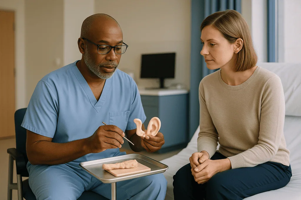
A horizontal incision is made along the lower edge of the rib cage. Beneath it lie the costal cartilages, the flexible segments that join the bony ribs to the breastbone. Cartilage from two to four adjacent ribs is typically removed, including the junction where neighboring cartilages fuse, which provides a broad flat plate for the main body of the ear. A longer, thinner strip from another rib becomes the helix. The surgeon takes care to leave the thin membrane lining the chest cavity intact; a tear there can let air into the chest, one reason the anesthesia team watches breathing closely during and after the harvest.
Then the carving begins. With scalpel blades and small gouges, the flat plate is thinned and sculpted into the antihelix and concha. The helix strip is bent around the rim and fixed in place with fine wire or permanent sutures. Cartilage does not bend willingly, so the surgeon scores it on one side or thins it to persuade it into a curve. A paper or film template traced from the opposite ear guides the size and position of every landmark.
A pocket is created under the skin where the ear belongs, sometimes after removing the remnant of the original ear. The framework slides in. Small suction drains are placed under the skin and left on gentle vacuum so the skin is pulled down into every groove of the cartilage. That suction is what gives the reconstructed ear its definition; without it, the skin would bridge across the hollows and the ear would look like a smooth pebble.
Ear reconstruction surgery stages: what each operation does
Nobody rebuilds an ear in one sitting when rib cartilage is used. The reason is biological, not scheduling. A newly buried framework needs months for blood vessels to grow into the skin around it before that skin can safely be cut and lifted again. Johns Hopkins describes rib cartilage reconstruction as a staged process with operations spaced months apart.
The two most widely taught approaches differ in how many stages they use. One classic method spreads the work across three or four operations: framework placement first, then relocation of the earlobe, then elevation of the ear from the head, and finally construction of the tragus and deepening of the bowl. A later refinement compresses the lobe transfer, tragus, and framework into a single first stage, leaving elevation as the second and often final operation.
- Stage one: rib harvest, carving, framework placement under the skin, and in the two-stage approach, repositioning of the earlobe and creation of the tragus. Extra cartilage is often banked under the chest or scalp skin for later use.
- Stage two: elevation. An incision is made around the back of the ear, and the framework is lifted off the head. The banked cartilage wedge is slid behind it to hold it out at the same angle as the other ear.
- Later stages, where used: fine adjustments to the lobe, tragus, or contours, and revision of scars.
The interval between stages is usually several months, long enough for swelling to settle and for the skin to heal firmly to the cartilage. Surgeons vary the plan for each person; someone with a partial ear remnant may need fewer steps, and someone with scarred skin from burns may need more. The plan should be laid out clearly before the first operation so the family knows roughly how many hospital visits lie ahead.
Skin coverage: how the framework is covered front and back
A perfect framework under poor skin is a failed reconstruction, which is why experienced teams talk about skin at least as much as cartilage. Three problems have to be solved: covering the front, covering the back once the ear is lifted, and dealing with scalp hair that may sit where the ear should be.
Covering the front is the job of the existing skin over the side of the head. It is thinned where necessary and drawn down by suction drains, as described above. If a child’s hairline is low, hair-bearing skin can end up on the upper ear. Some teams remove hair follicles later with laser treatment; others plan the framework position to avoid the hairline as far as possible.
The back is the harder problem. When the framework is lifted at the elevation stage, the raw surface behind it and the groove between ear and head need cover. Two tools are used. The first is a fascia flap, most often the temporoparietal fascia, a thin sheet of living connective tissue from just above and in front of the ear that is turned down over the back of the framework while still attached to its own blood supply. The second is a skin graft, a thin shaving of skin taken from elsewhere, commonly the scalp (where hair regrows and hides the donor site), the groin crease, or the opposite side of the chest incision, laid over the fascia and stitched in place.
Skin grafts survive only if they stay still and in contact with a bleeding surface, so bolster dressings are sewn over them for about a week. Grafts often look dark, purple, or patchy in the first days before settling; the team will tell you what shade to expect and what would concern them.
How painful is a rib cartilage graft?
Honest answer: the chest hurts more than the ear. Almost everyone who has had this operation says so. The ear itself is numb for a long time because small sensory nerves are divided when the pocket is made, and people often describe it as tight or strange rather than painful. The chest is different. Cartilage has been removed from between ribs that move with every breath, cough, and laugh, so the donor site aches with movement for days and is tender to pressure for weeks.
Anesthesia teams plan for this. During the operation, local anesthetic (a medicine that temporarily blocks nerve signals in one area) is commonly injected around the rib incision, and some centers place a thin catheter that delivers local anesthetic to the chest wall for a day or two afterward. Regular pain relief is given by mouth once eating resumes. Which medicines, at what schedule, is entirely a matter for the treating team and depends on age, weight, and other health conditions; nothing here should be read as a recommendation.
Deep breathing is encouraged from the first day even though it hurts, because shallow breathing lets the lower parts of the lungs collapse slightly and raises the chance of a chest infection. Children are often given a small toy or device to blow into for the same reason. Pressing a folded towel or pillow against the chest while coughing takes the edge off.
Most people describe the chest as manageable within one to two weeks and largely forgotten within a couple of months, though many notice a dull awareness at the harvest site for longer, especially when stretching or lying on that side. A permanent scar along the lower rib margin is expected, and a slight dip in the chest contour at the harvest site can persist, particularly if cartilage was taken before the chest finished growing.
What the following days and weeks usually look like
The first night is spent in hospital. A bulky head dressing protects the new ear, suction drains run from beneath the skin to small collection bottles, and the chest incision is covered. Nurses check the drains for volume and color; a sudden increase in bright red output is one of the signs they are watching for. Breathing is monitored because of the rib harvest.
Hospital stays for the first stage are typically a few days, until drains can be removed and pain is controlled with oral medicines. The head dressing usually stays on for around a week. During that time the person is asked to sleep propped up and to avoid lying on the operated side, because pressure on a fresh framework can cause the skin over it to break down.
The weeks that follow are quieter. The ear is swollen, numb, and often bruised; it looks nothing like the finished result and families should be warned of that. Stitches around the ear are usually removed or dissolve within one to two weeks. School or desk work is often resumed after a week or two, with contact sports, swimming, and anything that risks a knock to the ear held off for longer, commonly around six weeks, according to the team’s specific instructions. NHS guidance on plastic surgery generally notes that swelling and bruising settle over weeks and that scars keep maturing for many months.
The elevation stage, months later, brings a shorter hospital stay and a bolster dressing over the skin graft. The graft donor site on the scalp or groin is often described as the most uncomfortable part of that operation, similar to a graze. Between stages, life is normal, and the ear can be washed gently once the team confirms the incisions have sealed.
Risks and complications, in neutral terms
Every surgical program lists the same core risks for rib cartilage ear reconstruction, and it is worth understanding what each one actually means rather than skimming a consent form.
At the chest, the main concern is a breach of the lining of the chest cavity during harvest, which can allow air to leak around the lung (a pneumothorax). This is usually recognized during the operation and managed at the time; occasionally a chest drain or a short period of observation is needed. Longer term, removing cartilage from a growing chest can leave a visible contour change, and the scar can thicken.
At the ear, the most feared early complication is skin breakdown with exposure of the cartilage framework. Cartilage has no blood supply of its own and depends on the skin over it; if the skin dies or an infection develops, exposed cartilage can become infected and part of the framework may be lost. Small exposures are often covered with a local flap; larger ones may mean removing part of the framework and rebuilding later. Hematoma, a collection of blood under the skin, can lift the skin off the framework and blur the definition, which is why drains and dressings matter so much in the first days.
Over years, cartilage frameworks can partially resorb or warp, and the reconstructed ear does not grow in exactly the same way as a natural one. Scars can become raised, and skin grafts can look lighter or darker than surrounding skin. Sensation to the ear is reduced, sometimes permanently, so people need to be careful with cold and with heat from hair dryers.
Surgeons will not, and should not, quote a personal complication rate as a promise. What they can do is explain how often each problem is seen in published series, what they do to reduce it, and how they would manage it if it happened to you.
Prosthetic ear vs surgery, porous polyethylene, or waiting: how the options compare
Rib cartilage is not the only route, and a good consultation lays all the options out without steering. The table summarizes the main differences as described in mainstream clinical sources; none of it replaces an individual assessment.
| Option | What it is | Usual timing | Main trade-offs |
|---|---|---|---|
| Rib cartilage reconstruction | Framework carved from the patient’s own rib cartilage, covered with the patient’s skin | Usually school age or older, once ribs are large enough | Living tissue that heals and tolerates knocks; requires chest harvest and two or more operations; result depends heavily on carving and skin |
| Porous polyethylene framework | Pre-shaped manufactured scaffold covered with a fascia flap and skin graft | Can be done at a younger age because no rib growth is needed; often a single operation | No chest scar; framework cannot heal itself, so exposure, fracture, or infection may mean removal |
| Prosthetic ear | Silicone ear held by adhesive or by small implants anchored in bone | Any age; implants usually after early childhood | Excellent color match is possible without major surgery; must be removed daily, replaced every few years, and the remnant is usually removed to fit it |
| No reconstruction | Address hearing and leave the outer ear as it is | Any time; can be revisited later | No surgical risk; some people are content, others are not, and the option remains open |
The porous polyethylene and cartilage approaches have different philosophies. One favors a living framework that can be knocked and heal; the other favors avoiding the chest and finishing sooner. Neither has been shown in a definitive trial to be superior overall, and published comparisons describe trade-offs rather than a winner. Prosthetics suit some adults extremely well, particularly after cancer surgery or trauma where skin is compromised. A family choosing among these should expect the team to explain which options are realistic for that specific anatomy, and to accept whichever choice is made.
Rib cartilage for rhinoplasty and cartilage tympanoplasty: related searches, different operations
People researching ear reconstruction often stumble into two neighboring topics, because rib cartilage turns up in several parts of head and neck surgery. Neither is the subject of this article, but a brief orientation helps.
In rhinoplasty (surgery to reshape the nose), surgeons sometimes need cartilage to rebuild support after injury, previous surgery, or infection. Ear cartilage, taken from the bowl of a normal ear, is soft and curved, which makes it useful for small, flexible pieces. Rib cartilage is straight, strong, and plentiful, so it is favored when a long, rigid strut is needed. Its disadvantages are the chest incision and donor-site pain described above, and a tendency to warp over time as the carved piece dries and internal tensions settle; surgeons counter this by carving from the center of the rib and letting the piece sit before final shaping. Which graft is appropriate depends on what the nose needs, not on which is generally better, and that judgment belongs to the operating surgeon.
Cartilage tympanoplasty is an ear operation of a different kind. It repairs a hole in the eardrum using a thin slice of cartilage, usually from the tragus or the bowl of the same ear, not from the rib. People searching for its success rate should know that outcomes are usually reported as the proportion of eardrums that stay closed and the change in hearing, that figures vary with the size of the hole, the state of the middle ear, and the technique, and that no single percentage applies to everyone. An ear, nose, and throat specialist can explain what published series show for a situation like yours. It has nothing to do with rebuilding the outer ear, though a child with microtia and a closed canal may eventually encounter both kinds of surgeon.
What people often get wrong about rib cartilage ear reconstruction
It is cosmetic, so it is optional in the way a nose job is. Reconstructing a congenitally absent or traumatically lost body part is generally classed as reconstructive rather than cosmetic surgery. Insurers and health systems usually treat it that way, and the psychological weight of a visible difference in childhood is well recognized.
The new ear will hear. The outer ear funnels sound but is not where hearing happens. Reconstruction changes appearance and helps hold glasses or a hearing device; it does not open a closed canal or repair the middle ear. Hearing is addressed separately, sometimes with a bone-conduction device, sometimes with canal surgery, sometimes not at all if the other ear hears normally.
Earlier is always better. For rib cartilage, waiting is not delay for its own sake. Ribs that are too small give a framework that is too small, and the ear will not keep pace with the other side. The team weighs chest growth against the social pressures of school.
The ear will look finished after the first operation. It will look swollen, flat, and stuck to the head, because that is exactly what it is until elevation. Families who know this cope far better with the months in between.
Cartilage from the rib is dead tissue like a bone graft. Cartilage cells survive the transfer and continue to live inside the framework, drawing nutrients from surrounding tissue. That is why the framework tolerates minor injury and why it can, over decades, change shape slightly.
A photo gallery tells you what your result will be. Results depend on skin thickness, hairline, the size of the remnant, scarring, and healing. Published images show what is possible in other people, not what is promised to you.
Questions to ask your care team before agreeing to rib cartilage ear reconstruction
A first consultation is often overwhelming, and the useful questions surface in the car on the way home. Bring this list, and expect clear answers rather than reassurance.
- How many stages do you plan for my (or my child’s) ear, what happens at each, and how far apart are they usually spaced?
- Why do you recommend rib cartilage over a manufactured framework or a prosthesis for this anatomy, and what would make you change that recommendation?
- Which ribs will be used, what will the chest scar look like, and how do you protect the lining of the chest during harvest?
- How is the hairline handled if it sits low over the future ear position?
- How will pain in the chest be managed in hospital and at home, and who do we call if it is not controlled?
- Where will skin grafts be taken from at the elevation stage, and what will those donor sites look like afterward?
- What are the signs of skin breakdown or infection over the framework, and how quickly should we be seen if we notice them?
- How is the hearing plan coordinated with the reconstruction plan, and does the order of operations matter here?
- What activities are restricted after each stage, and for roughly how long?
- If part of the framework is lost, what would you do?
- How does a reconstructed ear change as a child grows, and what revisions are commonly needed later?
- Would you consider us reasonable to wait a year or two, or to decide not to operate at all?
Write the answers down. If you are told that a result is guaranteed, or that complications do not happen in this practice, treat that as a reason for a second opinion rather than a comfort. Good teams talk openly about what goes wrong because they have seen it and know how to handle it.
When to call your doctor after rib cartilage ear reconstruction
Most recoveries are uneventful, but a few problems need same-day attention because the skin over a fresh cartilage framework can deteriorate quickly. The team will give written instructions; these are the signs that mainstream surgical guidance, including NHS advice on recovery from plastic surgery, treats as reasons not to wait.
Call the surgical team or seek urgent care promptly if you notice any of the following:
- Sudden difficulty breathing, sharp chest pain that worsens with a breath, or a feeling that one side of the chest is not filling properly, particularly in the first days after rib harvest.
- Rapidly increasing swelling or tightness under the head dressing, or a drain that suddenly fills with bright red blood.
- Skin over the new ear turning dark purple, black, or white, or any area where cartilage appears to be showing through.
- Spreading redness, warmth, increasing pain, or pus at the ear, chest, or skin graft sites.
- Fever, chills, or feeling generally unwell in a way that is getting worse rather than better.
- A skin graft or bolster dressing that has lifted or slipped, or bleeding from a graft donor site that does not stop with gentle pressure.
- A direct blow to the reconstructed ear, even if it looks unchanged.
- Pain that is not controlled by the medicines prescribed, or a child who cannot be settled, will not drink, or is unusually drowsy.
Call emergency services rather than the clinic if breathing is difficult, chest pain is severe, or there is heavy bleeding. Between visits, a slow, steady improvement is what you are looking for. Anything moving in the wrong direction over a few hours deserves a phone call, and no reasonable team minds being asked. Every decision about treatment, revision, or timing rests with your treating clinicians, who know your anatomy and your history.
Frequently asked questions
How painful is rib cartilage graft pain compared with the ear itself?
The chest hurts considerably more than the ear. The new ear is largely numb because small nerves are divided when the skin pocket is made, while the rib harvest site moves with every breath and cough. Local anesthetic around the incision, sometimes delivered by a small catheter for a day or two, plus oral pain relief chosen by the treating team, usually keeps it manageable, and most people describe the chest as settling within one to two weeks.
At what age is rib cartilage ear reconstruction usually done?
Usually at school age, often around six years or older, according to Johns Hopkins, because the rib cartilage must be large enough to carve a framework the size of an adult-proportioned ear. Younger children are typically asked to wait. Adults can have the operation at any age if their health and the skin on that side of the head are suitable. The exact timing is a decision for the family and the surgical team together.
How many operations does ear reconstruction with rib cartilage take?
Two or more, spaced months apart. One widely used approach places the framework, repositions the earlobe, and shapes the tragus in the first operation, then elevates the ear from the head in a second. An older approach spreads the same steps across three or four shorter operations. Additional small revisions to scars or contours are common later. Your surgeon should outline the planned number of stages before the first one.
Does microtia ear reconstruction improve hearing?
No. The outer ear funnels sound but does not produce hearing, so building a new one does not change what a person hears. Hearing in microtia depends on whether the ear canal and middle ear formed normally, and is addressed separately, often with a bone-conduction hearing device or, in selected cases, canal surgery. Teams coordinate the two plans because operating on the canal and building the outer ear affect one another.
Which is better for rhinoplasty, ear cartilage or rib cartilage?
Neither is better in general; they suit different jobs. Ear cartilage is soft and curved, useful for small flexible grafts in the nose. Rib cartilage is straight, strong, and plentiful, favored when a long rigid strut is needed after injury or previous surgery, at the cost of a chest incision and a tendency to warp over time. The operating surgeon chooses based on what the nose needs.
What are the disadvantages of using rib cartilage?
The main drawbacks are a chest scar, donor-site pain for days to weeks, a small risk of injuring the lining of the chest cavity during harvest, and a possible dip in chest contour, especially when cartilage is taken from a growing child. Carved rib cartilage can also warp or partially resorb over years. The trade-off is a living framework that heals and tolerates knocks better than a manufactured one.
Prosthetic ear vs surgery: how do I decide?
It depends on anatomy, age, skin condition, and preference. A prosthetic ear offers a close color match without a chest harvest but must be removed and cleaned daily, is replaced every few years, and usually requires removal of the remnant. Reconstruction gives a permanent ear made of living tissue but involves multiple operations and scars. Both are legitimate choices; your team can explain which are realistic for your situation.
What is the success rate of cartilage tympanoplasty?
There is no single figure that applies to everyone. Cartilage tympanoplasty repairs a hole in the eardrum with a thin slice of ear cartilage, a different operation from outer ear reconstruction. Results are reported as the proportion of eardrums that remain closed and as hearing change, and they vary with hole size, middle ear health, and technique. An ear, nose, and throat specialist can discuss what published series show for your circumstances.
Will the reconstructed ear grow with a child?
Not in the same way a natural ear does. The framework is carved close to adult size, which is one reason surgeons wait until the ribs are large enough, so that the reconstructed ear does not end up noticeably smaller than the other ear later. Over years, cartilage can change shape slightly and skin can settle, so minor revisions in adolescence or adulthood are common and should be expected rather than seen as failure.
What can I do at home to protect the new ear?
Follow the team’s written instructions closely. In the early weeks that usually means sleeping propped up and off the operated side, keeping dressings dry and in place, avoiding contact sports, swimming, and hats that press on the ear until cleared, and protecting the numb ear from cold and from hair-dryer heat. Report any dark or pale patches of skin, spreading redness, or new swelling the same day rather than waiting for the next appointment.
References
- CDC – About Anotia and Microtia
- MedlinePlus Genetics – Craniofacial microsomia
- NHS – Plastic surgery
- MedlinePlus – Ear Disorders
This article is for general information only and is not a substitute for professional medical advice. Please consult a qualified doctor about your individual situation.

More from the Blog

How Bichectomy (Cheek Reduction) Is Performed: The Incision Inside the Mouth Explained
A bichectomy procedure, also called buccal fat removal, slims the lower cheeks by removing part of the buccal fat pad through a small incision…

Blood Thinners, Smoking and Rhytidectomy: What Surgeons Ask You to Stop Before a Facelift
Before a facelift, surgeons usually ask patients to stop anything that raises bleeding risk or starves healing skin of oxygen. That typically means blood-thinning…

Abdominal Etching: How Six-Pack Liposuction Works and Who It Actually Suits
Abdominal etching is a specialized liposuction technique that removes small amounts of fat in precise lines over the abdominal muscles to mimic a six-pack.…

Neck Lift Complications Explained: Hematoma, Nerve Weakness and Contour Irregularities
Neck lift complications are unwanted events caused by the surgery itself, as opposed to expected effects such as bruising and swelling. The three most…

What to Ask a Surgeon Before Prominent Ear Surgery: Technique, Symmetry and Timing
Before prominent ear surgery (otoplasty), ask the surgeon which technique they plan to use on your cartilage and why, how they measure and judge…

Questions to Ask Before a Rhytidectomy: Technique, Scars and What a Facelift Will Not Change
Before a rhytidectomy, ask which facelift technique the surgeon proposes and why, where the incisions will sit, how scars usually mature, what the operation…