

How Glaucoma Damages the Optic Nerve Silently and Why Treatment Aims at Eye Pressure
Key Takeaways
- Glaucoma is a disease of the optic nerve, and the retinal ganglion cell fibers it destroys do not regenerate, so lost vision does not return.
- Damage begins at the lamina cribrosa, the mesh where nerve fibers exit the eye, where elevated pressure interrupts nutrient transport and triggers cell death.
- Peripheral vision goes first and the brain fills in the gaps, which is why most people feel nothing until a substantial share of the nerve is already gone.
- Eye pressure is targeted because it is the only major risk factor that can be measured in minutes and changed with drops, laser or surgery.
- Normal tension glaucoma causes classic nerve damage at pressures within the usual 10 to 21 mmHg range, and lowering pressure further still slows it.
- The National Eye Institute estimates roughly half of the 3 million Americans with glaucoma are undiagnosed, making a dilated eye exam the single most protective step.
Glaucoma damages the optic nerve by slowly killing the retinal nerve fibers that carry images from the eye to the brain, usually without pain or early symptoms, so peripheral vision thins out unnoticed. Elevated or poorly tolerated eye pressure is the main modifiable cause, which is why treatment, whether drops, laser or surgery, concentrates on lowering pressure to slow further nerve loss.
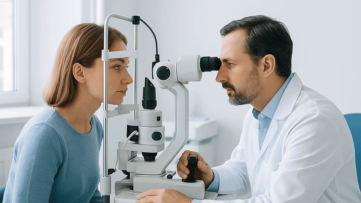
The routine eye test was supposed to be about new reading glasses. Instead the optometrist leaned back from the slit lamp, paused a second longer than usual, and said the words that sent a 58-year-old bookkeeper home with a referral letter and a head full of questions: the optic nerve looks a little hollowed out, and the pressure is on the high side. She could read the chart perfectly. Nothing hurt. How could something be wrong?
That gap between feeling fine and having a problem is the whole story of this disease, and it is why understanding how glaucoma damages the optic nerve matters more than almost any other fact in eye care. The nerve does not complain while it is being injured. It simply loses fibers, a few at a time, from the edges of your visual world inward.
This explainer walks through what is actually happening inside the eye, why eye pressure became the target of every proven treatment, what normal tension glaucoma means, and which questions deserve an honest answer from your care team.
What is glaucoma a disease of?
Glaucoma is a disease of the optic nerve, not of the lens, the cornea or the front of the eye where most people picture eye trouble. The optic nerve is the cable of more than a million nerve fibers that bundles together at the back of the eye and carries visual signals to the brain, according to the Cleveland Clinic. Those fibers are the long tails of retinal ganglion cells, the final relay neurons in the retina. When glaucoma destroys them, they do not grow back.
That one sentence explains why the condition is taken so seriously. The retina can be lasered, the lens can be replaced, the cornea can be transplanted. Nerve tissue in the central nervous system, which is what the optic nerve is, has no repair mechanism that medicine can currently switch on. Vision lost to glaucoma stays lost, which the NHS and MedlinePlus both state plainly.
Glaucoma is also not a single disease. It is a family of conditions that share one feature: a characteristic pattern of optic nerve damage, usually accompanied by matching blind spots in the visual field. Eye pressure is the most common driver and the only one that current treatment can reliably change, but it is a risk factor, not the definition. Some people have high pressure and no nerve damage for decades; others develop classic glaucoma with pressure that never leaves the normal range.
Scale matters too. The National Eye Institute (NIH) estimates that about 3 million people in the United States have glaucoma, and that roughly half of them do not know it. The Mayo Clinic lists it among the leading causes of blindness in people over 60. Most of that preventable loss happens in eyes that were never examined, not in eyes that were examined and treated.
How glaucoma damages the optic nerve, step by step
Picture the optic nerve head, the spot where all those fibers exit the eyeball, as a drain at the bottom of a sink. The fibers bend sharply through a sieve-like plate of connective tissue called the lamina cribrosa, a mesh of tiny holes that the nerve fibers must pass through on their way out. This is the weak point. Inside the eye, fluid pressure pushes outward on that mesh; behind it, pressure is much lower. The mesh bows and the holes distort.

Here is the mechanism as mainstream eye science currently describes it. Retinal ganglion cells depend on a constant two-way traffic of nutrients and signaling molecules along their fibers, a process called axonal transport. When the lamina distorts under pressure, that transport is pinched at the bottleneck. Starved of the survival signals they normally receive from the brain end of the fiber, the cells trigger their own programmed death. Reduced blood supply to the nerve head, which also varies with pressure, is thought to add a second insult.
Each lost cell leaves a gap. As thousands of gaps accumulate, the rim of nerve tissue around the central cup of the optic disc thins, and the cup looks larger. That “cupping” is what the optometrist saw in the opening scene, and it is one of the signs an ophthalmologist, a medical doctor specializing in eye disease, looks for when deciding whether glaucoma is present.
The process is relentless but slow in most forms of the disease. In chronic open-angle glaucoma the fibers are lost over years, which is why the NHS describes it as developing “slowly over many years.” In acute angle-closure, pressure can spike dramatically within hours and damage follows far faster. Same nerve, same bottleneck, different clock.
How can damage to the optic nerve affect vision, and why you don't notice?
Nerve fibers from the retina are arranged in orderly bundles, and the ones that pass through the top and bottom edges of the optic nerve head seem to be the most vulnerable. Those bundles happen to serve the outer parts of your visual field, especially the arcs above and below your straight-ahead gaze. So the first blind spots in glaucoma usually appear in the periphery, as the Mayo Clinic and NHS both describe, long before anything touches reading or recognizing faces.
Two things hide this from you. The first is that your two eyes overlap. A patch missing from the left eye’s field is often covered by the right eye, so with both eyes open the scene looks complete. The second is that the brain fills in gaps. It does this all the time with the normal blind spot where the optic nerve enters the eye, and it does the same with glaucoma defects. You do not see a black hole; you see a seamless picture that is simply missing information.
What people eventually notice, often late, is a kind of patchiness: missing a step on a staircase, bumping a shoulder in a doorway, not spotting a car approaching from the side. Difficulty adapting to dim light and glare can show up too. By the time central vision blurs, the MedlinePlus patient guidance notes that a large share of the nerve may already be gone.
This is the honest answer to why glaucoma is called the silent thief. It is not that the damage is invisible to instruments; a visual field test can map it clearly. It is that the damage is invisible to the person it is happening to, which is exactly why detection depends on examination rather than on symptoms.
Why does eye pressure matter so much in glaucoma?
The eye is a pressurized ball. A clear fluid called aqueous humor, not to be confused with tears, is produced continuously by a ring of tissue behind the iris, flows forward through the pupil, and drains out through a spongy mesh in the angle where the iris meets the cornea. Production and drainage are balanced, and the resulting intraocular pressure keeps the eye’s shape. The Cleveland Clinic gives roughly 10 to 21 millimeters of mercury (mmHg) as the typical range.
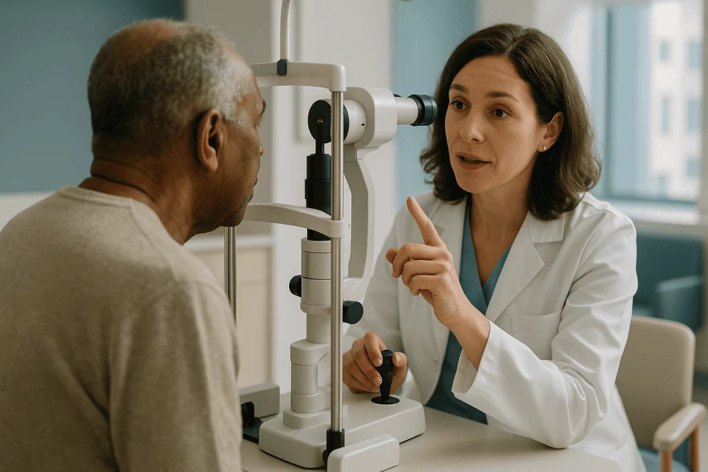
When drainage slows, pressure rises. Nothing about this hurts in chronic glaucoma, because the pressure rises gently and the eye adapts, but the lamina cribrosa and the nerve fibers passing through it feel every extra millimeter. Higher pressure means more distortion at the bottleneck, more interrupted transport, more cells lost per year.
Pressure earned its place at the center of treatment for a practical reason: it is the one factor that can be measured in a two-minute test and changed with medicine, laser or surgery. Age, family history and ancestry are risk factors no one can alter. Blood flow to the nerve is hard to measure and harder to influence. Pressure is both measurable and movable.
The evidence that moving it helps is the backbone of every national guideline. The Early Manifest Glaucoma Trial, a randomized study published in Archives of Ophthalmology and indexed on PubMed, compared early pressure-lowering treatment against observation in people with newly detected glaucoma and found that treated eyes progressed less often, with each unit of additional pressure reduction associated with a lower risk of worsening. Later trials have reinforced the same direction of effect. Treatment does not restore the nerve; it slows the rate at which the nerve is lost, and the earlier it starts, the more there is left to protect.
What is normal tension glaucoma?
Normal tension glaucoma, sometimes called low-tension glaucoma, is glaucoma in which the optic nerve shows the typical cupping and the visual field shows the typical defects, yet the measured eye pressure has never risen above the standard range. The National Eye Institute (NIH) and the Mayo Clinic both recognize it as a genuine, fairly common form of the disease, not a measurement error.
It forces an uncomfortable but important point: the optic nerve does not read a textbook. A pressure of 18 mmHg may be perfectly safe for one nerve and slowly destructive for another. Possible reasons under study include a more fragile lamina cribrosa, poor or unstable blood flow to the nerve head, low blood pressure overnight, and conditions such as migraine or cold hands and feet (Raynaud phenomenon) that hint at blood-vessel reactivity. Thin corneas can also make a tonometer under-read the true pressure, so some “normal” readings were never normal.
Diagnosis involves ruling out other causes of a damaged-looking nerve, which is why the ophthalmologist may ask about past episodes of very low blood pressure, severe blood loss or sleep apnea, and may order imaging of the brain and optic nerves if the pattern of field loss looks unusual.
Treatment still targets pressure. That surprises people, but the logic is sound: even a nerve that is damaged at normal pressure appears to do better when the pressure is pushed lower still, and lowering pressure is the only intervention with trial evidence behind it. Your care team sets an individual target pressure for each eye, based on how much damage exists and how fast it seems to be progressing, rather than aiming at a single population number.
Open-angle, angle-closure and other types compared
The word “angle” refers to the drainage angle between the iris and the cornea. Whether that angle is open or blocked is the main dividing line between glaucoma types, and it changes everything about how fast the disease moves and how it is treated.
| Type | What is happening at the drain | Typical pace | Usual first approach |
|---|---|---|---|
| Primary open-angle glaucoma | Angle is open but the drainage mesh works poorly | Slow, over years; no early symptoms | Pressure-lowering drops or laser to the drainage mesh |
| Angle-closure glaucoma | Iris physically blocks the angle; can be sudden or gradual | Acute form: hours; chronic form: years | Urgent pressure control, laser opening in the iris |
| Normal tension glaucoma | Angle open, pressure within range, nerve still vulnerable | Slow | Lower pressure further than the standard range |
| Secondary glaucoma | Drain impaired by another condition: inflammation, injury, diabetes, long-term steroid use | Varies with the cause | Treat the underlying cause plus pressure control |
| Congenital or childhood glaucoma | Drainage system did not form properly before birth | Present from infancy | Usually surgical, managed by a specialist team |
Primary open-angle glaucoma is by far the most common form in the United States and Europe, according to MedlinePlus. Angle-closure is more common in people of East Asian ancestry, in those who are farsighted, and in women, as the Mayo Clinic notes, because those eyes tend to have a shallower front chamber.
The acute form deserves its own flag. When the angle slams shut, pressure can climb to several times normal within hours. The eye becomes painful and red, vision blurs, halos appear around lights, and nausea or vomiting is common. The NHS treats this as a medical emergency because nerve damage can occur in a day rather than a decade.
Who is most at risk, and who is usually asked to wait?
Not everyone with high pressure has glaucoma, and not everyone with glaucoma needs treatment on day one. Sorting people into those groups is most of what a glaucoma clinic does.
Risk rises with age, particularly after 60, and earlier in some groups. The National Eye Institute (NIH) names Black or African American adults over 40, Hispanic or Latino adults over 60, and anyone with a parent or sibling with glaucoma as being at higher risk. Other factors listed by the Mayo Clinic include diabetes, high blood pressure, significant nearsightedness or farsightedness, thin central corneas, previous eye injury or surgery, and long-term use of corticosteroid medicines, including some eye drops and inhalers.
Three situations usually lead to a decision to treat now: established optic nerve damage with matching field loss, pressure high enough that damage is judged likely even without current loss, or evidence of progression between visits. Angle-closure that is detected on examination is also treated promptly even when symptom-free, because the goal is to prevent the acute attack.
Who is usually asked to wait? People with ocular hypertension, meaning pressure above the standard range but a healthy nerve and normal fields, are often monitored rather than treated, especially if the pressure is only modestly raised and the cornea is thick. “Glaucoma suspects”, a formal term for people whose nerve looks slightly unusual without clear damage, are commonly followed with repeat imaging and field tests over months or years. The idea is to treat the nerve, not the number, and to avoid committing someone to decades of drops for a condition they may never develop.
Whether you fall into the treat-now or watch-and-see group is a judgment for your ophthalmologist, weighing your risk profile, your test results and your own preferences.
How is glaucoma diagnosed, and what do the tests actually measure?
No single test diagnoses glaucoma. The ophthalmologist assembles a picture from several, and understanding what each one measures makes the appointment less mysterious.
Tonometry measures eye pressure, either with a puff of air or with a small probe that briefly touches the numbed cornea. A pachymetry reading measures corneal thickness, because a thick cornea inflates the pressure reading and a thin one deflates it. Gonioscopy uses a mirrored contact lens to look directly into the drainage angle and tell open-angle from angle-closure.
The nerve itself is examined two ways. In ophthalmoscopy, the doctor looks at the optic disc through a dilated pupil and judges the size of the cup relative to the disc and whether the rim is notched or pale. Optical coherence tomography, usually shortened to OCT, is a painless laser scan that measures the thickness of the nerve fiber layer in microns and compares it with a reference database. It can detect thinning before any visual field change appears.
Perimetry, the visual field test, maps what you can actually see. You stare at a central light and press a button when dim lights flash in your side vision. It is the only test that measures function rather than structure, and it is tiring; the NHS notes that results can vary from one visit to the next, which is why clinics often repeat it before trusting a change.
Diagnosis can take more than one visit. Many people are told they are a “suspect” and asked to return in a few months so the team can see whether anything is changing. That is not indecision; it is how a slow disease is distinguished from an unusual but stable nerve.
How treatment lowers eye pressure: drops, laser and surgery
Every proven glaucoma treatment works on the same plumbing: it either turns down the tap, meaning aqueous production, or opens the drain. The choice between them depends on glaucoma type, how far the nerve has been damaged, other medical conditions and what the person can realistically manage.
Eye drops are usually the first line in open-angle glaucoma. Prostaglandin analogues increase outflow through an alternative drainage route in the eye wall. Beta blockers, carbonic anhydrase inhibitors and alpha agonists reduce fluid production. Rho kinase inhibitors improve flow through the main drainage mesh. Each class has its own side-effect profile, from darker lashes and iris color with prostaglandins to slowed heart rate or breathing effects with beta blockers in susceptible people, which is why the prescribing clinician asks about asthma, heart conditions and other medicines. Decisions about which drop, how often, and whether to combine them belong entirely to that clinician.
Laser trabeculoplasty applies gentle laser energy to the drainage mesh to improve its function. The NHS describes it as a common option for open-angle glaucoma, performed in clinic with numbing drops. For angle-closure, a laser iridotomy makes a tiny opening in the iris so fluid can bypass the block.
Surgery is usually reserved for eyes where drops and laser have not achieved the target. Trabeculectomy creates a controlled new drainage channel under the conjunctiva. Drainage implants place a small tube that shunts fluid to a reservoir. A newer group of procedures grouped as minimally invasive glaucoma surgery uses tiny stents or shunts, often combined with cataract surgery, and aims for a smaller pressure drop with fewer complications.
None of these repairs the optic nerve. They all buy time for the fibers that remain.
What the following weeks and months usually look like
Starting glaucoma treatment feels anticlimactic, and that is normal. Nothing improves, because nothing can; the measure of success is that nothing gets worse, which takes patience to appreciate.
After drops are started, the clinic typically wants to see you within a few weeks to check that the pressure has fallen toward the target and that the eye tolerates the medicine. Mild stinging, redness or a gritty feeling in the first days is common with many drops and often settles; persistent redness, swelling, or a change in breathing or heart rate are reasons to call rather than wait. Technique matters more than people expect: one drop, lids closed for a minute, gentle pressure on the inner corner to keep the medicine in the eye rather than down the tear duct.
After laser trabeculoplasty, the eye may be slightly sore and light-sensitive for a day or two, and the pressure-lowering effect is assessed over the following weeks because it builds gradually. After trabeculectomy or an implant, expect more frequent visits in the first month, because the surgeon is adjusting the new drainage pathway, sometimes with small procedures in clinic, and watching for the pressure dropping too low rather than too high.
Once the pressure is stable, follow-up settles into a rhythm. The National Eye Institute (NIH) recommends a dilated exam at least every one to two years for people at higher risk, and treated glaucoma usually means visits at the shorter end of that range or more often, with a visual field test and OCT repeated periodically to look for drift. Progression, if it happens, prompts a lower target and a change in approach. Stability earns longer gaps between visits. Either way, the appointments do not end; glaucoma is managed for life, not for a course of treatment.
How to prevent glaucoma: what the evidence actually supports
The honest answer is that glaucoma cannot be prevented in the way an infection can be prevented by a vaccine. Age, genetics and eye anatomy set most of the risk. What can be prevented, in the great majority of cases, is the vision loss, and the single most effective tool for that is a dilated eye examination at the recommended interval, which the CDC and the National Eye Institute (NIH) both emphasize because so much glaucoma is undiagnosed.
Knowing your family history comes next. A first-degree relative with glaucoma raises your risk several-fold, and the Mayo Clinic suggests telling your eye care professional so that screening starts earlier and happens more often.
Protecting the eye from injury has real value, because blunt trauma can damage the drainage angle and lead to secondary glaucoma years later. Safety eyewear for racket sports, power tools and home repair is not glamorous advice, but it is evidence-based.
Steroid awareness matters too. Long-term corticosteroid use, whether as eye drops, inhalers, skin creams near the eye or tablets, raises pressure in a proportion of people, and anyone on them for months should mention it at eye checks.
Regular moderate aerobic exercise is associated with modestly lower eye pressure, which the Mayo Clinic mentions as a reasonable habit, though no trial has shown it prevents glaucoma. Caffeine in large amounts can briefly raise pressure; drinking a liter of fluid quickly can do the same; sleeping with the head slightly elevated lowers overnight pressure a little. These are small levers. Supplements marketed for “eye pressure” or “optic nerve support” have no evidence from controlled trials that they prevent or slow glaucoma, and nothing on a shelf replaces the examination.
What people often get wrong about glaucoma
“My vision is fine, so my eyes are fine.” This is the most dangerous misconception, and it comes straight from how glaucoma damages the optic nerve: from the periphery inward, with the brain patching the gaps. Normal reading vision says nothing about the health of the nerve.
“High pressure means I have glaucoma.” Ocular hypertension is a risk factor, not the disease. Many people with raised pressure never develop nerve damage, and clinicians often monitor rather than treat them.
“Normal pressure means I cannot have glaucoma.” Normal tension glaucoma exists and is common. A pressure reading alone, especially a single air-puff test, cannot rule the disease in or out.
“Treatment will improve my sight.” It will not. Drops, laser and surgery slow loss; the nerve fibers already gone are gone. Expecting improvement leads people to conclude the treatment is not working and to stop it, which is exactly the wrong lesson.
“Glaucoma is painful.” Acute angle-closure is. Chronic open-angle glaucoma, the form most people have, is painless from start to finish.
“It only affects older people.” Risk rises with age, but glaucoma occurs in children, young adults with injury or inflammation, and middle-aged people with a strong family history.
“Once the pressure is down, I can stop the drops.” The pressure is down because of the drops. Stopping them without discussing it with the prescribing clinician usually returns the eye to where it started. If side effects or cost of daily use are the problem, that is a conversation worth having, because alternatives such as laser exist for some people.
“Cannabis treats glaucoma.” Smoked cannabis lowers eye pressure for a few hours, as the National Eye Institute (NIH) acknowledges, but the effect is too brief and the side effects too substantial for it to be a practical treatment, and no major guideline recommends it.
Questions to ask your care team
A glaucoma visit is often short and heavy with numbers. Arriving with specific questions turns it into a conversation about your own eyes rather than a lecture about the disease. These are the ones that experienced patients tend to find most useful.
- Which type of glaucoma do I have, and is the drainage angle open or narrow?
- How much optic nerve damage is there in each eye, and is it the same in both?
- What target pressure have you set for me, and why that number?
- Are you treating me now, or monitoring me as a suspect, and what would change your mind?
- What side effects of this treatment should make me call you rather than wait for the next visit?
- How will I know whether the disease is progressing, and how often will you repeat the visual field and OCT?
- Is laser an option for me instead of, or alongside, drops?
- Which of my other medicines or health conditions could affect my eye pressure or my treatment?
- Should my parents, siblings or children be examined, and from what age?
- Is it safe for me to drive, and is there anything about my visual field I should know for work or sport?
- What would an emergency look like for my type of glaucoma, and where should I go?
Ask for your pressure readings and the result of the field test in plain terms, and consider keeping a note of them. People who understand their own numbers tend to use their treatment more consistently, and consistency is the quiet variable that determines how much vision a slow disease actually takes.
Every one of these answers will be specific to your eyes. The treating team holds the full picture, and the final decisions sit with them and with you.
When to call your doctor
Most of glaucoma care is unhurried. A few situations are not, and knowing the difference protects the nerve.
Seek emergency care the same day, through an emergency department or an urgent eye clinic, if you develop sudden severe pain in one eye, a red eye with blurred vision, rainbow-colored halos around lights, headache on that side, or nausea and vomiting with any of these. The NHS and Mayo Clinic describe this combination as the picture of acute angle-closure glaucoma, in which pressure can damage the nerve within hours. Do not wait to see whether it passes.
Contact your eye care team promptly, within a day or two, if you notice a sudden new blind spot, a curtain or shadow across part of your vision, a sudden drop in sight in one eye, or a marked change in how you see after a change of treatment. Some of these point to problems other than glaucoma, such as a retinal detachment, which also need fast attention.
Call during office hours if drops cause persistent redness, swelling, itching or stinging that does not settle after the first week; if you notice a change in breathing, an unusually slow pulse, dizziness or fatigue after starting a new drop; if you have missed doses for more than a few days because of supply, side effects or forgetfulness; if you are starting a corticosteroid medicine in any form; or if you are pregnant, planning pregnancy or breastfeeding, because some glaucoma medicines need review.
After glaucoma surgery, report increasing pain, worsening redness, discharge, or a sudden decrease in vision immediately; infection in a new drainage pathway is uncommon but serious. Whatever the symptom, the people who know your eyes are the ones to judge it. Do not adjust, stop or add any treatment on your own.
Frequently asked questions
How can damage to the optic nerve affect vision in glaucoma?
Glaucoma kills nerve fibers in a pattern that first removes patches of side vision, usually the arcs above and below your central gaze. Because the other eye and the brain fill in the missing areas, early loss goes unnoticed. As fibers continue to die, the blind areas enlarge and merge, producing tunnel vision, and only in advanced disease does central sight for reading and faces fade.
What is glaucoma a disease of, exactly?
It is a disease of the optic nerve, the bundle of more than a million fibers connecting the retina to the brain. Raised eye pressure is the most common cause, but the diagnosis rests on characteristic damage to the nerve and matching visual field defects, not on the pressure reading alone. The lens, cornea and surface of the eye are not the problem.
What is normal tension glaucoma?
Normal tension glaucoma is optic nerve damage with typical glaucoma features in an eye whose measured pressure has never exceeded the standard range. Proposed explanations include a fragile nerve head, poor blood flow, overnight low blood pressure and thin corneas that mask true pressure. Treatment still lowers pressure, because trial evidence shows these nerves also do better at lower pressures.
How to prevent glaucoma if it runs in my family?
You cannot prevent the disease itself, but you can usually prevent the vision loss. Tell your eye care professional about your family history so screening starts earlier and repeats more often. The National Eye Institute (NIH) advises a dilated exam every one to two years for higher-risk groups. Protect your eyes from injury and mention any long-term steroid use.
What are glaucoma optic nerve damage symptoms I might notice?
In chronic open-angle glaucoma, usually none for years, which is the central problem. Later, people describe missing steps, bumping into things on one side, trouble in dim light or increased glare. Acute angle-closure is different: sudden eye pain, redness, blurred vision, halos around lights and nausea, which needs emergency care the same day.
Does lowering eye pressure bring back lost vision?
No. Nerve fibers destroyed by glaucoma do not regrow, and no current treatment restores them. Lowering pressure slows or halts further loss, protecting the vision you still have. That is why clinicians judge success by stable visual field and OCT results over time rather than by any improvement, and why staying on treatment matters even when nothing seems to change.
Can you have high eye pressure without glaucoma?
Yes. This is called ocular hypertension: pressure above the usual range with a healthy optic nerve and normal visual fields. It raises the risk of developing glaucoma but is not the disease. Many people are monitored rather than treated, especially if the pressure is only mildly raised and the cornea is thick, with the decision resting on the ophthalmologist’s assessment.
Why do glaucoma eye drops have to be used every day?
Because they work only while they are in the eye. Drops either reduce fluid production or improve drainage, and each effect wears off within hours to a day depending on the class. Missing doses lets pressure drift back up, and the nerve is exposed during every gap. If daily use is difficult, the prescribing clinician can discuss alternatives such as laser treatment.
Is glaucoma surgery a last resort?
Not always. Traditional surgery such as trabeculectomy is usually reserved for eyes where drops and laser have not reached the target pressure or where damage is advancing quickly. Minimally invasive procedures, often combined with cataract surgery, may be considered earlier for mild to moderate disease. The sequence depends on your type of glaucoma, your nerve’s condition and your surgeon’s judgment.
How often should I have my eyes checked for glaucoma?
The National Eye Institute (NIH) recommends a dilated eye examination at least every one to two years for people at higher risk, including adults over 60, Black or African American adults over 40, Hispanic or Latino adults over 60, and anyone with a family history or diabetes. If you already have glaucoma, your care team sets the interval, often more frequent.
References
- Glaucoma | National Eye Institute (NIH)
- Glaucoma | NHS
- Glaucoma | MedlinePlus
- Glaucoma: Symptoms, Causes, Types and Treatment | Cleveland Clinic
This article is for general information only and is not a substitute for professional medical advice. Please consult a qualified doctor about your individual situation.

More from the Blog

Caring for Someone After Eye Surgery: Drop Schedules, Shields and the First Night Back
Caring for someone after eye surgery mostly means keeping to the written drop schedule without touching the eye, making sure the protective shield goes…

Macular Hole, Macular Pucker and Macular Edema: How the Main Macular Diseases Differ
The main types of macular disease damage the small patch of central retina in different ways. A macular hole is a small gap in…

Preparing for Cataract Surgery: Lens Measurements, Medication Review and Drop Schedules
Cataract surgery preparation usually involves a pre-assessment visit where the eye is measured to calculate the power of the replacement lens, a review of…

Does DCR Surgery Stop the Watering? Realistic Results and Why a Few Need a Second Look
For most adults with a confirmed blockage of the nasolacrimal duct, DCR surgery relieves or greatly reduces watering, because it creates a new drainage…
Ahead of Eye Angiography: Allergies, Pregnancy, Kidney History and Bringing a Driver
Eye angiography preparation is mostly about information, not fasting. Before the test, tell your eye team about any allergy or past dye reaction, whether…

Getting Ready for Cataract Treatment: Medication Review, Drops and Arranging a Companion
Preparing for cataract treatment usually means attending a pre-assessment visit, giving the care team a complete list of your medicines (including blood thinners, prostate…