

How Gynecologic Laparoscopy Is Performed: Small Incisions, Gas and the Camera Explained
Key Takeaways
- The first incision in a gynecologic laparoscopy is placed at the navel because the abdominal wall is thinnest and most predictable there, and it is typically only 1 to 1.5 cm long.
- Carbon dioxide is chosen as the inflating gas because it does not burn near electrical instruments and dissolves readily into blood so the lungs can clear it.
- Shoulder-tip pain after keyhole surgery comes from leftover gas irritating the diaphragm, which shares a nerve pathway with the shoulder, and it usually settles within a day or two.
- The NHS estimates minor complications in about 1 to 2 of every 100 laparoscopies and serious complications in roughly 1 in 1,000.
- Recovery is set by the work done inside, not the size of the scars: about 5 days after a diagnostic look, 1 to 2 weeks after minor treatment, and up to 12 weeks after major surgery.
- A planned laparoscopy can be converted to an open operation for safety, which is a contingency you consent to in advance rather than a sign that something went wrong.
A gynecologic laparoscopy procedure is keyhole surgery of the pelvis performed under general anesthesia. The surgeon makes a small incision near the navel, fills the abdomen with carbon dioxide gas to create working room, and inserts a thin lighted camera. One to three further tiny cuts admit instruments for diagnosis or treatment. Most people go home the same day, with recovery ranging from days to several weeks depending on what was done.
The consent form is on the kitchen table, and the word that keeps snagging is “gas.” Small cuts she can picture. A camera, fine. But being inflated like a beach ball while asleep sounds like something from a cartoon, not an operating room. Her surgeon explained it in eight minutes; she has been turning it over for three days.
That reaction is common, and it is reasonable. A gynecologic laparoscopy procedure asks you to trust a method you cannot watch and will not remember. The best antidote is to understand exactly what happens between the anesthetic taking hold and the moment you wake in recovery, feeling oddly full and wondering why your shoulder aches.
What follows is that walk-through: the incisions, the gas, the camera, the hours and weeks afterward, and the questions worth asking before you sign. None of it replaces the conversation with your own team, but it should make that conversation easier to have.
What is a gynecologic laparoscopy procedure?
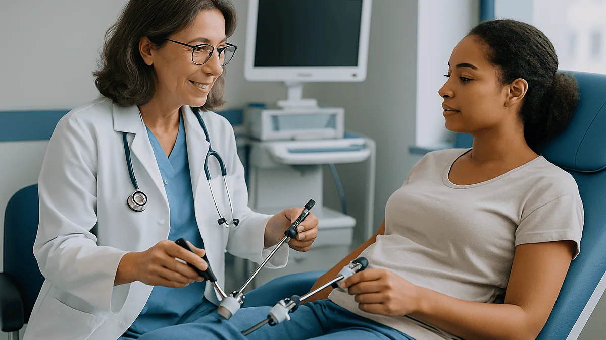
Laparoscopy is surgery performed through a few small openings in the abdominal wall rather than one long cut. The word comes from Greek roots meaning “to look into the flank,” which is still a fair description. A laparoscope, a slim tube containing a lens and a light source, goes in through an opening near the navel and sends a magnified image to a screen. The surgeon operates while watching that screen, not by looking directly into the body.
In gynecology the targets are the organs of the pelvis: the uterus, the two fallopian tubes, the ovaries and the lining tissue around them. Because these organs sit low and are surrounded by bowel, seeing them clearly from the outside is hard. Ultrasound shows shapes and fluid but not surface detail. Laparoscopy shows the actual tissue in color, at magnification, which is why it remains the standard way to confirm certain diagnoses, such as endometriosis, a condition in which tissue similar to the womb lining grows outside the womb (NHS).
Two broad versions exist. A diagnostic laparoscopy is mainly a look: the surgeon inspects, may take a small tissue sample, and stops. An operative laparoscopy is a look plus a repair, removing a cyst, freeing scar tissue, treating endometriosis patches or removing a fallopian tube. The incisions, gas and camera are the same in both; what differs is how long it takes and how much your body has to heal afterward.
One point matters more than any other for what follows: the small cuts on your skin are not the whole story. Inside, the amount of surgery can be modest or considerable, and your recovery tracks the inside work, not the size of the bandages.
What happens during a laparoscopy in gynecology, and why it's chosen
Gynecologists reach for laparoscopy in three main situations. The first is unexplained pelvic pain or painful periods that have not responded to simpler measures and where imaging has not given an answer. The second is a known problem that needs treatment: an ovarian cyst, fibroids in certain positions, a suspected ectopic pregnancy (a pregnancy growing outside the womb, usually in a tube), or endometriosis identified on scans. The third is fertility investigation, where the surgeon checks whether the tubes are open and the pelvis is free of adhesions, the bands of scar tissue that can tether organs together (MedlinePlus).
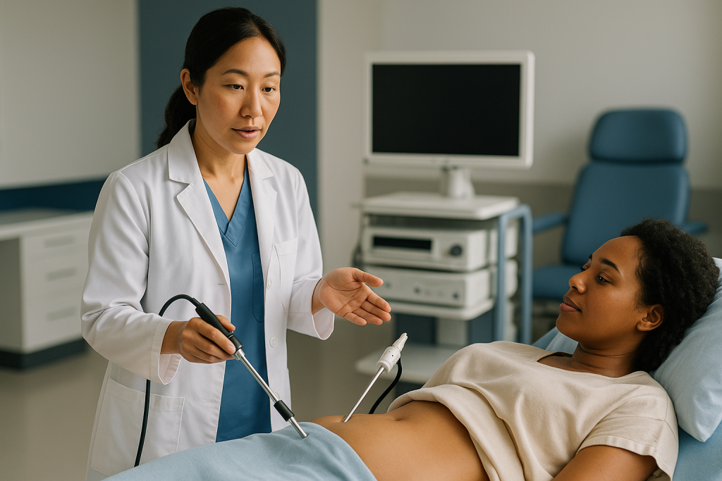
Why not simply open the abdomen? Because the trade-off usually favors the smaller approach. A traditional open operation, called a laparotomy, requires a cut long enough for the surgeon’s hands to enter. That means more tissue divided, more pain, longer hospital stays and a bigger scar. Laparoscopy uses instruments as thin as a pencil, so the abdominal wall is disturbed far less. The NHS notes that recovery tends to be quicker and hospital stays shorter compared with open surgery for the same problem, though not every situation suits the keyhole route (NHS).
The other advantage is the view. A camera sitting inside the pelvis, a few centimeters from the ovary, sees detail a surgeon’s naked eye at arm’s length cannot. Small, flat endometriosis lesions in particular are easier to spot on a magnified screen. Diagnosis and treatment can happen in one anesthetic: if the surgeon finds a lesion during the look, it can often be treated then and there, provided you consented to that possibility beforehand. That is why the pre-operative conversation about “what will you do if you find X” is so valuable.
Who is gynecologic laparoscopy usually for, and who is asked to wait?
The typical candidate is someone with a pelvic problem that imaging, examination and time have not resolved, and who is fit enough for a general anesthetic. That covers a wide age range, from teenagers with severe period pain to people well past menopause with an ovarian cyst that needs assessing.
Fitness for anesthesia is the main gate. Laparoscopy places specific demands on the heart and lungs that open surgery does not: the gas raises pressure inside the abdomen, which pushes on the diaphragm and makes the lungs work harder, and the head-down tilt used during pelvic surgery shifts blood toward the chest. For most people this is trivial. For someone with severe lung disease, advanced heart failure or very high body weight, the anesthetist may recommend extra assessment or a different plan (Cleveland Clinic).
Several groups are commonly asked to wait or reconsider:
- People with an active infection, chest cold or fever on the day, because anesthesia risks rise and healing suffers.
- Those whose blood-thinning medicines have not yet been reviewed by the prescribing clinician, since bleeding control matters when working through small ports.
- Anyone whose symptoms have not yet had a reasonable trial of non-surgical management, where guidelines suggest trying that first.
- People with extensive previous abdominal surgery, where dense scar tissue may make the first entry unsafe; the surgeon may plan a different entry technique or an open approach.
Pregnancy is not an automatic bar. Laparoscopy is used for ectopic pregnancy and occasionally for ovarian problems during an ongoing pregnancy, but it requires specific planning. Whether to proceed, wait, or choose another route is a judgment your treating team makes with you, weighing your particular anatomy, history and goals. No article can substitute for that individual assessment.
Getting ready: what happens in the days before surgery
Preparation is mostly about making the anesthetic safe and the entry predictable. You will have a pre-assessment visit or call. Expect questions about medicines, allergies, previous operations, smoking, and whether you have had problems with anesthesia before. Blood tests, and sometimes an ECG, are ordered based on age and health, not routinely for everyone.

Fasting is the instruction people most often get wrong. A general anesthetic relaxes the muscle that keeps stomach contents down, so an empty stomach protects your lungs. Your team will give specific timings for food and clear fluids; follow theirs, not a friend’s. The NHS advises that you will be told when to stop eating and drinking, typically several hours before (NHS).
Medicines deserve a direct conversation. Some, especially those that thin the blood or affect blood sugar, may need adjusting around surgery, but only the clinician who prescribes them should make that call. Do not stop anything on your own initiative because you read that surgeons prefer it. Bring a written list to the pre-assessment and ask what to do with each item.
Practical steps that genuinely help:
- Arrange someone to take you home and stay the first night, since you cannot drive or be alone safely after a general anesthetic.
- Remove nail polish and jewelry; monitoring clips need bare skin and nails.
- Shower with plain soap the morning of surgery; some units provide an antiseptic wash.
- Plan loose clothing for going home, because the abdomen will feel bloated.
If you smoke, stopping even briefly beforehand improves oxygen levels and wound healing; your team can direct you to support. And if you develop a cold, cough or fever in the final days, telephone the unit rather than turning up and hoping. Postponement is disappointing but safer.
Going to sleep: anesthesia, positioning and the first minutes
Gynecologic laparoscopy is almost always performed under general anesthesia, meaning you are fully unconscious and a breathing tube supports your lungs. Regional techniques are used for some other keyhole procedures, but the head-down tilt and the pressure of the gas on the diaphragm make it uncomfortable to be awake, so general anesthesia is standard (MedlinePlus).
In the anesthetic room a small plastic cannula goes into a vein on the back of your hand. Monitors are attached: sticky pads for heart rhythm, a cuff for blood pressure, a clip on your finger for oxygen. You breathe oxygen through a soft mask, the anesthetic drug is given through the cannula, and consciousness fades within about a minute. The next thing you know will be the recovery room.
What happens after you are asleep is precise and a little unglamorous. The team positions you on your back with your legs supported in padded stirrups, allowing access to the vagina as well as the abdomen. A soft tube may be passed into the bladder to keep it empty, since a full bladder sits in the surgeon’s view and can be injured. Through the vagina the surgeon often places a slim instrument that gently holds the cervix, the neck of the womb, so the uterus can be tilted forward or back during the operation. This is why some people notice light vaginal spotting afterward even when nothing was done to the uterus itself.
The skin is cleaned with antiseptic and sterile drapes are arranged. The anesthetist confirms you are deeply asleep, muscles fully relaxed, and only then does the surgeon pick up the scalpel. Before any incision, the whole team runs through a safety checklist confirming your name, the planned operation, allergies and equipment, a practice the WHO promotes worldwide to reduce surgical errors (WHO).
The small incisions: where they go and how big they really are
The first cut is nearly always at or just inside the navel. The reason is anatomical: at the umbilicus the layers of the abdominal wall are thinnest and fused together, so the distance from skin to the inside of the abdomen is shortest and most predictable. The NHS describes this incision as roughly 1 to 1.5 centimeters, about half an inch, hidden in the natural fold of the belly button (NHS).
Through this opening the surgeon places a port, a hollow tube with a valve that lets instruments pass in and out without letting the gas escape. The camera lives in this port for most of the operation. A diagnostic laparoscopy may need only one further port; an operative case usually needs two or three, placed low on the abdomen, typically just above the pubic hairline in the midline and off to each side, below the level of the hip bones (MedlinePlus). These side ports are often only 5 millimeters, roughly the width of a pencil.
Placement is not casual. Before each additional cut the surgeon watches from inside with the camera, pressing on the skin from outside to see the abdominal wall dimple, and looking for the large blood vessels that run down each side of the lower abdomen. The instrument is then introduced under direct vision, so its tip is seen entering the space rather than poked in blind.
Sometimes a port needs to be enlarged, for example to remove a cyst inside a bag or to bring out a fibroid in pieces. That is why one incision may end up longer than the others. Each cut is closed at the end with a stitch or two, surgical glue or a small dressing. Most fade to faint lines; the navel one is usually invisible within the fold.
The gas: why the abdomen is filled with carbon dioxide
Here is the part that unsettles people, and it deserves an unhurried explanation. In its resting state the abdomen has no empty space. Bowel, bladder and pelvic organs lie against one another and against the abdominal wall like clothes packed in a suitcase. Insert a camera into that and you see a wall of pink tissue a millimeter from the lens. To work, the surgeon needs a cavity.
So gas is introduced through the first port or through a fine needle passed at the navel before the port goes in. The gas lifts the abdominal wall away from the organs like a tent, creating a dome of working space several centimeters high. Surgeons call this pneumoperitoneum, literally “air in the peritoneal cavity.” The pressure is controlled by a machine that keeps it at a set level, adding gas as small amounts leak around instruments (Cleveland Clinic).
Why carbon dioxide rather than ordinary air? Three properties make it the standard choice. It does not burn, which matters when electrical instruments are cutting and sealing tissue inside a sealed space. It dissolves readily into blood, so any small amount that enters a vein is carried to the lungs and breathed out rather than forming a lasting bubble. And the body already handles carbon dioxide constantly, so the lungs simply clear the extra by breathing slightly faster, something the anesthetist adjusts while you sleep.
At the end the ports are opened to let the gas escape, and the surgeon presses gently on the abdomen to expel as much as possible. Some always remains. Trapped under the diaphragm, it irritates a nerve that shares a pathway with the shoulder, which is why the most famous after-effect of laparoscopy is pain at the tip of the shoulder rather than the belly. It clears as the gas is absorbed over a day or two, according to the NHS (NHS).
The camera and instruments: how the surgeon sees and works
The laparoscope is a rigid metal tube, usually 5 or 10 millimeters across, carrying a chain of lenses and a bundle of fiber-optic strands that pipe light from an external source into the pelvis. At the outer end a high-definition camera converts the image into a signal displayed on one or more screens mounted around the table. The surgeon, assistant and scrub nurse all watch the same picture. The magnification means a structure the size of a grain of rice fills a hand-sized area on the monitor (Johns Hopkins Medicine).
Some scopes have an angled tip, letting the surgeon look around corners by rotating the instrument, which is useful for seeing behind the uterus where endometriosis often hides. Fogging is the everyday enemy: a cool lens entering a warm, moist abdomen mists over instantly, so the scope is warmed or wiped with anti-fog solution repeatedly.
The working instruments are long, thin shafts with tiny jaws at the tip, controlled by handles outside the body. The family includes:
- Graspers to hold tissue steady.
- Scissors, sometimes carrying an electrical current to seal small vessels as they cut.
- Energy devices that clamp a vessel or tissue bridge and seal it with heat before dividing it.
- Suction and irrigation tubes to wash the field and clear blood or fluid.
- Needle drivers for placing stitches inside the abdomen, a skill that takes surgeons years to make fluent.
Everything the surgeon does is done in mirror-image geometry, watching a screen while moving hands that pivot around a fixed point at the abdominal wall. Move your hand left and the tip goes right. Depth perception on a flat screen relies on shadows and experience. This is why laparoscopic training is long and structured, and why surgical teams often audit their own results.
Step by step: the gynecologic laparoscopy procedure from first cut to last stitch
Put together, a typical operative case runs like this. After anesthesia, positioning and the safety checklist, the surgeon makes the navel incision and establishes the gas, either with a fine needle first or by cutting down directly and inserting the port under vision. The camera goes in and the surgeon performs a systematic survey: liver edge, bowel surfaces, then the pelvis with the table tilted head-down so the bowel slides away toward the chest and out of the way.
The survey is unhurried on purpose. Each ovary is lifted and its underside inspected. The tubes are traced from uterus to tip. The pouch behind the uterus, the most common site for endometriosis, is examined in close-up. Photographs or video are recorded for your notes. In a diagnostic case, if a tissue sample is needed the surgeon takes a small piece with biopsy forceps and controls any bleeding with gentle pressure or a touch of energy.
Operative steps follow. A cyst might be peeled from the ovary and dropped into a retrieval bag that is drawn out through a port, so its contents never touch the abdominal wall. Adhesions are divided with scissors. Endometriosis patches are excised or destroyed with heat, depending on depth and location. If a tube must be removed, it is sealed and cut free with an energy device. Throughout, the anesthetist tracks the gas pressure and adjusts breathing.
At the end the pelvis is washed and checked for bleeding, the gas is released, ports are withdrawn one at a time with the camera watching for oozing, and the incisions are closed. Duration varies with the task: the NHS gives 30 to 60 minutes for a diagnostic look, while MedlinePlus quotes 30 to 90 minutes overall, and complex operative cases can run longer (NHS; MedlinePlus). Your surgeon will estimate for your situation.
Laparoscopy versus open surgery: how the two approaches compare
The choice between keyhole and open surgery is not about which is “better” in the abstract but which fits the job. The table summarizes typical differences described by mainstream sources; individual cases vary, and your team weighs these against your anatomy and diagnosis.
| Feature | Laparoscopy (keyhole) | Laparotomy (open) |
|---|---|---|
| Incisions | One near navel plus one to three others, mostly 0.5 to 1.5 cm | One cut, often 10 cm or more, across the lower abdomen |
| How the surgeon sees | Magnified camera image on a screen | Direct vision and touch |
| Need for gas | Yes, carbon dioxide | No |
| Typical hospital stay | Often same day or one night | Usually several nights |
| Return to usual activity | Days to a few weeks depending on what was done | Commonly six weeks or more |
| Best suited to | Diagnosis, cysts, endometriosis, tubal surgery, many hysterectomies | Very large masses, extensive scarring, some cancers, emergencies with heavy bleeding |
Sources: NHS, Cleveland Clinic.
Two caveats keep the table honest. First, a laparoscopy can become an open operation. If the surgeon meets dense scar tissue, unexpected bleeding or a mass too large to handle safely through ports, the safest step is to convert to a larger incision. This is not a failure; it is a planned contingency you will be asked to consent to beforehand. Second, “faster recovery” is an average, not a guarantee. Someone who has extensive endometriosis excised through four tiny ports has had major internal surgery and will feel it, whatever the scars suggest.
Laparoscopy recovery time: what the first days and weeks usually look like
You wake in recovery with an oxygen mask, a dry mouth from the breathing tube and a sensation of fullness in the abdomen. Nurses check your pain, sickness and wound sites, and you are usually offered a drink within an hour or two. Most people having a diagnostic or minor operative laparoscopy go home the same day; more extensive surgery may mean one night in hospital (NHS).
The first 48 hours are dominated by three things. Shoulder-tip pain from residual gas, which improves with walking and changing position and typically settles within a day or two. Bloating and a tender belly, especially around the navel. And tiredness from the anesthetic, which is why the NHS advises not driving, operating machinery or making important decisions for at least 24 hours (NHS). Light vaginal bleeding for a few days is common if an instrument was placed on the cervix.
How long before you feel yourself depends on what was done inside. The NHS gives these typical ranges:
- Diagnostic laparoscopy only: normal activities within about 5 days.
- Treatment of a minor condition, such as removing a small cyst or mild endometriosis: 1 to 2 weeks.
- Major surgery, such as a hysterectomy or removal of extensive disease: up to 12 weeks.
Within those windows, gentle walking from day one helps the gas disperse and reduces the risk of blood clots. Showering is usually fine once dressings allow; soaking baths wait until wounds have sealed. Heavy lifting and strenuous exercise are typically held back until your team clears them. Pain relief after discharge is prescribed by your team; take it as directed and ask them rather than adjusting it yourself.
Emotional recovery is real too. A brief low mood or teariness in the first week is common after any anesthetic. It usually lifts; if it does not, mention it at follow-up.
Risks and alternatives, in plain clinical language
Laparoscopy is considered a safe procedure, but no operation is free of risk, and the discussion beforehand should be candid. The NHS summarizes the picture this way: minor complications occur in an estimated 1 or 2 out of every 100 cases, and serious complications in about 1 in 1,000 (NHS).
Minor problems include infection or bruising at a port site, nausea and vomiting after the anesthetic, and the gas-related shoulder pain already described. These are uncomfortable rather than dangerous and usually settle with simple care.
Serious complications are rare but worth naming because knowing them is what makes the red-flag list meaningful. They include injury to a nearby organ such as the bowel, bladder or ureter (the tube from kidney to bladder); damage to a major blood vessel, which is why entry at the navel is done so carefully; a blood clot in the leg or lung; a severe allergic reaction to the anesthetic; and, very rarely, gas entering a blood vessel. Some of these are recognized and repaired during the operation; others declare themselves in the following days, which is why fever, worsening pain or difficulty passing urine after discharge should never be waited out (MedlinePlus).
Alternatives depend on the reason for surgery. For pelvic pain and suspected endometriosis, options often tried first include anti-inflammatory pain relief and hormonal treatments that suppress the menstrual cycle; these manage symptoms rather than confirm a diagnosis, and the choice sits with you and your prescribing clinician (NHS). For simple cysts, watchful waiting with repeat ultrasound is common. For some conditions, an open operation or a different keyhole route entirely may be advised. Ask your surgeon to walk through what happens if you choose to do nothing for now, since that too is a legitimate option in many non-urgent situations.
What people often get wrong about keyhole gynecologic surgery
“Small scars mean small surgery.” The most common misconception, and the one that leads to overdoing it in week one. The incisions describe how the surgeon got in, not what was done inside. Four half-inch cuts can hide two hours of intricate excision. Judge your recovery by the internal work your surgeon described, not by your bandages.
“The gas is pumped into my stomach.” It is not. The carbon dioxide goes into the peritoneal cavity, the space around the organs, never into the stomach or bowel. Your digestive system is untouched by the gas itself, although it may be sluggish for a day or two after any anesthetic.
“Shoulder pain means something has gone wrong.” Almost always the opposite. Pain at the shoulder tip is the classic sign of a little leftover gas irritating the diaphragm, and it clears as the gas absorbs. What should prompt a call is pain that is worsening rather than easing, or pain accompanied by fever, vomiting or a swollen, hard abdomen.
“If they do not find anything, the pain is imaginary.” A normal-looking pelvis rules out visible disease; it does not rule out pain. Conditions such as adenomyosis, where lining tissue grows within the wall of the womb, or pain arising from muscles, nerves or bowel can all produce a clear laparoscopy. A negative result redirects the search rather than ending it.
“Laparoscopy always avoids a big cut.” Usually, but conversion to open surgery is a planned safety valve, discussed at consent. Rates vary with the complexity of the case and cannot be promised in advance.
“I can drive myself home; it was only keyhole.” The anesthetic, not the incisions, is the issue. General anesthesia impairs judgment and reaction time well after you feel awake. The NHS advises against driving for at least 24 hours, and longer if you are still taking strong pain relief (NHS).
Questions to ask your care team before a gynecologic laparoscopy
A good consent conversation is a dialogue, and arriving with written questions changes its quality. These are the ones that most often shape a decision or smooth a recovery.
- Is this planned as a look only, or will you treat what you find? Ask specifically what you would do if you discover endometriosis, a cyst or adhesions, and what you would not do without waking me to discuss.
- How many incisions do you expect, and where? Knowing in advance stops the surprise of a fourth dressing.
- What would make you convert to an open operation, and how often does that happen in cases like mine?
- Will an instrument be placed on my cervix, and should I expect vaginal bleeding afterward?
- How long do you estimate the operation will take, and will I go home the same day?
- Which of my regular medicines should I take, pause or adjust, and who will confirm that for me? Direct this to the prescriber if the surgeon is unsure.
- What pain relief will I go home with, and who do I contact if it is not enough?
- When can I shower, bathe, return to work, exercise, lift and have sex? Get ranges tied to what you are having done.
- How and when will I get the results, including any tissue samples?
- What are the alternatives, including waiting, and what happens to my condition if I choose one of those?
- Who do I call out of hours if I develop a fever or worsening pain? Write the number down before you leave.
Take someone with you if you can; two sets of ears catch more. And if an answer leaves you uneasy, it is entirely reasonable to ask for time to think or for a second appointment. Surgery that is not urgent can wait for a decision you are comfortable with.
When to call your doctor after laparoscopy: red-flag signs
Most recoveries are uneventful, and a steady, day-by-day improvement is the expected pattern. What matters is the direction of travel. Discomfort that is a little better each morning is normal. Anything moving the other way deserves a phone call, and some symptoms deserve urgent attention the same day.
Contact your surgical team or seek urgent care if you notice any of the following, which mainstream sources list as warning signs after laparoscopy (NHS; MedlinePlus):
- A fever or chills, or feeling generally unwell in a way that is getting worse.
- Abdominal pain that is increasing rather than easing, or a belly that becomes swollen, hard or very tender.
- Persistent vomiting, or inability to keep fluids down.
- Redness, warmth, spreading swelling or pus-like discharge at any incision, or a wound that opens.
- Heavy vaginal bleeding, soaking pads, or bleeding that is increasing rather than tailing off.
- Pain, burning or difficulty when passing urine, or passing very little urine.
- Pain, swelling or tenderness in a calf, which can signal a blood clot.
Call emergency services immediately if you develop chest pain, sudden shortness of breath, coughing up blood, fainting, or confusion. These can indicate a clot that has traveled to the lungs or internal bleeding, and both need hospital assessment without delay.
Shoulder-tip pain on its own, mild bloating, a small amount of spotting and tiredness are all expected and not on this list. When in doubt, ring the number you were given; teams would far rather hear about a false alarm than miss a real problem. Every judgment about what your symptoms mean, and what to do next, rests with the clinicians who know your operation and your history.
Frequently asked questions
What happens during a laparoscopy for endometriosis?
The surgeon inspects the pelvis with a camera and, if endometriosis patches are seen, usually removes or destroys them in the same operation, provided you agreed to that beforehand. Tissue samples may be sent to the laboratory to confirm the diagnosis. The look-and-treat approach means one anesthetic covers both steps, but the extent of treatment depends on how much disease is found and what you consented to.
How long does a gynecologic laparoscopy procedure take?
A diagnostic laparoscopy typically takes 30 to 60 minutes according to the NHS, and MedlinePlus gives a range of 30 to 90 minutes overall. Operative cases that remove cysts, extensive endometriosis or the uterus can run considerably longer. Your surgeon will give an estimate for your specific operation, and time in the anesthetic and recovery rooms adds to the total you spend away from the ward.
Why does my shoulder hurt after laparoscopy when the surgery was in my pelvis?
Because a little carbon dioxide gas remains in the abdomen and collects under the diaphragm, irritating a nerve that also carries sensation from the shoulder tip. The brain misreads the signal as shoulder pain. It is expected, not a complication, and typically fades within a day or two as the gas is absorbed. Walking and changing position help it disperse. Pain that worsens instead should be reported.
Is laparoscopy shoulder pain from gas dangerous?
No, on its own it is a normal after-effect and not a sign of harm. The gas is gradually absorbed into the bloodstream and breathed out through the lungs. What would need attention is shoulder or abdominal pain that keeps increasing, or pain alongside fever, vomiting, a swollen hard belly or breathlessness. Those combinations should prompt a call to your surgical team the same day.
What is a typical laparoscopy recovery time?
It depends on what was done inside rather than on the size of the incisions. The NHS gives about 5 days to return to normal activities after a diagnostic laparoscopy, 1 to 2 weeks after treatment of a minor condition, and up to 12 weeks after major surgery such as a hysterectomy. These are typical ranges, not promises, and your own team will tailor advice on driving, work and exercise.
Will I have a scar after keyhole gynecologic surgery?
Yes, but small ones. The navel incision usually sits within the fold of the belly button and becomes hard to see. Side ports of around half a centimeter typically fade to faint marks over months. If one port had to be enlarged to remove a cyst or fibroid, that scar may be slightly longer. Keeping wounds clean and protected from sun in the first months helps them fade.
Can a laparoscopy be done without general anesthesia?
In gynecology it is almost always performed under general anesthesia. The head-down positioning and the pressure of the gas on the diaphragm are uncomfortable while awake, and full muscle relaxation makes the surgery safer and easier. Your anesthetist will assess your fitness for a general anesthetic beforehand and discuss any concerns, including previous reactions or existing heart and lung conditions.
Why is carbon dioxide used instead of air to inflate the abdomen?
Three reasons. Carbon dioxide does not support combustion, which matters because electrical cutting and sealing instruments are used inside a closed space. It dissolves quickly in blood, so any small amount that enters a vein is carried to the lungs and exhaled rather than persisting as a bubble. And the body already regulates carbon dioxide constantly, so the anesthetist can clear the extra simply by adjusting your breathing.
Can I drive home after a laparoscopy?
No. General anesthesia impairs reaction time and judgment for longer than you feel drowsy, and the NHS advises against driving for at least 24 hours afterward. You need someone to collect you and ideally to stay with you the first night. If you are still taking strong pain relief in the following days, ask your team when it is safe to drive again.
What if the surgeon finds nothing during a diagnostic laparoscopy?
A normal-looking pelvis is useful information: it rules out visible conditions such as endometriosis, cysts and adhesions. It does not mean your pain is imagined. Causes that laparoscopy cannot see include adenomyosis within the womb wall, pelvic floor muscle pain, nerve-related pain and bowel conditions. Your team will use the result to redirect investigation and management rather than to close the case.
References
- NHS: Laparoscopy (keyhole surgery)
- MedlinePlus: Pelvic laparoscopy
- Cleveland Clinic: Laparoscopy
- NHS: Endometriosis: Treatment
This article is for general information only and is not a substitute for professional medical advice. Please consult a qualified doctor about your individual situation.

More from the Blog

Completing Vaginal Infection Treatment: Irritants to Avoid and When Symptoms Settle
After vaginal infection treatment, most people notice itching, burning and discharge easing within a few days, with thrush symptoms typically settling within one to…

Can Prolapse Come Back After Surgery? Why Recurrence Happens and What Revision Involves
Yes, pelvic organ prolapse can come back after surgery, either at the repaired site or in a neighboring part of the vagina, sometimes years…
Preparing for Robotic Hysterectomy: Blood Thinners, Fasting and Bowel or Skin Preparation
Preparing for a robotic hysterectomy usually means a pre-operative assessment, a full review of every medicine and supplement you take, and written instructions from…

Excision vs Ablation for Endometriosis: How the Two Surgical Techniques Differ
Excision removes endometriosis tissue by cutting it out at its full depth so a pathologist can examine it, while ablation destroys the surface of…

Preparing for a PCOS Evaluation: Why Tracking Cycles, Weight Changes and Symptoms Helps
Preparing for a PCOS evaluation means bringing a dated record of your menstrual cycles (first day of each period, cycle length, skipped months), a…
What a Hysteroscopy Can Show: Polyps, Fibroids, Adhesions and How Findings Guide the Plan
A hysteroscopy lets a clinician look directly inside the uterus with a thin camera, so it can show endometrial polyps, fibroids that bulge into…