

How Jaw Cyst Removal Surgery Protects Nearby Teeth and Nerves While the Cyst Comes Out
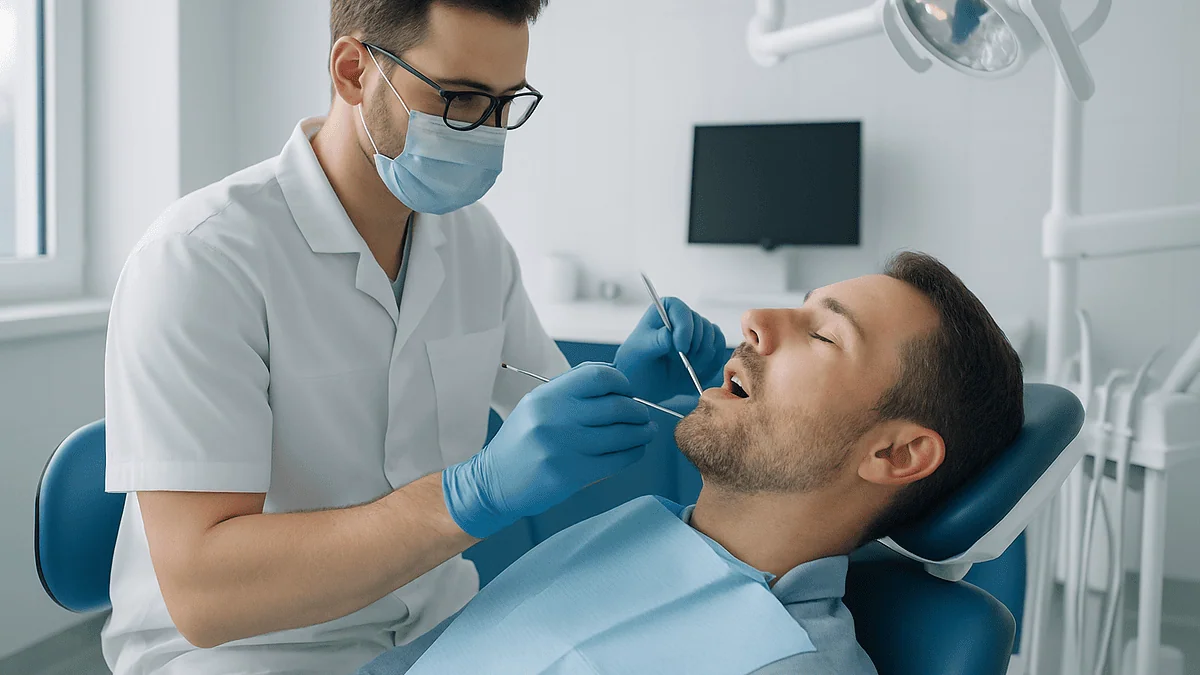
Key Takeaways
- Most jaw cysts are odontogenic, arising from tooth-forming tissue, and are found on routine X-rays before they cause any pain.
- The inferior alveolar nerve supplies feeling to the lower lip and chin, and surgeons protect it by dissecting along the cyst lining with blunt instruments rather than cutting through bone toward it.
- Large cysts pressing on the nerve or developing teeth are often shrunk first through marsupialization or decompression, then removed in a smaller second operation.
- Neighboring teeth are checked for vitality before surgery so living roots are preserved and dead ones are treated with root canal therapy or removed.
- Every removed cyst is examined by a pathologist because keratocysts and ameloblastoma can mimic simple cysts yet need longer follow-up or wider treatment.
- Soft tissue healing takes up to about two weeks, but the bony cavity refills over many months, so hard foods and contact sports are often restricted until follow-up X-rays confirm healing.
Jaw cyst removal surgery protects nearby teeth and nerves through planning and technique: three-dimensional imaging maps the cyst against tooth roots and the nerve canal, the surgeon lifts the cyst lining away from those structures rather than cutting through them, and large cysts near the nerve may first be shrunk through a small drainage opening. Every choice is tailored by the treating team to the cyst's size, position and biopsy result.
The X-ray was meant to be routine. A dark, rounded shadow sat under a lower molar, quiet and painless, and the dentist paused a beat too long before speaking. Most people first learn they have a jaw cyst exactly this way, not because something hurt, but because a picture taken for another reason revealed a hollow that should not be there.
What follows is usually a referral to an oral surgeon and a conversation about jaw cyst removal surgery. The questions that keep people awake are rarely about the cyst itself. They are about the tooth next door, the nerve that runs through the lower jaw, and whether the lip and chin will feel normal afterward.
Those are the right questions. The answer lies less in the removal and more in what the surgeon does to leave everything else undisturbed.
What a jaw cyst is, and why the neighbors matter more than the cyst
A cyst is a closed, fluid-filled sac lined by a thin layer of cells. In the jaw, most cysts are odontogenic, meaning they arise from tissue that was involved in forming teeth. Left alone, they grow slowly by pressure, pushing bone outward and thinning it the way a balloon presses against a paper bag.
Three types account for most of what oral surgeons see. A radicular cyst develops at the tip of a tooth root after the tooth’s nerve has died, usually from decay or an old injury. A dentigerous cyst forms around the crown of a tooth that never erupted, most often a lower wisdom tooth. An odontogenic keratocyst grows from leftover tooth-forming cells and has a habit of extending along the jaw and returning after removal, which is why the Mayo Clinic notes it needs careful long-term follow-up.
The cyst is almost never the dangerous part. Its position is. The lower jaw carries the inferior alveolar nerve, a sensory cable running inside a bony tunnel from the wisdom-tooth area to a small exit hole below the premolars, where it becomes the mental nerve and supplies feeling to the lip and chin. The upper jaw sits directly beneath the maxillary sinus, the air pocket behind the cheek. Tooth roots pass within a millimeter or two of both.
So the real skill in jaw cyst removal surgery is separating a fragile sac from structures it has spent months or years wrapping itself around. The cyst comes out. Everything around it is supposed to stay exactly where it was and keep working.
How jaw cyst removal surgery actually works, step by step
The procedure usually happens under local anesthetic, sometimes with sedation, and in larger cases under general anesthesia; the treating team decides based on the cyst’s size and the person’s health. What happens next follows a fairly consistent sequence.

First comes access. The surgeon makes an incision in the gum, well away from the delicate edge that hugs the teeth, and lifts a flap of tissue to expose bone. Over a large cyst, that bone may already be as thin as an eggshell. Where it is still solid, a small window is created with a fine drill under constant water cooling so heat never reaches the nerve or roots.
Then comes the part that protects the neighbors. Rather than cutting the cyst out, the surgeon slides a blunt, curved instrument between the cyst lining and the bony wall and peels the sac free in one piece. This is called enucleation, which simply means removing the whole sac intact. Because the lining is a natural boundary, working along it keeps the surgeon on the safe side of roots and nerve.
Once the sac is out, the cavity is inspected, irrigated and checked for any remaining lining. If a tooth root is bathed in the cavity, its tip may be sealed. If an unerupted tooth caused the cyst, it is usually removed at the same time, though in younger patients it may be left to erupt.
The flap is closed with dissolving stitches. The bone hollow fills with blood, and over months the body remodels that clot into new bone, generally without any graft. The removed tissue goes to a pathology laboratory, for reasons the next sections explain.
Enucleation, marsupialization or decompression: how the surgeon chooses
Not every cyst is removed the same day it is found. The Mayo Clinic describes several approaches, and the choice turns on one question: how much is at risk if the whole sac comes out right now?
Enucleation is the straightforward option for small and medium cysts that sit a comfortable distance from the nerve canal. Marsupialization is different in spirit. The surgeon opens the cyst, stitches its lining to the gum so it becomes a pouch open to the mouth, and lets pressure drain. Without pressure, the cyst shrinks over months while bone grows back behind it, moving the nerve and roots out of harm’s way. Decompression is a cousin of marsupialization that uses a small tube or stent to keep a drainage opening patent. Curettage, scraping the bony wall after enucleation, is added when the pathology suggests a cyst likely to return.
| Approach | What happens | Typically chosen when | Trade-off |
|---|---|---|---|
| Enucleation | Whole sac removed in one sitting | Small or medium cyst, clear margin from nerve and roots | Single procedure; requires enough safe bone |
| Marsupialization | Cyst opened and kept as a pouch to drain and shrink | Large cyst pressing on nerve, sinus or developing teeth | Slower; needs daily rinsing and follow-up |
| Decompression | Small tube keeps an opening for drainage | Same situations, when a stable opening is needed | Often followed by a second, smaller enucleation |
| Enucleation with curettage | Sac removed, bony wall scraped | Cysts with a known tendency to recur | More bone removed; reduces leftover lining |
A two-stage plan can feel like a delay, but it is often the single biggest protective choice available. A cyst that has thinned the nerve canal to a thread can be shrunk until there is bone between the sac and the nerve again, turning a risky operation into a routine one.
Protecting the inferior alveolar nerve during lower jaw cyst removal
The inferior alveolar nerve is the structure people worry about most, and rightly so. It provides feeling, not movement, so damage does not cause drooping, but numbness or tingling of the lower lip and chin is a real and recognized risk of any surgery in the back of the lower jaw. The NHS notes that after wisdom tooth removal, such nerve injury is usually temporary but can occasionally be permanent, and the same anatomy applies to jaw cyst removal surgery.

Protection begins before the first incision. On a three-dimensional scan, the surgeon traces the nerve canal and measures how much bone separates it from the cyst. If the answer is essentially none, that alone may tip the decision toward marsupialization first.
During the operation, several habits keep the nerve safe. The bony window is placed on the side of the jaw away from the canal wherever the anatomy allows. Instruments are blunt and the motion is gentle and sweeping, never stabbing, because a healthy nerve slides away from a curved instrument but can be nicked by a sharp one. Drilling stops as soon as the lining is visible. Where the cyst has grown around the nerve, the surgeon works from the bone side toward the nerve, leaving the nerve’s own protective sheath intact.
Afterward, the nerve often needs time to recover from stretching and bruising even when it was never cut. That is why a numb lip in the first days is monitored rather than treated as a complication. The NHS describes altered sensation after lower jaw surgery as lasting weeks to months in most cases where it occurs. Your surgeon will map and document the numb area at each visit so recovery can be tracked honestly.
How dentigerous cyst surgery and other jaw cyst removal protects the teeth next door
A cyst rarely grows in empty space. Roots of neighboring teeth often jut into the cavity like stalactites into a cave, and a surgeon who scrapes carelessly can strip the fine blood vessels and nerve fibers that enter each root at its tip.
Before surgery, the dentist or surgeon usually tests whether those teeth are still alive by checking their response to cold or a gentle electric stimulus. This is called a pulp vitality test, and it tells the team which teeth need protection and which have already lost their nerve. A tooth that is dead and sits at the center of a radicular cyst is generally the cause of the cyst; it is treated with root canal therapy beforehand, or removed if it cannot be saved. Healthy teeth whose roots merely happen to touch the cyst are treated with what amounts to surgical politeness.
That politeness has specific forms. The cyst lining is peeled off the root surface rather than the root being scraped. The root tip is left alone unless it has already lost its nerve, in which case the surgeon may trim and seal it, a step called an apicoectomy. The gum incision is placed to preserve the blood supply to the bone around those roots.
Dentigerous cyst surgery adds a particular question: what about the tooth that caused it? In an adult, an impacted wisdom tooth wrapped in a cyst is usually removed with the sac. In a child or teenager, a cyst around a developing permanent tooth is often marsupialized so the tooth can keep erupting into its proper place. The Mayo Clinic lists both removal and preservation as standard options, and the choice rests with the treating team.
Why the scan before surgery matters as much as the surgery
Most jaw cysts are first spotted on a plain dental X-ray, often a panoramic radiograph, the wide picture that shows both jaws and all the teeth in a single sweep. MedlinePlus describes this view as useful for detecting cysts and impacted teeth, but it has a limit: it is flat. A cyst may appear to sit beside the nerve canal when it actually surrounds it.
That is why surgeons planning anything near the nerve or sinus often add a cone-beam computed tomography scan, usually shortened to CBCT. Think of it as a low-dose three-dimensional dental scan that can be rotated on a screen. On it, the surgeon can see whether the cyst has pushed the nerve canal downward, whether the bony wall between cyst and sinus is intact, and how thick the outer jaw bone remains.
Those measurements answer practical questions. Where should the bony window go? Is there enough safe bone to enucleate today, or should the cyst be shrunk first? Will the jaw be weak enough after removal that a person should avoid hard foods for a while? Could a root be so deeply involved that the tooth is unlikely to survive?
Imaging also gives a baseline. Bone refills a cyst cavity from the edges inward over many months, and follow-up X-rays are compared with the original to confirm that healing is proceeding and that nothing has returned. For cysts with a known tendency to recur, such as keratocysts, the Mayo Clinic advises long-term imaging follow-up; the interval is set by the treating team.
If your surgeon orders a scan you were not expecting, that is a sign of planning, not of trouble.
Who jaw cyst removal surgery is usually for, and who is asked to wait
Surgery is usually recommended when a cyst is growing, has already weakened bone, has become infected, is displacing teeth, or when the biopsy or imaging appearance raises the possibility of a lesion that behaves more aggressively. A cyst does not have to hurt to qualify; many are removed precisely because they are silent and would otherwise keep expanding.
People are commonly asked to wait, or to take a staged path, in a few situations. Someone with an active infection in the cyst is often treated for that first, since inflamed tissue bleeds more and tears more easily near nerves. A child with a cyst around a developing tooth may have marsupialization rather than removal so the tooth can erupt. A very large cyst hugging the nerve canal is frequently shrunk before the definitive operation.
General health shapes timing too. Uncontrolled diabetes slows bone and gum healing. Certain medicines that affect bone turnover or bleeding need to be reviewed with the prescribing clinician before jaw surgery, and no one should stop or change a prescribed medicine on their own. Smoking impairs wound healing and raises the risk of infection in the socket, which is why surgeons ask people to stop or pause around the operation.
Some cysts are simply watched. A tiny, stable cyst discovered on X-ray in someone with other pressing medical needs may be photographed and re-imaged rather than operated on. Watchful waiting is a legitimate plan when the risks of surgery outweigh the risks of the cyst, but it carries a responsibility: the follow-up appointments have to actually happen.
None of these are rules a reader can apply to themselves. They are the considerations a surgical team weighs, and the decision always belongs to that team and the patient together.
Why the removed tissue always goes to the lab
Every jaw cyst removed by a surgeon is sent for histopathology, the microscopic examination of tissue by a pathologist. This is not a formality. Several jaw lesions look nearly identical on an X-ray and behave very differently in the body.
A radicular cyst, once its cause is removed, almost never returns. An odontogenic keratocyst, which can look the same on a plain film, has a lining that can leave behind microscopic daughter cysts, and the Mayo Clinic notes it may recur and warrants follow-up over years. Ameloblastoma, described by MedlinePlus as a usually benign but locally aggressive tumor that most often arises in the lower jaw near the molars, can mimic a cyst on imaging yet may require wider removal. Rarely, a cyst-like shadow turns out to be something that needs a different specialist entirely.
The pathology report therefore changes the plan after surgery, not before it. A benign radicular cyst may need only a healing check. A keratocyst may prompt a discussion about additional treatment of the bony wall and a longer imaging schedule. An unexpected diagnosis leads to referral and further imaging.
Results typically take days to a couple of weeks, depending on the laboratory and whether special stains are needed; your team will tell you the expected window. It is entirely reasonable to ask for the report to be explained in plain language, and to ask what the diagnosis means for follow-up.
One reassurance is worth stating clearly. The overwhelming majority of jaw cysts are benign, and pathology mainly confirms that. The lab step exists so that the rare exception is caught early, when it is small.
Jaw cyst surgery recovery time: what the first days and weeks usually look like
Recovery from jaw cyst removal surgery tends to follow the pattern of other oral surgery in the same area, and the NHS gives up to two weeks as the typical window to recover fully from lower wisdom tooth removal. Smaller cysts usually mean the shorter end; large cysts or those requiring a second stage mean longer.
The first two to three days bring swelling that peaks around the second day, some bruising of the cheek, and stiffness on opening the mouth. Pain is usually managed with over-the-counter or prescribed pain relief chosen by your surgeon; the plan is theirs to set. A little oozing of blood-tinged saliva is normal on day one. Soft, cool foods and gentle salt-water rinses after the first day are the standard advice.
By the end of the first week, swelling has usually receded and most people are back to normal work and light activity. Dissolving stitches loosen and disappear over one to two weeks. Numbness of the lip, if present, may still be there and is monitored rather than alarmed over at this stage.
The bone itself heals far more slowly than the gum. X-rays taken at follow-up visits show the cavity filling from the edges over roughly six to twelve months for medium cysts, longer for large ones, according to the surgical literature the Mayo Clinic summarizes; the treating team sets the imaging schedule. During that time, a large cavity in the lower jaw leaves the bone weaker, and surgeons often advise avoiding very hard foods and contact sports.
People who had marsupialization follow a different rhythm: daily rinsing of the pouch, periodic checks as the cyst shrinks, and often a smaller second procedure months later.
Risks of jaw cyst removal surgery and how surgeons reduce them
Every operation carries risk, and honest counseling names them. For jaw cyst removal surgery, the list is short but specific.
Nerve injury has been covered above: altered sensation of lip, chin or tongue, usually temporary, occasionally lasting, more likely with large cysts in the back of the lower jaw. Careful imaging, blunt dissection and staging are the counters.
Bleeding is generally modest, because the cyst has already displaced the vessels that would otherwise run through that bone. People taking blood-thinning medicines need a plan agreed between the surgeon and the prescriber; the medicine itself is never changed without that conversation.
Infection of the cavity can occur, more often when a large clot fills a big hollow. Sterile technique, thorough irrigation and sometimes a short course of antibiotics prescribed by the surgeon address this. Smoking and poor oral hygiene raise the risk.
Fracture of the jaw is rare but possible when a cyst has hollowed out much of the bone. Surgeons sometimes choose marsupialization specifically to let bone thicken first, or place a supporting plate when the remaining bone is thin.
Sinus involvement applies to upper jaw cysts. If the thin floor between cyst and sinus opens, it is usually closed at the time of surgery, and nose-blowing is restricted for a period.
Damage to neighboring teeth, from loss of vitality to loosening, is minimized by preoperative vitality testing and by dissecting along the cyst lining rather than the root.
Recurrence depends on cyst type; keratocysts are the notable example, which is why pathology and follow-up imaging matter. None of these risks is a reason to avoid needed surgery. They are the reason planning takes longer than the operation.
Are there alternatives to jaw cyst removal surgery?
People often ask whether a cyst can be treated without an operation. The honest answer is that a true jaw cyst has a lining that will keep producing fluid, and no rinse, tablet or supplement dissolves that lining. There is no evidence that herbal remedies, oil pulling or dietary changes shrink an established jaw cyst, and presenting them as alternatives would be misleading.
Real alternatives exist, and they fall into three groups.
The first is treating the cause. A small radicular cyst at the tip of a tooth root, caught early, sometimes resolves after root canal therapy alone, because removing the infected pulp removes the stimulus that keeps the sac inflamed. The Mayo Clinic notes that treatment depends on the lesion type, and for very small lesions a dentist may treat the tooth and re-image rather than operate. If the shadow does not shrink over follow-up, surgery is reconsidered.
The second is extraction alone. When a cyst surrounds the crown of an impacted wisdom tooth and is small, removing the tooth and its attached sac in a single, ordinary extraction may be all that is needed, since the sac comes out attached to the crown.
The third is watchful waiting, appropriate for tiny, stable, asymptomatic cysts in people for whom surgery carries unusual risk. It is not a passive choice. It means scheduled X-rays and a clear threshold agreed in advance for when growth would trigger surgery.
Marsupialization and decompression are sometimes described as alternatives, but they are better understood as gentler forms of surgery, chosen to protect nerves and developing teeth rather than to avoid an operation. Whichever path is proposed, the reasoning should be explained and the decision shared with the treating team.
Special considerations for children and teenagers
Jaw cysts in young people raise a particular concern: the jaw is still growing and permanent teeth are still forming under the gum. A cyst that displaces a developing tooth can push it far from its intended path, and the last thing anyone wants is to remove a healthy adult tooth that simply had the misfortune of sitting inside a cyst.
This is why marsupialization is used more freely in children. Opening the cyst relieves pressure, and the displaced tooth often drifts back toward its natural position and erupts on its own as the cavity fills with bone. The Mayo Clinic describes this approach as an option when preserving a tooth or reducing surgical extent matters, and orthodontists may later guide the tooth into place.
Preparation is mostly about comfort and clarity. Children do best when they know, in age-appropriate terms, what will happen: a sleepy medicine or numbing, a sore jaw for a few days, soft foods, and a follow-up visit. Sedation or general anesthesia is common for younger children so the surgeon can work precisely without a frightened patient moving. The anesthesia team assesses each child individually; families should share any history of breathing problems, previous reactions or medicines.
Recovery follows the adult pattern but often faster, since young bone heals quickly. Parents are asked to watch the same red flags as adults, plus a child’s reluctance to drink, which can lead to dehydration.
Follow-up is longer, not shorter, in this age group. The surgeon and dentist track eruption of the involved tooth and the refilling of bone over months, and any cyst with a tendency to recur is imaged for years. Nothing about medicines is settled by a magazine article; every dose and choice belongs to the prescribing clinician.
What people often get wrong about jaw cyst removal surgery
Myths gather around any procedure people fear, and jaw cysts attract more than their share. A few deserve correction.
“If it doesn’t hurt, it isn’t a problem.” Most jaw cysts are painless until they become infected or large enough to thin the bone. Pain is a late signal, not an early one.
“A cyst means cancer.” The Mayo Clinic and MedlinePlus both describe the vast majority of jaw cysts and tumors as benign. Pathology exists to confirm that, and to catch the rare exception early.
“They will have to remove all the teeth near it.” The opposite is the goal. Vitality testing, root canal therapy where a tooth has already died, and dissection along the cyst lining are all designed to keep neighboring teeth.
“A numb lip afterward means the surgeon cut the nerve.” Stretching and bruising of an intact nerve commonly cause temporary numbness. The NHS describes such symptoms after lower jaw surgery as usually resolving over weeks to months, though permanent change can occur.
“The hole will need a bone graft.” Most cyst cavities refill with the body’s own bone from the clot. Grafts are the exception, used for very large defects or when an implant is planned.
“A two-stage plan means the surgeon isn’t confident.” Marsupialization before enucleation is frequently the most protective choice for a large cyst near the nerve, chosen deliberately.
“Once it’s out, it’s over.” For most cysts, essentially yes. For keratocysts and a few others, follow-up imaging over years is part of the treatment, not an afterthought.
Believing the myths tends to produce two bad outcomes: people delay care because it does not hurt, or they panic over a diagnosis that is almost always benign. Neither serves the jaw.
Questions to ask your care team before jaw cyst surgery
A good consultation answers most of these before you ask. Bringing the list ensures none are missed, and it signals that you want to share the decision.
- What type of cyst do you suspect, and what would change if the pathology shows something else?
- How close is the cyst to the nerve canal, the sinus or the roots of my teeth on the scan?
- Are you planning to remove it in one stage or to shrink it first, and why?
- Which of my teeth have been tested for vitality, and what happens to any that are not alive?
- Will the tooth that caused the cyst be removed, treated with a root canal or kept?
- What kind of anesthesia are you recommending, and who will manage it?
- What are the chances of numbness in my lip or chin in my particular case, and how will you monitor it?
- How long should I plan to be off work, and when can I eat normally?
- Will I need a bone graft or a plate, and how weak will the jaw be while it heals?
- What is the follow-up schedule, including X-rays, and for how many years?
- Which of my current medicines need to be discussed with the doctor who prescribes them before surgery?
- What signs after the operation should make me call you the same day?
Write the answers down or ask permission to record the conversation. Surgical consultations pack a great deal into a short time, and memory under stress is unreliable. If an answer is unclear, ask for it again in plainer words; a team that plans carefully will not mind explaining carefully. Every choice on this list, from staging to anesthesia to medicines, remains the treating team’s to make with you.
When to call your doctor after jaw cyst removal
Most recoveries are uneventful: swelling that peaks and fades, soreness that responds to the pain relief your surgeon set, stitches that dissolve on their own. Certain signs, though, should prompt a same-day call to the surgical team or, if they cannot be reached, urgent medical care.
Bleeding that does not stop after twenty to thirty minutes of firm pressure with gauze, or that soaks through repeated pads, needs attention. So does swelling that keeps increasing after the third day, or swelling that spreads toward the eye, down the neck or under the tongue, particularly if it makes swallowing or breathing feel difficult. Any difficulty breathing is an emergency.
A fever that develops after the first day, a foul taste or pus from the wound, or pain that worsens rather than eases after day three can indicate infection of the cavity. A sudden bad taste with a sensation of air or liquid passing into the nose after upper jaw surgery suggests a sinus opening and should be reported.
Numbness of the lip or chin in the first days is expected in some people, but numbness that appears newly, expands or is accompanied by weakness of the face should be assessed. A clicking, shifting or grinding sensation in the jaw, or a change in how the teeth meet, could signal a fracture through weakened bone and needs prompt review.
After marsupialization, call if the opening closes, drainage stops abruptly with return of swelling, or the tube is lost.
Do not adjust, stop or add any medicine on your own in response to these symptoms, including pain relief or blood thinners. Describe what you see and let the team direct the next step. Trust the pattern of a normal recovery, and trust your instinct when the pattern breaks.
Frequently asked questions
What is odontogenic cyst removal, and is it different from jaw cyst removal surgery?
Odontogenic cyst removal is jaw cyst removal surgery for cysts that arose from tooth-forming tissue, which describes most jaw cysts. The surgeon exposes the cyst through the gum and bone, peels the lining out intact, and sends it for pathology. The term simply specifies the cyst’s origin; the protective techniques for nerves and teeth are the same.
How long is jaw cyst surgery recovery time?
Soft tissue recovery typically takes up to two weeks, the window the NHS gives for comparable lower wisdom tooth surgery, with swelling peaking around day two and most people back to normal activity within a week. The bone cavity refills far more slowly, over many months, and follow-up X-rays confirm healing. Large or staged cysts take longer; your surgeon sets expectations for your case.
What is marsupialization of a jaw cyst?
Marsupialization of a jaw cyst means opening the cyst and stitching its lining to the gum so it becomes a pouch that drains into the mouth. Without internal pressure, the cyst shrinks over months while bone grows back around nerves and tooth roots. It is chosen for large cysts near the nerve canal or developing teeth, and is often followed by a smaller removal later.
Will dentigerous cyst surgery mean losing the tooth involved?
Often, but not always. In adults, an impacted wisdom tooth wrapped in a dentigerous cyst is usually removed with the sac. In children and teenagers, the cyst is often marsupialized so the developing permanent tooth can erupt naturally. The Mayo Clinic lists both approaches as standard, and the treating team decides based on the tooth’s position and value.
Will my lip be numb after jaw cyst removal surgery?
It can be, especially after removal of a large cyst in the back of the lower jaw near the inferior alveolar nerve. The NHS describes such numbness after lower jaw surgery as usually temporary, resolving over weeks to months, though it can occasionally be permanent. Your surgeon will document the numb area at each visit and track recovery.
Can a jaw cyst be treated without surgery?
A true jaw cyst has a fluid-producing lining that no medicine, rinse or supplement dissolves, so established cysts generally need surgery. Exceptions exist: a small radicular cyst may resolve after root canal therapy of the causative tooth, and tiny stable cysts can be monitored with X-rays. Any non-surgical plan requires scheduled follow-up agreed with the treating team.
Does a jaw cyst mean I have cancer?
Almost never. The Mayo Clinic and MedlinePlus describe the great majority of jaw cysts and tumors as benign. The removed tissue is still examined by a pathologist because a few lesions, such as odontogenic keratocysts and ameloblastoma, look like simple cysts on X-ray but need longer follow-up or wider treatment. Pathology confirms the diagnosis rather than implying doubt.
Will the hole in my jaw need a bone graft?
Usually not. After the cyst is removed, the cavity fills with a blood clot that the body gradually remodels into new bone over months. Grafts are reserved for very large defects, cases where the jaw is at risk of fracture, or when a dental implant is planned in that area. Your surgeon will explain if your cavity is an exception.
How does the surgeon know where the nerve is before operating?
A panoramic X-ray shows the general position, but for cysts near the nerve canal surgeons commonly add a cone-beam CT scan, a three-dimensional dental scan that shows exactly how much bone separates the cyst from the nerve, sinus and tooth roots. Those measurements decide where the bony window goes and whether to shrink the cyst first.
Can a jaw cyst come back after removal?
It depends on the type. Radicular cysts rarely return once the causative tooth is treated or removed. Odontogenic keratocysts can recur because their lining leaves microscopic remnants, which is why the Mayo Clinic recommends long-term imaging follow-up for them. The pathology report tells your team which category applies and how long to keep checking.
References
This article is for general information only and is not a substitute for professional medical advice. Please consult a qualified doctor about your individual situation.

More from the Blog
Conscious Sedation in Dentistry: What You Feel, Remember and Need to Arrange
Conscious sedation in dentistry uses medication to make you deeply relaxed and drowsy while you stay awake enough to breathe on your own and…

Healing After Gingival Aesthetics: Soft Diet, Gentle Brushing and How the Gum Line Settles
Gum reshaping healing is usually quick compared with other dental surgery. Soreness and mild swelling are common for a few days, and the surface…
Living With Aesthetic Dental Work: Staining, Biting Habits and Long-Term Upkeep
Maintaining cosmetic dental work rests on three habits: protecting the bond line where restoration meets tooth, controlling biting forces such as grinding, nail-biting and…

Composite Bonding Results: Blending With Natural Teeth and What the Resin Cannot Change
Composite bonding results depend on how carefully tooth-colored resin is shade-matched, layered, and polished onto natural enamel; done well, it can disguise small chips,…
After Dental Trauma Care: Splints, Follow-Up Visits and How the Tooth Is Monitored
Dental trauma recovery usually involves a flexible splint worn for about two to four weeks depending on the injury, a soft diet, careful hygiene…

Who Is a Candidate for Laser Dentistry? Gums, Small Cavities and Sensitivity Explained
Laser dentistry candidates are usually adults and older children who need gum treatment (deep-pocket cleaning, reshaping, small soft-tissue procedures), have small or early cavities…