Jaw Cysts Treatment
Jaw cysts are fluid-filled lesions in the jawbone or soft tissues that may damage teeth, nerves, or bone if untreated. Treatment usually involves diagnosis, removal, and pathology review.
Quick answer
Jaw cyst treatment is the surgical management of fluid-filled, tissue-lined sacs that develop in the jawbone or the soft tissues of the mouth, including the dentigerous cyst that forms around an unerupted tooth. It typically involves imaging to define the lesion, removal (enucleation) or gradual decompression, management of any involved tooth, and microscopic examination of the tissue to confirm the diagnosis and guide follow-up.
Jaw Cysts and the Dentigerous Cyst: Understanding the Diagnosis
Jaw cysts are sacs of fluid or semi-solid material, lined by living tissue, that develop inside the jawbone or in the soft tissues of the mouth. Almost all of them are benign. Treatment has three parts: confirming what the lesion actually is, removing it or shrinking it when removal is indicated, and examining the tissue under a microscope so that nothing is left to assumption. Among these lesions, the dentigerous cyst, a sac that forms around the crown of a tooth that has failed to erupt, is one of the most frequently treated, and much of what applies to it applies to jaw cysts in general.
Most people hear the word cyst for the first time when a dentist points at a dark shadow on a panoramic X-ray taken for another reason entirely: a routine check-up, an orthodontic assessment, implant planning, or an evaluation before wisdom tooth removal. That is normal. Jaw cysts often grow slowly and without pain, which is why the diagnosis can feel like it came out of nowhere. Other patients arrive with symptoms, swelling of the jaw or gums, a tooth that has drifted or loosened, repeated infection, a persistent ache that ordinary dental treatment does not settle, or altered sensation in the lip or chin.
Benign does not mean harmless to ignore. A cyst can expand inside the bone for years, thinning the jaw, dissolving the roots of neighbouring teeth, pushing teeth out of position, or pressing on the sensory nerves that run through the lower jaw. Some cysts become infected and cause pain, drainage, fever or facial swelling. And in a small number of cases, a lesion that looks like a simple cyst on imaging turns out to be a different condition that needs a different plan. This is why the diagnosis is never finished at the X-ray stage: careful imaging, sensible surgical planning and pathology review together make up good jaw cyst care.
This page explains what a dentigerous cyst is, how it differs from other jaw cysts, what the honest answers are to the questions people worry about most, how treatment is actually performed, and what recovery looks like. It also explains what influences the outcome, because two cysts that look identical on a basic X-ray can need quite different treatment.
What Is a Dentigerous Cyst?
A dentigerous cyst is a fluid-filled sac that develops around the crown of a tooth that has not erupted into the mouth, most often a lower wisdom tooth or an upper canine. The cyst attaches to the tooth at the neck, where the enamel of the crown meets the root, and encloses the crown itself. On an X-ray it usually appears as a well-defined dark area surrounding the crown of the buried tooth. Older classification lists sometimes index the term in reverse, as cyst dentigerous, but the lesion is the same.
The cyst arises from the dental follicle: the soft-tissue sleeve that surrounds every developing tooth before it erupts. When fluid accumulates between this follicle and the crown, the follicle stretches into a cyst. Because impacted wisdom teeth are common, the dentigerous cyst is common too. It is frequently diagnosed in adolescents and younger adults, when unerupted teeth are being assessed, but it can be found at any age, including decades after a tooth quietly failed to erupt. Its most characteristic behaviour is silent growth: it can displace the involved tooth deep into the jaw, tilt neighbouring teeth, and slowly resorb their roots without causing any pain at all.
What causes dentigerous cysts?
Dentigerous cysts are caused by fluid collecting between the crown of an unerupted tooth and the follicle that surrounds it; the precise trigger is not fully understood. Obstructed eruption seems to play a role: the tooth cannot come through, pressure builds, and the follicle expands. A recognised variant, the dentigerous cyst of inflammatory origin, develops when infection at the root of a baby tooth irritates the follicle of the permanent tooth developing beneath it. None of this is caused by anything you did. Oral hygiene, diet and lifestyle do not create a dentigerous cyst, although hygiene matters a great deal once treatment begins.
How serious is a dentigerous cyst?
A dentigerous cyst found early is usually not a serious condition: it is benign and it responds well to a properly planned operation. Its significance grows with its size. A large cyst can hollow out a substantial portion of the jaw, thin the bone enough to raise the risk of fracture, resorb the roots of healthy teeth, and grow close to the nerve that supplies feeling to the lower lip and chin. There is one further reason it is taken seriously: in rare cases, other lesions can develop from the same lining, which is why the removed tissue is always examined by a pathologist rather than simply discarded.
What happens if you leave a dentigerous cyst?
Left untreated, a dentigerous cyst tends to keep enlarging, because its lining continues to produce fluid. What starts as a small shadow on an X-ray can, over years, displace the impacted tooth far from its original position, damage the roots of teeth you want to keep, weaken the jawbone, and expand toward the nerve canal or the maxillary sinus. An untreated cyst can also become infected, converting a quiet lesion into a painful, swollen one that is harder to operate on. A lesion that might once have needed a limited outpatient procedure can end up requiring staged surgery, extraction of additional teeth and bone grafting. Watchful waiting is occasionally reasonable for very small, stable lesions under specialist supervision, but “leaving it” without a plan is not a neutral choice.
How do you get rid of a dentigerous cyst?
A dentigerous cyst is removed surgically; no medication, mouthwash or home remedy will dissolve it. The standard operation is enucleation, removal of the entire cyst lining, usually together with the impacted tooth it surrounds, since the tooth is often unsalvageable and leaving it risks recurrence. In carefully selected younger patients, the surgeon may instead open and decompress the cyst so that it shrinks and the buried tooth is given a chance to erupt into a useful position, sometimes with orthodontic help. Which approach is right depends on the cyst’s size, the position of the tooth, the patient’s age, and how close the lesion sits to nerves and neighbouring roots.
What Jaw Cyst Treatment Involves
Jaw cyst treatment is the medical and surgical management of tissue-lined lesions in the jawbone, around the teeth, or in nearby oral tissues. Whatever the cyst type, the pathway follows the same logic: define the lesion with imaging, remove or decompress it when indicated, and confirm the diagnosis with pathology. The details, anaesthesia, incision, tooth management, grafting, are then tailored to the individual case.
A jaw cyst is not simply a pocket of fluid. It is lined by tissue that can continue producing fluid and expanding unless it is dealt with. Some cysts, like the dentigerous type, relate to impacted teeth. Others form at the tip of a tooth root after longstanding infection of the dental pulp. Some arise from developmental tissue left behind during tooth formation. A smaller number occur in the soft tissues of the mouth rather than inside the bone, and these are handled differently.
The most common operation is enucleation: the surgeon separates the cyst lining from the surrounding bone and removes it, in one piece where possible. For large cysts, or cysts sitting hard against important nerves, the surgeon may first perform marsupialisation or decompression. These techniques create a small, maintained opening into the cyst so that the internal pressure drops and the lesion shrinks gradually over months. Bone regenerates around the shrinking cavity, and a second, smaller operation removes what remains. Staged treatment demands patience, but in the right patient it reduces the risk of nerve injury, jaw fracture and unnecessary tooth loss.
Treatment often includes more than the cyst itself. An associated tooth may need extraction, root canal treatment or root-end surgery. A large bony defect may be grafted. A communication with the maxillary sinus may need closure. Follow-up imaging confirms that the bone is healing. And when a cyst has unusual features, has come back after previous treatment, or resembles a tumour on the scan, the case may be reviewed by several specialists together before anyone operates. The aim is never just removal: it is understanding why the lesion developed and reducing the chance of seeing it again.
Types of Jaw Cysts: Odontogenic and Other Lesions
Odontogenic cysts arise from the tissues involved in tooth development, or from infection related to the teeth, and they account for most cysts of the jaws. Non-odontogenic cysts and cyst-like lesions make up the remainder. Knowing the type matters, because the type determines how aggressively the lesion behaves, how completely it must be removed, and how long it should be followed afterwards.
Radicular cyst
A radicular cyst, also called a periapical cyst, develops at the tip of a tooth root after chronic infection or death of the dental pulp, typically the late consequence of deep decay, a failed filling or an old injury. It is the everyday workhorse of jaw cyst surgery. Treatment addresses both the cyst and its cause: the diseased tooth is managed with root canal treatment, root-end surgery or extraction, depending on whether it can realistically be saved, and the cyst lining is removed. A residual cyst is the same lesion left behind after the offending tooth was extracted; it can persist and grow for years in an apparently healed extraction site.
Odontogenic keratocyst
The odontogenic keratocyst is a distinct cystic lesion known for growing along the length of the jaw rather than ballooning outward, which lets it reach a considerable size before anyone notices, and for a higher tendency to recur than most other jaw cysts. It demands more from the surgeon: complete removal of a lining that is thin and tears easily, sometimes adjunctive treatment of the bone cavity, and disciplined long-term imaging follow-up even after an apparently clean operation. When multiple keratocysts are found, particularly in a younger patient, the specialist will also consider whether they form part of an inherited condition, because that possibility changes both surveillance and the wider medical assessment.
Other cysts of the jawbone
Several other lesions follow the same evaluation pathway. The lateral periodontal cyst sits alongside the root of a living tooth. The nasopalatine duct cyst arises from developmental tissue in the front of the upper jaw, behind the upper front teeth. The traumatic bone cyst is an empty or fluid-containing cavity without a true lining, often found in younger patients and usually treated with a simple surgical exploration that triggers healing. Cyst-like spaces can also accompany benign jaw tumours, which is one more reason imaging alone is never accepted as the final word.
Gum cyst and other soft-tissue cysts of the mouth
A gum cyst, clinically a gingival cyst, is a small, usually painless swelling in the gum tissue itself rather than in the bone beneath it. Not every cyst in mouth tissue involves the jawbone: mucoceles caused by blocked or damaged minor salivary glands, and cysts of salivary gland origin, sit entirely in the soft tissues of the lips, cheeks or floor of the mouth. These are generally simpler to treat than bony lesions, but a persistent or unusual soft-tissue swelling still deserves removal and pathology review rather than repeated draining, because appearance alone does not confirm what it is.
Symptoms and Who May Need Treatment
Many patients with jaw cysts have no symptoms at all in the early stages. Because the jawbone yields gradually, a cyst can become surprisingly large before it causes discomfort, one reason panoramic X-rays and three-dimensional dental imaging are so valuable when a lesion is suspected. Cysts are regularly discovered during implant planning, orthodontic work-ups and pre-extraction assessments in people who feel entirely well.
Symptoms appear when the cyst enlarges, becomes infected or starts affecting its neighbours. You might notice swelling in the jaw or gums, a visible bulge inside the mouth, tenderness, repeated dental infection, a bad taste from drainage, or pain on chewing. Teeth may loosen, drift or fail to erupt on schedule. If the cyst sits near a sensory nerve, you may feel numbness or tingling in the lip, chin, cheek or tongue. A cyst in the upper jaw can produce pressure or symptoms around the sinus.
Treatment is generally recommended when a cyst is growing, causing symptoms, threatening nearby teeth, associated with infection, blocking a tooth from erupting, weakening the jaw, or when its appearance is uncertain enough that tissue diagnosis is needed. It is also usually addressed before dental implants, orthodontic treatment or larger rehabilitation such as full mouth reconstruction, because an untreated lesion sitting under new dental work compromises everything built on top of it.
Are cysts in the jaw common?
Yes, jaw cysts are among the most frequent lesions found inside the jawbones, and dentists encounter them regularly on routine imaging. The radicular cyst related to infected teeth and the dentigerous cyst related to impacted teeth make up the bulk of everyday cases. Being told you have one places you in familiar territory for any oral and maxillofacial surgery service; these are routine operations with well-established techniques, even though each case still deserves individual planning.
How Dangerous Are Jaw Cysts?
The honest answer is that most jaw cysts are benign, slow-growing and very treatable, and that they still deserve respect, because the damage they do is quiet and cumulative. The specific worries people search for deserve direct answers.
Can a jaw cyst be a cancerous jaw tumor?
A jaw cyst is very rarely a cancerous jaw tumor; cysts are by definition benign, and the overwhelming majority behave that way. Two caveats keep specialists careful. First, some benign but locally aggressive jaw tumours can mimic a cyst on an X-ray, and the treatment for a tumour differs from the treatment for a cyst. Second, malignant change within a cyst lining has been reported, though it is exceptional. This is exactly why every removed cyst goes to pathology and why lesions with warning features, rapid growth, irregular borders, unexplained numbness, significant bone destruction, or recurrence after treatment, may be biopsied before definitive surgery. Blood tests do not settle the question: tumour marker blood tests used in other areas of medicine have no role in diagnosing a jaw cyst; the diagnosis rests on imaging and tissue examination.
Can a jaw cyst go away on its own?
True jaw cysts do not reliably disappear on their own, because the lining keeps producing fluid. There are limited exceptions: a small inflammatory lesion at a tooth root can resolve once the tooth is properly root-treated, and occasional small lesions remain stable for years. But an established cyst, particularly a dentigerous cyst around an impacted tooth or a keratocyst, should be expected to persist or enlarge. Hoping it resolves is not a strategy; monitoring it under specialist supervision, with repeat imaging at defined intervals, is a legitimate one for selected small lesions.
Can a jaw cyst kill you?
A jaw cyst itself is not a life-threatening condition. The realistic risks of neglect are different: progressive bone loss, tooth loss, nerve involvement and a weakened jaw that can fracture under normal chewing forces. The one scenario that becomes genuinely serious is deep infection: an infected cyst can, in uncommon cases, spread into the facial and neck spaces, and infections in that territory are treated as urgent in any hospital. Timely treatment of the cyst removes that pathway before it exists.
Diagnosis: How Jaw Cysts Are Identified
Diagnosis begins with a detailed examination and history. The specialist asks about pain, swelling, previous infections, root canal treatment, impacted teeth, trauma and prior oral surgery, then examines the mouth, the bite, the teeth near the lesion and the sensation of the lip and chin. Teeth adjacent to the cyst may be tested for vitality, mobility and periodontal health, because a dead tooth points toward one diagnosis and a living one toward another.
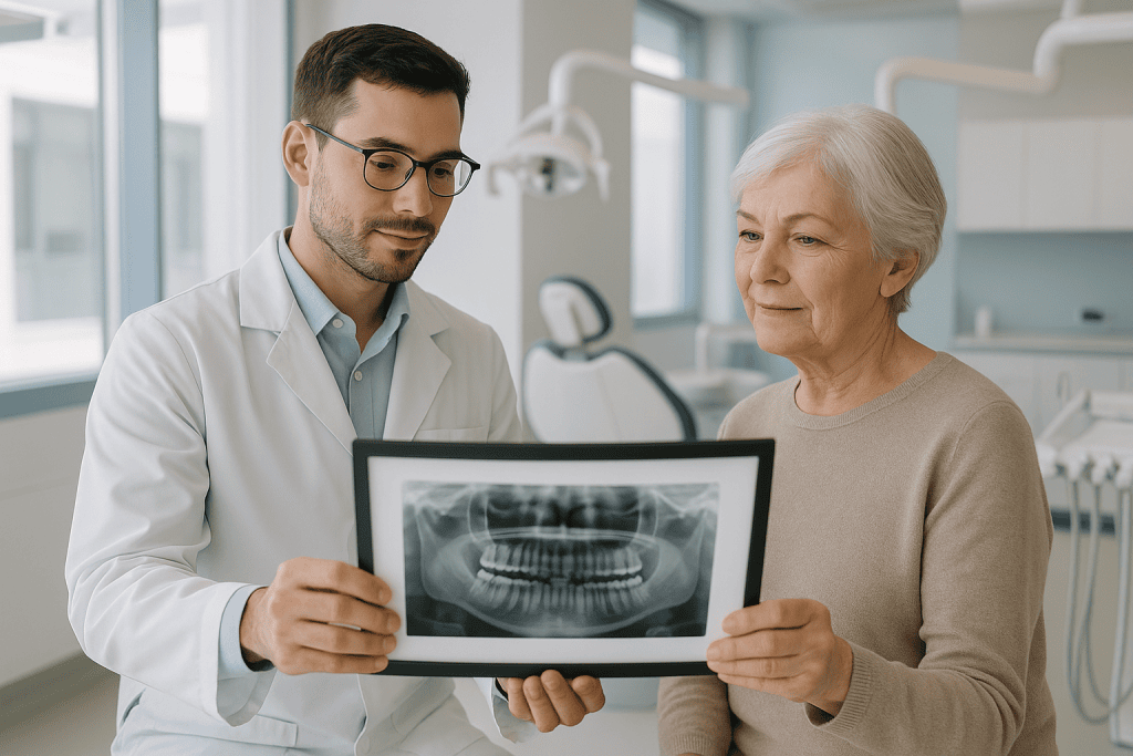
Imaging then defines the lesion. A panoramic X-ray shows the general size and location. Cone beam CT or medical CT adds the third dimension: how much bone remains, where the tooth roots sit, how close the lesion lies to the inferior alveolar nerve or the maxillary sinus, and whether the cyst has expanded or perforated the jawbone. MRI is reserved for selected complex cases where soft-tissue involvement or the lesion’s contents need clarification. In many patients, imaging plus the clinical picture makes the diagnosis strongly probable: a well-defined dark area around the crown of an impacted wisdom tooth is a classic dentigerous cyst appearance.
Probable is not the same as proven. Different jaw cysts can look alike on a scan, and some tumours mimic cysts, so the definitive diagnosis comes from pathology after biopsy or removal. If warning features are present, a biopsy may be done first; otherwise the removed lining itself provides the answer. It is also worth saying plainly that a cyst in the jaw has nothing in common with cysts elsewhere in the body: an ovarian cyst, for instance, is a different entity managed by an entirely different speciality, and having one does not predispose you to the other.
How Jaw Cyst Treatment Is Performed
Good jaw cyst surgery starts well before the operating room. Existing records, X-rays, scans and dental history are reviewed first, so the team can judge complexity, decide whether further imaging or a biopsy is needed, and plan the sequence of care. This review usually happens before surgery is scheduled, so the number of procedures, the anaesthesia plan and the expected recovery are understood in advance rather than discovered on the day.
Anaesthesia and setting
The procedure is performed under local anaesthesia, sedation or general anaesthesia depending on the cyst’s size and location, the anticipated duration, and your comfort and medical history. A small cyst is often removed as an outpatient procedure under local anaesthesia. Larger cysts, lesions close to nerves or the sinus, multiple lesions, or cases combined with bone grafting are usually better managed with sedation or general anaesthesia in a hospital setting, where anaesthesiology support and medical monitoring are integrated into the plan. If active infection is present, it may be controlled first, with antibiotics or drainage as the surgeon prescribes, because inflamed tissue bleeds more, heals less predictably and blurs surgical planes.
Enucleation, step by step
When the plan is complete removal, the operation typically follows this sequence:
- The surgical field is prepared and anaesthetised, and an incision is made in the gum or oral mucosa over the affected area.
- A small window is created in the bone to expose the cyst, sized to give access without sacrificing more bone than necessary.
- The cyst lining is carefully separated from the bony cavity and removed, ideally in one piece, or in deliberate sections where anatomy demands it.
- Any involved tooth is managed: extracted, root-treated, or its root tip removed, depending on whether keeping it is realistic and safe.
- The cavity is cleaned and inspected, bony edges are smoothed, nearby nerves are protected, and any opening into the sinus is closed if the upper jaw is involved.
- A graft is placed if indicated, the wound is closed with sutures, and the removed tissue is sent to pathology.
Marsupialisation and decompression
When a cyst is large, or immediate removal would endanger the nerve, risk a jaw fracture or cost teeth worth keeping, the surgeon may open the cyst instead of excising it. A small opening is created and kept patent with a device or dressing, so the internal pressure falls and the cyst shrinks over the following months while new bone forms around it. You will be taught how to keep the opening clean and what signs of blockage or infection to watch for, and you will attend regular reviews so the shrinkage can be tracked on imaging. Once the lesion is small enough, a second, more contained procedure removes the residual lining. In younger patients with a dentigerous cyst, this staged approach sometimes allows the buried tooth to erupt into a usable position instead of being extracted: a result no single-stage operation can offer. The trade-off is time and discipline: staged treatment only works for patients who keep the opening clean and keep their appointments.
Bone grafting and reconstruction
Not every cyst cavity needs grafting. Many defects fill naturally with new bone over time, particularly in younger patients and smaller lesions. Grafting with bone or a bone substitute earns its place when the defect is large, when the remaining bone is structurally weak, or when future dental implants are planned and the site needs predictable volume. The decision is driven by size, location, infection status and your dental goals, not by a default policy.
Pathology: the step that finishes the diagnosis
Everything removed is examined under a microscope. The pathologist confirms the cyst type and identifies any feature that changes the follow-up plan. If the result differs from what the imaging suggested: a keratocyst where a dentigerous cyst was expected, for example, or a cystic tumour where a cyst was expected: the plan is adjusted: closer surveillance, further surgery, or review by a multidisciplinary board. This step is why no reputable service skips pathology, however typical the lesion looked.
Operating time varies with all of the above: a small cyst is dealt with quickly, while a large lesion near the nerve, combined with extraction and grafting, takes considerably longer. Most patients go home the same day; some complex cases involve a short hospital stay. Whatever the setting, at least one early postoperative review is built into the plan, with the exact interval set by the operation performed and how healing progresses.
Why Early Evaluation Matters
Because jaw cysts grow silently, delay converts easy problems into harder ones. As a cyst expands it thins the jawbone, undermines the support around teeth, and makes complete removal technically more demanding. A lesion that once needed a limited outpatient procedure may later need staged surgery, extractions, grafting and reconstruction: a heavier operation with a longer recovery, for the same underlying diagnosis.
Delay also raises the odds of infection. An infected cyst causes real misery, pain, swelling, drainage, fever, difficulty opening the mouth, and repeated infections progressively damage bone and soft tissue, complicating the eventual surgery. Infections in the jaw and facial spaces can occasionally spread beyond the mouth, which is the one way a fundamentally benign lesion becomes a genuinely urgent problem.
Nerve involvement is a further argument for acting early. In the lower jaw, cysts grow toward the inferior alveolar nerve, which supplies sensation to the lower lip and chin. Pressure on this nerve, or surgery conducted right against it, can cause temporary, and less commonly, lasting, sensory changes. A cyst removed while it is still clear of the nerve simply carries less of this risk than the same cyst removed two years later.
Finally, early evaluation separates the routine from the exceptional. Most jaw cysts are benign, but imaging alone is not definitive, and pathology is what provides certainty. When a more aggressive lesion is caught early, treatment can be more focused and less extensive than it would be after years of unchecked growth.
Benefits of Jaw Cyst Treatment
For most patients, treatment protects function and stops a silent lesion from doing progressive damage. The gains are concrete:
| Benefit | What It Means for You |
|---|---|
| Accurate diagnosis | Imaging plus pathology review confirms the cyst type and whether further treatment or extended follow-up is needed. |
| Protection of teeth and bone | Removing or decompressing the cyst relieves pressure on tooth roots, preserves jaw structure and lets bone heal. |
| Reduced infection risk | Treatment resolves the source of chronic infection, drainage, swelling and recurrent flare-ups. |
| A clean foundation for dental work | Implants, orthodontics and prosthetic treatment can proceed on healthy bone rather than over an untreated lesion. |
| Relief from symptoms | Pain, swelling, pressure and chewing difficulty typically improve gradually as healing progresses. |
| Long-term surveillance | A defined follow-up schedule catches recurrence early, especially for cyst types known to return in some patients. |
Recovery Timeline After Jaw Cyst Treatment
Recovery depends on cyst size, location, infection status, whether a tooth was removed, and whether the procedure was straightforward enucleation, staged decompression or a larger reconstruction. The broad pattern looks like this:
| Time Period | What to Expect |
|---|---|
| Day 1 | Numbness from the anaesthetic wears off gradually. Mild bleeding, swelling and soreness are normal. Soft foods and prescribed medications begin. |
| First week | Swelling usually peaks and then subsides. Oral hygiene instructions matter most in this window. Light daily activities resume; strenuous exercise waits. |
| First month | Soft-tissue healing is well advanced. The pathology result is reviewed and the surgeon confirms whether further care or imaging is needed. |
| Three to six months | Bone healing continues. Follow-up imaging typically shows gradual filling of the cavity, depending on the original defect and whether grafting was used. |
| Longer term | Some cyst types need periodic monitoring. Implant planning, orthodontics or other dental rehabilitation proceeds once healing is sufficient. |
The first days reward simple discipline. Cold compresses and prescribed medication manage swelling and discomfort. A soft diet spares the wound; hard, sharp or very hot foods disturb it. Vigorous rinsing, smoking and heavy exertion all interfere with early healing and are best avoided for as long as your surgeon specifies, smoking in particular slows bone and soft-tissue repair. Gentle but consistent oral hygiene around the site, exactly as instructed, does more for the result than anything else in your control.
If a decompression device was placed, your responsibilities extend further: keeping the opening clean, irrigating it as taught, recognising the signs of blockage or infection, and attending every scheduled review. The staged approach only delivers its advantages when the interim phase is properly maintained.
Follow-up is part of the treatment, not an optional extra. Sutures are checked or removed, the pathology result is discussed, and repeat imaging is scheduled, often at several months, to confirm that bone is filling the cavity. Cysts with recurrence potential, the odontogenic keratocyst above all, warrant imaging for years even when everything feels normal, because recurrence is typically silent, exactly as the original lesion was. After the initial postoperative period, long-term surveillance can usually be coordinated with your own dentist or oral surgeon, with a clear written schedule of what to check and when.
What Influences the Outcome
The first factor is the correct diagnosis. Two lesions can look identical on a basic X-ray yet behave completely differently over time: a dentigerous cyst and a keratocyst around the same impacted tooth demand different surgical margins and different follow-up. High-quality imaging and rigorous pathology make sure the treatment matches the biology of the lesion, which matters most for recurrent cysts, large lesions and anything with unusual radiographic features.
Size and location come next. A small cyst away from nerves and major roots is a contained problem. A large cyst wrapping around multiple teeth, or extending toward the mandibular nerve canal, the maxillary sinus or the nasal floor, is a planning exercise. In complex anatomy, the best outcome often comes from a staged plan rather than one aggressive operation; decompression is a considered choice, not a lesser one, when nerve function, bone strength or valuable teeth are at stake.
The state of the neighbouring teeth shapes the plan. If an infected tooth caused the cyst, the source must be dealt with, saving the tooth is possible when the root structure, periodontal support and endodontic prognosis are favourable, and extraction is the more durable answer when they are not. That judgement should weigh both the surgery and your restorative future: a tooth heroically saved but doomed within a few years serves nobody.
Your general health affects healing. Active infection increases swelling and wound complications, and sometimes justifies a short delay before definitive surgery. Smoking, uncontrolled diabetes, immune suppression, poor oral hygiene and certain medications can each slow healing or raise complication risk. A careful medical history lets the team adapt the anaesthesia, the timing and the aftercare rather than discovering problems mid-course; any adjustment to your regular medication is decided by your treating doctors, never assumed.
Surgical technique carries obvious weight: complete removal of the lining, protection of the nerve, sound management of the involved tooth and careful wound closure all contribute measurably to healing. Where grafting is used, patient selection and infection control decide whether it helps. Grafting is not automatically better: it is better when it serves a defined functional or dental goal.
The last factor is you. Diet, hygiene, medication adherence, activity restrictions, smoking avoidance and attendance at follow-up visits are all in the patient’s hands, and they meaningfully influence the result. Patients with decompression devices carry the largest share of this responsibility. And for cyst types that can recur, keeping the long-term imaging appointments after symptoms have long since vanished is the single most commonly neglected part of the plan.
How Jaw Cyst Care Is Organised at Acibadem
At Acibadem, jaw cyst care is built around coordinated evaluation rather than a single procedure. The pathway can involve oral and maxillofacial surgery, dental imaging, radiology, pathology, anaesthesiology, endodontics, prosthodontics and orthodontics, depending on what the individual case requires. Complex situations, imaging that suggests an aggressive lesion, a recurrent cyst, or a case where removal and later dental rehabilitation must be planned together, can be discussed across specialities before anyone operates, so the plan reflects more than one perspective.
Diagnostic depth is where this structure earns its keep. Three-dimensional imaging clarifies the relationship between the cyst and the nerve canal, the sinus and the tooth roots; digital planning helps anticipate the defect after removal and whether tooth preservation, decompression, grafting or staged care fits best; and pathology services provide the microscopic confirmation that separates one cyst type from another and sets the follow-up intensity. The specific tools vary by hospital and case, but the purpose is constant: higher diagnostic confidence, more accurate surgery, less avoidable risk.
The hospital setting itself matters for some patients. Anyone who needs sedation or general anaesthesia, or who has conditions such as diabetes, heart disease, anticoagulant use or immune suppression, benefits from having anaesthesiology and medical support integrated into the surgical plan rather than bolted on. Patients who are not ideal candidates for office-based oral surgery are managed with a level of monitoring that a dental office cannot provide.
The pathway ends the way it began, with clarity. Once the early postoperative reviews are complete, the pieces of the episode are drawn together: sutures checked, the pathology result explained in plain terms, and the future imaging schedule written down so that long-term surveillance can be carried forward by your own dentist or oral surgeon without anything being lost along the way. Not every jaw cyst is treated the same way, and the value of a comprehensive centre lies precisely in making those distinctions carefully and explaining them in language you can use.
Monitoring or Surgery: How the Decision Is Made
A jaw cyst diagnosis deserves attention, but it rarely means extensive surgery. Most cysts, including the common dentigerous cyst, are handled with a single well-planned procedure and a sensible follow-up schedule. The decision between monitoring and operating rests on a handful of factors: whether the lesion is growing, whether it threatens teeth, bone or nerve, whether infection has occurred, whether the diagnosis is secure without tissue, and what dental work is planned for the same region. A very small, stable, radiographically typical lesion may reasonably be watched with periodic imaging; anything growing, symptomatic, structurally threatening or diagnostically uncertain is better treated.
What makes the difference in the long run is not the operation alone but the completeness of the pathway around it: imaging that defines the problem honestly, surgery matched to the lesion’s biology rather than a standard recipe, pathology that closes the diagnostic loop, and follow-up that continues for as long as the cyst type demands. A lesion treated that way protects the teeth, preserves the jawbone, and lets whatever dental future you are planning proceed on solid ground.
Preparation
- Evaluation usually includes dental examination, panoramic X-ray, and sometimes CT imaging to assess the cyst size and nearby nerves or teeth. Your dentist or oral and maxillofacial surgeon will review medications, infection risk, and anesthesia options. Antibiotics or dental treatment may be recommended before surgery if there is active infection.
Aftercare
- After cyst removal, swelling, mild bleeding, and discomfort are common for a few days and are managed with prescribed medicines and cold compresses. Patients should follow oral hygiene instructions, avoid smoking, and eat soft foods as advised. Follow-up visits and pathology results help confirm healing and rule out recurrence.
Frequently Asked Questions
What affects the cost of jaw cyst treatment?
The main factors are the type and size of the cyst, its position in the jaw, whether it is close to nerves or sinuses, the need for biopsy or pathology, anaesthesia type, hospital setting, and any additional dental or reconstructive treatment.
How can I get a personalised quote?
You can request a free consultation and share your dental records, panoramic X-ray, CBCT or other scans, medical history, and any previous pathology results. A specialist team can then review your case and provide a personalised treatment plan and quote.
Is pathology review included in the treatment plan?
Pathology review is commonly recommended after removal or biopsy to confirm the diagnosis. Whether it is included in a package or billed separately depends on the hospital and the individual treatment plan, so it should be clarified before travel.
Will I need more than one procedure?
Some jaw cysts can be treated in one surgical session, while larger or complex lesions may require decompression, later removal, tooth treatment, or bone reconstruction. The specialist will explain the likely pathway after reviewing imaging and examination findings.
Can travel and accommodation be included in the quote?
Some international patient programmes can coordinate airport transfers, accommodation guidance, interpretation, appointments, and follow-up planning. These services may be included or arranged separately depending on the package.
Is this information medical or financial advice?
No. This is general educational information. A specialist consultation is needed to confirm the diagnosis, choose the safest treatment option, and provide a personalised cost estimate.
Medically reviewed by the Acıbadem International Medical Board August 30, 2026
See our medical review board →
Update history
- PublishedJune 8, 2026
- Medical review approvedAugust 30, 2026
- Last content updateSeptember 8, 2026
Trusted care for international patients
From the Acibadem Blog
How Jaw Cyst Removal Surgery Protects Nearby Teeth and Nerves While the Cyst Comes Out
Jaw cyst removal surgery protects nearby teeth and nerves through planning and technique: three-dimensional imaging maps the cyst against tooth roots and the nerve…