

How Neuroendoscopic Surgery Reaches Deep Brain Areas Through a Keyhole or the Nose
Key Takeaways
- An endoscope moves the surgeon's 'eye' inside the head, which is why a coin-sized burr hole or a nostril can replace a large skull opening for selected deep targets.
- The pituitary gland sits directly behind the sphenoid sinus, so endoscopic endonasal surgery reaches it with no external incision, according to Mayo Clinic.
- Endoscopic third ventriculostomy treats obstructive hydrocephalus by opening a new fluid pathway instead of implanting a shunt, but the NHS notes the opening can close in some patients and follow-up continues long term.
- All neuroendoscopic procedures are done under general anesthesia; the 'awake' technique belongs to a different, open operation.
- MedlinePlus gives a typical hospital stay of 3 to 7 days for brain surgery and describes full recovery in weeks to months, with route and diagnosis shaping where you fall in that range.
- A clear, watery nasal drip after skull-base surgery can indicate a cerebrospinal fluid leak and should be reported the same day, per Johns Hopkins guidance.
Neuroendoscopic surgery uses a thin, lighted tube with a camera to reach deep parts of the brain through a small skull opening or through the natural passages of the nose, rather than a large opening in the skull. The surgeon watches a magnified image on a screen and passes fine instruments alongside the camera. It is performed under general anesthesia, and suitability depends on the location and type of the problem, decided by the treating team.
The scan is up on the screen, and the surgeon’s pen is resting on a shadow the size of a grape, sitting almost in the geographic center of the head. The patient’s first question is the one nearly everyone asks: “How do you even get there?” The honest answer, for a growing number of deep targets, is that you don’t open the roof. You go through a window.
Understanding how neuroendoscopic surgery works starts with a simple idea borrowed from keyhole surgery elsewhere in the body: put a camera where the surgeon’s eyes cannot go. A rod no wider than a drinking straw carries light in and a picture out. Fine instruments travel beside it. The route may be a coin-sized opening in the skull or, for some targets at the base of the brain, the nostrils themselves.
This explainer walks through what happens in the operating room, who is usually offered this approach, what the days afterward tend to look like, and which warning signs matter once you are home.
How neuroendoscopic surgery works: the endoscope explained
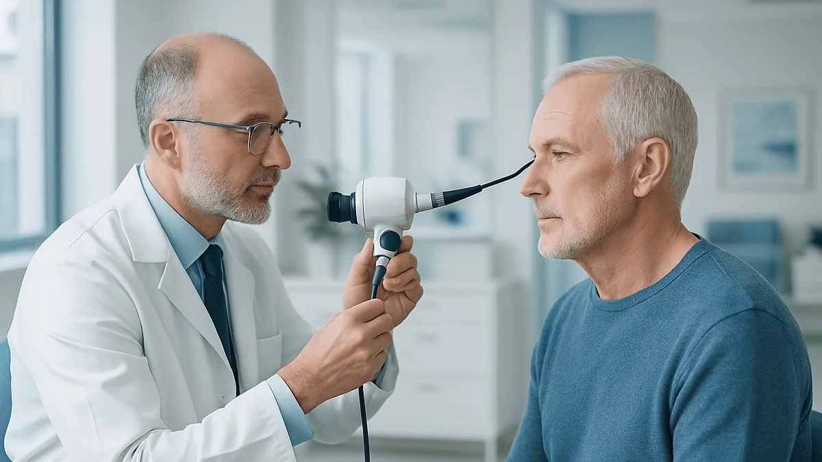
An endoscope is a slim tube with a light source and a lens at its tip; in the brain, it is usually rigid and roughly the thickness of a pencil, though flexible versions exist for winding fluid spaces. A camera on the outer end sends the image to a high-definition monitor, so the surgeon operates while looking up at a screen rather than down into the wound.
That single shift changes almost everything about the operation. Because the light and the lens travel to the target, the surgeon no longer needs a wide corridor to see. Traditional open brain surgery relies on a surgical microscope positioned outside the head, which needs a straight, unobstructed line of sight, so the opening must be large enough to admit light and give the surgeon a view down a cone. An endoscope brings the “eye” inside, past the outer structures, and can even look sideways or around corners with angled lenses.
Working channels matter just as much as the camera. Some endoscopes have built-in ports through which tiny graspers, scissors, suction, and irrigation lines pass. In other techniques, particularly through the nose, the endoscope is held in one hand or on a fixed arm while separate instruments enter through the same or an adjacent passage.
The brain itself does not feel pain, which is why the procedure is about protecting tissue rather than numbing it. The goal of the endoscopic route is to disturb as little normal brain, bone, and skin as possible on the way to the problem. MedlinePlus describes this family of techniques as an alternative to a wider craniotomy for selected conditions, noting that the choice depends on what is being treated and where it sits.
Two doors into the skull: the keyhole and the nose
Surgeons speak of “corridors,” meaning the path from skin to target. For neuroendoscopy there are two main ones, and the anatomy of the problem decides which is used.

The keyhole corridor begins with a small incision in the scalp and a burr hole, which is a round opening drilled in the skull about the width of a coin. Through that opening, the endoscope is guided into one of the brain’s ventricles, the fluid-filled chambers deep inside the brain, or along a natural plane between structures. Cysts, some ventricular tumors, and blockages in the flow of cerebrospinal fluid, the clear liquid that cushions the brain and spinal cord, are typical targets.
The nasal corridor takes advantage of the fact that the pituitary gland, a pea-sized hormone center, sits in a bony pocket directly behind the nose. By passing the endoscope through a nostril and opening the thin wall of the sphenoid sinus, an air pocket at the back of the nasal cavity, the surgeon arrives at the underside of the skull with no external incision at all. Mayo Clinic describes this transsphenoidal approach as the usual route for pituitary tumors that require surgery.
Each door has its own logic. The keyhole gives access to the center of the brain from above; the nose gives access to the floor of the skull from below. Neither is “better” in the abstract. A cyst floating in a ventricle cannot be reached through the nose, and a pituitary tumor rarely justifies a route through the top of the head. The corridor is chosen for the target, not the other way round.
What actually happens in the operating room, step by step
The day rarely feels dramatic from the patient’s side, because it begins and ends asleep. Once general anesthesia has taken effect, the head is positioned and often held gently in a padded frame so it cannot shift. Many teams then register the head to a navigation system, which is a computer that matches the patient’s preoperative MRI or CT scan to their real anatomy, much as a satellite map overlays a moving car. This lets the surgeon see, in real time, where the tip of the instrument is relative to the scan.
For a keyhole approach, a small strip of hair may be trimmed, the scalp is opened, and the burr hole is made. A protective sheath is slid in, and the endoscope follows. Warm saline flows continuously to keep the view clear and to maintain gentle pressure inside the fluid space.
For a nasal approach, no skin is cut. An ear, nose, and throat surgeon often works alongside the neurosurgeon, opening the passage and, at the end, reconstructing the floor of the skull, sometimes using a flap of the patient’s own nasal lining to seal it.
The central part of the operation, whether removing a tumor piece by piece, draining a cyst, or opening a new pathway for fluid, is done while watching the monitor. When the work is complete, instruments are withdrawn, the small opening is closed, and the patient is woken in a recovery area where nurses check alertness, pupils, limb strength, and speech at frequent intervals. MedlinePlus notes that close neurological monitoring in the first hours is standard after any brain operation.
Do they put you to sleep for endoscopic brain surgery?
Yes. Neuroendoscopic procedures, whether through a keyhole or the nose, are performed under general anesthesia, which means you are fully unconscious, breathing through a tube placed by the anesthesiologist, and monitored continuously. MedlinePlus lists general anesthesia as standard for brain surgery. The brain cannot be operated on safely with a moving, awake patient when a rigid scope sits inside a fluid space or beside major vessels.
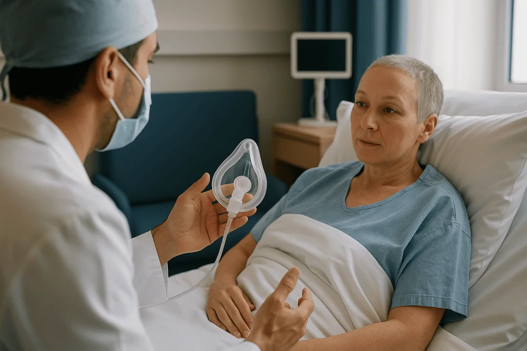
People sometimes confuse neuroendoscopy with “awake craniotomy,” a specialized open technique used for certain tumors near speech or movement areas, in which the patient is woken mid-operation to answer questions. That is a different operation with a different purpose. Endoscopic surgery does not involve being woken partway through.
What to expect around the anesthetic:
- A preoperative visit or phone call with the anesthesia team to review your medical history, allergies, and any medicines you take. Decisions about which medicines to continue or pause belong to that team and your prescribing clinicians, not to a leaflet.
- Instructions on fasting before surgery, because an empty stomach reduces the risk of inhaling stomach contents during anesthesia.
- A sore throat afterward from the breathing tube, and often a feeling of grogginess for the rest of the day.
Recovery from the anesthetic itself is usually measured in hours, but recovery from the surgery is measured in days to weeks, and the two should not be confused. Nausea is common on the first day and is managed by the recovery nurses. If you have had trouble with anesthesia before, tell the team early; it changes how they plan, not whether the operation can happen.
Endoscopic endonasal surgery: reaching the pituitary through the nose
Endoscopic endonasal surgery is the formal name for operating through the nostrils. “Endonasal” simply means inside the nose. The most common reason is a pituitary adenoma, a usually noncancerous growth of the pituitary gland that may press on the optic nerves or overproduce hormones.
Why the nose works so well here comes down to geography. The pituitary sits in the sella turcica, a saddle-shaped hollow in the bone at the base of the skull, and the back wall of the sphenoid sinus is the front wall of that hollow. Open a few millimeters of thin bone and the gland is directly ahead. Mayo Clinic describes the transsphenoidal approach as the method used for most pituitary tumors that need surgery, with the endoscope allowing the surgeon to see the tumor and surrounding structures in magnified detail.
The camera’s wide, close-up view matters for two reasons. The optic nerves cross just above the gland, and the carotid arteries, the main blood supply to the front of the brain, run along either side. Seeing those boundaries clearly helps the surgeon judge how much can be safely removed.
Beyond the pituitary, the same corridor can reach some tumors of the skull base such as certain meningiomas, chordomas, and craniopharyngiomas, though these are more complex operations and are considered case by case.
Afterward, the nose feels congested, and patients are usually asked not to blow it, sneeze with the mouth closed, or strain, because pressure can disturb the repair at the skull base. Johns Hopkins notes that a clear, watery drip from the nose after this surgery should be reported promptly, since it may signal a cerebrospinal fluid leak.
Endoscopic third ventriculostomy for hydrocephalus
Hydrocephalus is a buildup of cerebrospinal fluid inside the brain’s ventricles, which raises pressure and can cause headache, vomiting, vision changes, and unsteadiness. For decades the standard treatment has been a shunt, a thin tube that drains fluid from the brain to the abdomen through a valve under the skin. Endoscopic third ventriculostomy, usually shortened to ETV, offers a different idea for some patients: instead of adding hardware, make a small opening so the brain drains itself.
The name describes the technique. “Third ventricle” is one of the deep fluid chambers; “-ostomy” means creating an opening. Through a burr hole, the endoscope is passed into a lateral ventricle, then through a natural gap into the third ventricle. On its floor lies a thin membrane. The surgeon makes a small hole in it, often widening the hole with a tiny balloon, so fluid can bypass a blockage lower down and be absorbed normally.
The NHS explains that ETV is an alternative to shunt surgery when the cause is an obstruction, and that it avoids the long-term risks of shunt infection and mechanical failure. It is not suitable for every form of hydrocephalus. When the problem is poor absorption rather than a blockage, opening a new channel does not help, and the NHS notes the procedure can fail to keep the pathway open in a proportion of patients, who may then need a shunt.
Because the effects of a blocked or reopened pathway can develop over time, the National Institute of Neurological Disorders and Stroke emphasizes ongoing follow-up after any hydrocephalus treatment. A successful ETV changes the plumbing; it does not end the relationship with the neurosurgery team.
Who neuroendoscopic surgery is usually for, and who is usually asked to wait
The short list of common indications is fairly consistent across guidelines and major centers:
- Pituitary tumors that press on the optic pathway or drive hormone excess, when a specialist team judges surgery is needed rather than medical treatment or observation.
- Obstructive hydrocephalus in adults and children old enough for their fluid-absorbing membranes to have matured, where ETV is considered.
- Colloid cysts and some other cysts inside the ventricles that block fluid flow.
- Selected tumors within or beside the ventricles, where the endoscope can reach for biopsy or removal.
- Certain skull-base tumors accessible from below, in experienced hands.
Being asked to wait is not a rejection. Several groups are commonly observed rather than operated on:
Small pituitary tumors found incidentally on a scan taken for another reason, without symptoms or hormone effects, are often followed with repeat imaging. Mayo Clinic describes watchful waiting as an option for these. Some hormone-producing pituitary tumors respond to medicines that shrink them or control hormone output; in those cases endocrinologists usually lead, and surgery is reserved for when medicine is not enough or not tolerated.
Infants with hydrocephalus are a special case. The NHS notes that ETV is less likely to succeed in very young babies, so shunts remain more common early in life, with ETV sometimes considered later.
People whose problem simply sits in the wrong place, such as a tumor deep in the substance of the brain rather than in a fluid space, are usually steered toward other approaches. The deciding factors are anatomy, the biology of the lesion, the patient’s overall health, and the team’s judgment, weighed together at a multidisciplinary meeting rather than by any one rule.
Neuroendoscopy vs open craniotomy: a side-by-side
Neither approach replaces the other. Open craniotomy, in which a section of skull is temporarily removed to expose the brain, remains the workhorse for many tumors, aneurysms, and injuries. The table below summarizes the typical differences as described by MedlinePlus, Mayo Clinic, and the NHS. It is a guide to the questions worth asking, not a scorecard.
| Feature | Neuroendoscopic (keyhole or nasal) | Open craniotomy |
|---|---|---|
| Entry | Burr hole or nostril | Section of skull lifted and replaced |
| View | Camera inside; panoramic, angled lenses | Microscope outside; straight line of sight |
| Typical targets | Ventricular cysts, hydrocephalus, pituitary and some skull-base tumors | Tumors within brain tissue, aneurysms, trauma, many others |
| Brain handled | Minimal retraction | May require retraction to open a corridor |
| Anesthesia | General | General (occasionally awake technique) |
| Hospital stay | Varies with procedure; MedlinePlus gives 3 to 7 days for brain surgery generally | Same general range, often at the longer end |
| Main specific risks | Cerebrospinal fluid leak (nasal route), fluid-pathway reclosure (ETV) | Larger wound, bone-flap issues, longer scalp healing |
Two honest caveats belong beside any table like this. First, “smaller opening” does not automatically mean “smaller operation”; a skull-base tumor removed through the nose can be a longer and more delicate procedure than a straightforward craniotomy. Second, the person best placed to weigh these columns for your case is the surgeon who has seen your scans, because the factors that matter most, such as how a tumor wraps around a vessel, do not appear in any general comparison.
Risks and alternatives in plain language
Every operation on or near the brain carries risk, and the endoscopic route changes the pattern of risk rather than removing it. The list below reflects complications described by MedlinePlus, Mayo Clinic, Johns Hopkins, and the NHS for these procedures.
- Bleeding inside the brain or fluid spaces, which can be harder to control through a narrow corridor and may occasionally require conversion to an open approach.
- Infection, including meningitis, an infection of the membranes around the brain, which is more likely when there is a fluid leak.
- Cerebrospinal fluid leak, mainly after nasal surgery, where the seal at the skull base does not hold; it may need bed rest, a temporary drain, or a return to surgery.
- Hormone disturbance after pituitary surgery, most often a temporary problem with water balance called diabetes insipidus, which causes excessive thirst and urination and is watched for closely in the first days.
- Damage to nearby structures, including vision changes, particularly with lesions close to the optic nerves.
- Failure of the intended effect, such as an ETV opening closing over, or incomplete tumor removal that requires further treatment.
- General anesthetic and surgical risks such as blood clots in the legs and chest infections.
Alternatives depend entirely on the diagnosis. For pituitary tumors they may include observation with repeat scans, hormone-controlling medicines, or focused radiation. For hydrocephalus the main alternative to ETV is a shunt. For some cysts and tumors, a conventional craniotomy is the alternative, and for others no treatment at all, with monitoring, is a reasonable choice.
Neutral language matters here because fear and false reassurance are both unhelpful. A good consultation will name the specific risks that apply to your anatomy, estimate how likely they are for you, and describe what would be done if each occurred.
How long does it take to recover from endoscopic brain surgery?
People searching for endoscopic brain surgery recovery time usually want one number, and the honest answer is a range that depends on the route, the diagnosis, and the person. MedlinePlus, describing brain surgery broadly, gives a typical hospital stay of 3 to 7 days and says full recovery can take several weeks to months, longer if there were complications or if the underlying condition itself caused deficits before surgery.
Within that broad picture, some patterns hold:
After a keyhole procedure such as ETV or a cyst removal, the incision is small and scalp healing is quick. Fatigue and headache are the main complaints, and most people are walking the day after surgery. The NHS notes that hospital stays after ETV are generally shorter than after shunt placement, because no hardware is tunneled under the skin.
After endoscopic endonasal surgery, the nose rather than the head dominates the early recovery. Congestion, crusting, reduced sense of smell, and a dull headache are common. Johns Hopkins describes several weeks of nasal healing and advises avoiding nose blowing and heavy lifting during that time. Hormone levels are checked repeatedly, and some people need temporary hormone replacement while the gland recovers.
Return to work is highly individual. Someone with a desk job after an uncomplicated pituitary operation may feel ready in a few weeks; a person whose hydrocephalus caused months of unsteadiness may need a longer period of rehabilitation. Driving is usually restricted until the team confirms it is safe, and any seizure after surgery extends that restriction under most licensing rules.
None of these timelines is a promise. They are the shapes most recoveries take, offered so that a slow week does not feel like failure and a fast one does not tempt you to skip follow-up.
The first days and weeks: what recovery usually looks like
Day zero is spent in a recovery or high-dependency area. Nurses perform neurological checks every hour or so at first, asking your name, the date, and where you are, shining a light in your eyes, and testing grip strength. This can feel relentless when you want to sleep; it is the single most important safety net of the first night, because it catches bleeding or swelling early.
Days one to three tend to bring the return of appetite and the first walks along the corridor. Early mobility is encouraged because lying still raises the risk of blood clots in the legs; compression stockings or calf pumps are common. After nasal surgery, saline rinses may be started to keep the passages clear, following the team’s specific instructions. After pituitary surgery, blood and urine tests track sodium levels and urine output, because water-balance problems most often appear in this window and again about a week later.
Discharge usually comes within the 3-to-7-day range MedlinePlus gives for brain surgery generally, with a written plan covering wound care, activity limits, medicines, and who to call.
Weeks one to four at home are about pacing. Fatigue is the most under-anticipated symptom; many people describe needing an afternoon rest even when they feel mentally fine. Headaches gradually fade. Scalp stitches or staples, if used, are removed at a clinic visit. Nasal crusting peels away over several weeks.
A follow-up scan and clinic appointment usually fall within the first one to three months, depending on the condition. For hydrocephalus, the National Institute of Neurological Disorders and Stroke stresses that monitoring continues long term, since drainage pathways can change. For pituitary conditions, an endocrinologist typically joins the follow-up to reassess hormone function once the gland has settled.
What people often get wrong about neuroendoscopy
“It’s the most complicated neurosurgery procedure.” Searchers often ask which neurosurgery is hardest, and neuroendoscopy is sometimes assumed to top the list because it sounds high-tech. Complexity in neurosurgery is driven by the target, not the tool. A routine ETV is a relatively short, standardized operation; an endoscopic removal of a skull-base tumor wrapped around the carotid artery is among the most demanding procedures any surgeon performs. Both are “neuroendoscopy.” The label tells you the route, not the difficulty.
“Small hole means small risk.” The opening is smaller, but the surgeon is still working millimeters from vessels and nerves. MedlinePlus lists the same core risks, bleeding, infection, swelling, seizures, for brain surgery regardless of approach. The risk profile shifts; it does not vanish.
“Through the nose means it’s really an ENT operation.” An ear, nose, and throat surgeon often helps open the corridor, but the work at the pituitary or skull base is neurosurgery, and the two specialties usually operate together.
“Endoscopic surgery is always the modern choice, so open surgery must be outdated.” Craniotomy remains the right operation for most tumors within brain tissue and for many vascular problems. Being offered an open approach is not a sign of an old-fashioned team; it is usually a sign that the anatomy demands it.
“Once the ETV is done, hydrocephalus is over.” The NHS is clear that the new opening can close in some patients, which is why follow-up continues and why returning symptoms must be taken seriously.
“Recovery is a weekend.” Anesthetic grogginess clears in a day. Healing of the skull base, hormone recovery, and fatigue run on a timescale of weeks, as the sources above describe.
Questions to ask your care team before endoscopic pituitary surgery or any neuroendoscopic procedure
A consultation goes better with a written list. These questions are phrased so that the answers give you something concrete, and they apply whether you are facing endoscopic pituitary surgery, an ETV, or a keyhole cyst removal.
- Which corridor are you planning, keyhole or nasal, and what about my anatomy makes that the right one?
- What is the goal of this operation for me: complete removal, partial removal to relieve pressure, biopsy, or restoring fluid flow?
- What would happen if you needed to change to an open approach during surgery, and how often does that happen for this type of case?
- Which two or three specific complications are most relevant to my situation, and what would be done about each?
- What are the alternatives, including observation, medicine, radiation, or a shunt, and why do you favor surgery now rather than waiting?
- Who else will be in the operating room, such as an ENT surgeon or an endocrinologist, and who leads my follow-up afterward?
- What blood tests or hormone checks will I need in the first weeks, and how will I know the results?
- What activity restrictions apply, for how long, and when can I drive, fly, exercise, and return to work?
- Which of my current medicines should I discuss with the anesthesia team or my prescribing doctor before surgery?
- What is the follow-up imaging plan, and what would prompt an earlier scan?
- If this is for a child: how will you prepare them for the day, who can stay with them, and what will the first night look like?
Write the answers down or ask permission to record them. Mayo Clinic and the NHS both suggest bringing a companion to consultations for exactly this reason; two people hear more than one. If an answer is “it depends,” ask what it depends on. That single follow-up often reveals the real decision your team is weighing.
When to call your doctor after neuroendoscopic surgery
Most recoveries are uneventful, but a small number of complications need prompt attention, and the early signs can be subtle. The list below draws on guidance from MedlinePlus, Johns Hopkins, and the NHS. If any of these occur, contact the surgical team or seek emergency care without waiting for the next appointment.
Seek care urgently for:
- A clear, watery drip from the nose or a salty or metallic taste at the back of the throat after nasal surgery, especially when leaning forward, which may indicate a cerebrospinal fluid leak.
- Fever with a stiff neck, severe headache, sensitivity to light, or confusion, which can signal meningitis.
- A headache that is severe, rapidly worsening, or different from the postoperative ache you were told to expect, particularly with vomiting or drowsiness.
- New weakness, numbness, difficulty speaking, or facial drooping.
- Sudden change in vision, such as loss of part of the visual field or double vision.
- A seizure of any kind.
- Extreme thirst with very frequent, pale urination after pituitary surgery, which may reflect a water-balance disturbance that needs blood tests.
- Redness, swelling, warmth, or discharge at a scalp incision, or the wound edges separating.
- Calf pain and swelling, chest pain, or sudden shortness of breath, which can indicate a blood clot.
- After ETV or any hydrocephalus surgery, a return of the symptoms that led to treatment, such as headache with vomiting, unsteadiness, sleepiness, or, in children, irritability and a bulging soft spot.
Make a non-urgent call for persistent nausea, low mood that does not lift, sleep disruption, or a sense of smell that has not begun to return after several weeks; these are common and manageable, and your team would rather hear about them than not. Keep the emergency contact number from your discharge paperwork somewhere visible, and make sure whoever is staying with you knows where it is and what the red flags are.
Frequently asked questions
How long does it take to recover from endoscopic brain surgery?
Most people leave the hospital within the 3-to-7-day range MedlinePlus gives for brain surgery, and full recovery typically takes several weeks to a few months depending on the route and diagnosis. Keyhole procedures such as ETV often have the shortest hospital stays; nasal skull-base surgery involves several weeks of nasal healing and hormone monitoring. Fatigue is usually the last symptom to settle. Your team will set individual limits on driving, lifting, and work.
Do they put you to sleep for endoscopic surgery on the brain?
Yes, neuroendoscopic surgery through a keyhole or the nose is performed under general anesthesia, so you are fully unconscious and monitored throughout. The ‘awake craniotomy’ some people have read about is a different open technique used for tumors near speech or movement areas. Expect a sore throat from the breathing tube and grogginess for the rest of the day, followed by frequent neurological checks in the recovery area.
What is the most complicated neurosurgery procedure?
There is no single answer, because difficulty depends on the target rather than the technique. Endoscopic removal of a skull-base tumor wrapped around the carotid artery ranks among the most demanding operations, while an endoscopic third ventriculostomy is comparatively standardized. Open procedures such as complex aneurysm repair or brainstem tumor surgery are equally challenging. The word ‘neuroendoscopy’ describes the route into the head, not how hard the operation is.
What is endoscopic endonasal surgery used for?
Endoscopic endonasal surgery is used mainly to remove pituitary tumors and some tumors of the skull base by passing a camera and instruments through the nostrils and the sphenoid sinus. Mayo Clinic describes this transsphenoidal approach as the usual route for pituitary tumors needing surgery. It avoids an external incision and gives a magnified view of the optic nerves and carotid arteries that border the gland.
How does endoscopic third ventriculostomy differ from a shunt?
An endoscopic third ventriculostomy creates a small opening in the floor of the third ventricle so cerebrospinal fluid can bypass a blockage and be absorbed naturally, whereas a shunt is an implanted tube and valve that drains fluid to the abdomen. The NHS notes ETV avoids the infection and mechanical-failure risks of shunts but suits only obstructive hydrocephalus, and the opening can close in some patients.
Is endoscopic brain surgery recovery time shorter than after a craniotomy?
Often, but not always. The smaller opening usually means faster scalp healing and less pain at the wound, and the NHS reports shorter stays after ETV than after shunt surgery. Recovery from a complex skull-base tumor removed through the nose, however, can take as long as an open operation because of skull-base healing and hormone recovery. The underlying condition shapes recovery more than the route does.
What does endoscopic pituitary surgery feel like afterward?
The nose dominates the early days: congestion, crusting, a reduced sense of smell, and a dull headache are common, while the head itself has no incision. Johns Hopkins describes several weeks of nasal healing during which nose blowing, straining, and heavy lifting are avoided. Blood and urine tests track hormone and water balance, and some people need temporary hormone support while the gland recovers under an endocrinologist’s care.
Can children have neuroendoscopic surgery?
Yes, neuroendoscopy is widely used in children, particularly for hydrocephalus, cysts, and some tumors in the fluid spaces. The NHS notes that ETV is less likely to succeed in very young infants, so shunts remain more common early in life. Preparation focuses on age-appropriate explanation, a parent staying close, comfort measures, and clear guidance on the warning signs of returning pressure, such as irritability, vomiting, or unusual sleepiness.
What are the main risks of neuroendoscopic surgery?
The core risks are the same as for any brain surgery, bleeding, infection, swelling, and seizures, as MedlinePlus lists, plus route-specific ones. Nasal surgery carries a risk of cerebrospinal fluid leak and hormone disturbance; ETV carries a risk that the new opening closes. Injury to nearby vision or blood-vessel structures is possible with lesions close to them. Your surgeon should name the risks specific to your anatomy.
Who decides whether I am suitable for a keyhole or nasal approach?
The treating neurosurgical team decides, usually after a multidisciplinary meeting that includes radiologists and, for pituitary conditions, endocrinologists. Suitability rests on where the problem sits, what it is likely to be, whether it is causing symptoms or hormone effects, and your general health. Some people are advised to wait and have repeat scans instead. The decision is individual and should be explained to you in plain terms.
References
- MedlinePlus – Brain surgery
- NHS – Hydrocephalus: Treatment
- NIH – National Institute of Neurological Disorders and Stroke: Hydrocephalus
This article is for general information only and is not a substitute for professional medical advice. Please consult a qualified doctor about your individual situation.

More from the Blog

Is Everyday Life With HSP Manageable? Stairs, Driving, Work and Pacing Strategies
For most people, daily life with HSP (hereditary spastic paraplegia) is manageable, though it usually needs adjusting over time. HSP causes gradually increasing stiffness…

Trigger Assessment Before Migraine Treatment: What Your Neurologist Wants Tracked
A migraine trigger assessment is a structured period, usually several weeks to a few months, of recording attacks alongside sleep, meals, stress, menstrual cycle,…

What to Ask Your Neurologist When an Autonomic Neuropathy Plan Involves Several Specialists
When several specialists share an autonomic neuropathy plan, ask your neurologist who coordinates the whole plan, which symptom each clinician owns, how test results…

Spinal Cord Disorder Follow-Up: Which Bladder, Bowel or Weakness Changes Need an Urgent Call
After a spinal cord disorder, call your care team the same day for any new loss of bladder or bowel control, difficulty passing urine,…

Fetal Surgery or Repair After Birth for Spina Bifida: What Does Each Pathway Involve?
Fetal surgery for spina bifida closes the spinal opening before birth, usually between about 19 and 26 weeks of pregnancy, through an incision in…
Daily Life With ALS: Assistive Devices, Nutrition and Energy Conservation Explained
Daily life with ALS changes gradually as motor neurons weaken muscles, but much of it can be planned for and managed. Assistive devices such…