

How Orofacial Pain Is Diagnosed: Jaw Movement, Bite, Muscle and Nerve Examination

Key Takeaways
- Orofacial pain diagnosis relies chiefly on history and hands-on examination; imaging and tests are added only to answer a specific question the exam raises.
- Temporomandibular disorders are the most common non-dental cause of chronic facial pain, affecting an estimated 11 to 12 million US adults according to NIDCR.
- Jaw clicking without pain or restricted opening is common and, per NIDCR, generally does not need treatment on its own.
- There is no lab test for trigeminal neuralgia; the Mayo Clinic notes MRI is used to exclude other causes such as multiple sclerosis or a compressing vessel.
- Tenderness on pressing a muscle is only diagnostic when it reproduces the patient's familiar pain, not simply when it hurts.
- Atypical odontalgia is toothache with no dental cause, and recognising it early prevents cascades of root canals and extractions that do not relieve the pain.
Orofacial pain is diagnosed mainly through a detailed history and a hands-on examination rather than a single test. Clinicians measure how the jaw opens and moves, check how the teeth meet, press the chewing muscles to find tender or referring spots, and test facial sensation and nerve function. Imaging, tooth vitality tests or blood tests are added only when the examination points toward a specific cause.
The tooth has been checked twice. The X-ray looks clean. The filling was replaced anyway, just in case. And the ache in the left cheek, the one that flares when she chews a bagel or wakes her at four in the morning, is still there. Somewhere between the third appointment and the first sleepless week, the question changes from “which tooth is it?” to “what if it isn’t a tooth at all?”
That pivot is exactly where orofacial pain diagnosis begins. Orofacial pain is the umbrella term for pain in the mouth, jaw and face, and the region is crowded: teeth, sinuses, a hinge joint that moves thousands of times a day, chewing muscles thick as a thumb, and the trigeminal nerve, the large sensory nerve that supplies feeling to the whole face.
The good news is that the examination that sorts these apart is low-tech, methodical and surprisingly revealing. This article walks through what the examiner is actually doing with your jaw, your bite, your muscles and your nerves, and what the results can and cannot tell you.
What does an orofacial pain diagnosis actually involve?
Most people expect a scan. What they get, at least at first, is a conversation that can run twenty minutes before anyone touches their face. That is not a delay; it is the single most informative part of the visit. Pain from a cracked tooth, an irritated jaw joint, a strained chewing muscle and an inflamed nerve each has a characteristic story: where it starts, what sets it off, how long an episode lasts, whether it throbs, burns, stabs or aches.
The examiner will usually ask you to point to the pain with one finger, then describe it in your own words. They will want to know whether it wakes you, whether cold drinks, chewing, talking or touching the skin trigger it, whether the jaw clicks or locks, and whether you grind or clench. Headache history, neck pain, sleep, stress and recent dental work all matter, because the face shares nerve pathways with the head and neck and pain routinely travels between them.
Only after the history narrows the field does the physical examination begin. It follows a predictable sequence: watching the jaw move and measuring how far it opens, checking how the upper and lower teeth meet, pressing specific muscles and joint landmarks, then testing the nerves of the face for altered sensation or trigger points. Each step is designed to either provoke the patient’s own pain or fail to, and that reproduction is the key evidence.
Tests come last, and selectively. The Mayo Clinic describes temporomandibular joint (TMJ) assessment as listening to and feeling the jaw while you open and close, observing range of motion, and pressing around the joint to find discomfort, with X-rays, CT or MRI reserved for when the examination suggests a structural problem. The same principle applies across orofacial pain: the exam decides whether a test is worth ordering.
What are the common causes of facial pain?
Ask a dentist, a neurologist and an ear, nose and throat surgeon what causes facial pain and you will hear three different lists. All of them are right, which is precisely why diagnosis needs a structured approach. Broadly, the causes fall into four families, and the examination is built to test each in turn.
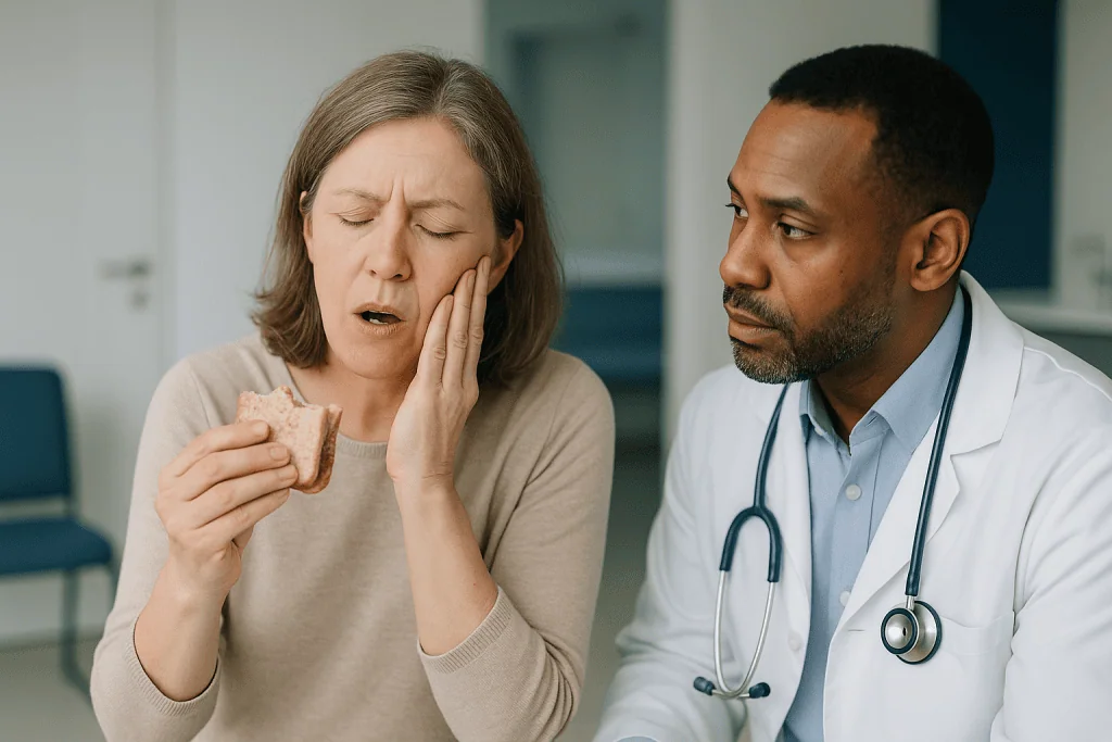
Dental and oral causes come first because they are the most frequent and the most fixable. Tooth decay reaching the pulp, a cracked tooth, gum infection, an impacted wisdom tooth or an ulcer can all produce pain that seems far larger than its source. Dental pain typically responds to a specific tooth being tapped, cooled or bitten on.
Musculoskeletal causes centre on the temporomandibular joints, the two hinges just in front of the ears, and the muscles that close the jaw. Temporomandibular disorders (TMD) are the umbrella term for pain and dysfunction in these joints and muscles, and they are the most common non-dental source of chronic facial pain according to the National Institute of Dental and Craniofacial Research (NIDCR).
Neuropathic causes arise from the nerves themselves rather than the tissues they supply. Trigeminal neuralgia, a condition of brief electric-shock pains along the trigeminal nerve, is the best known. Persistent neuropathic pain after dental procedures or injuries, and burning mouth syndrome, a burning sensation with no visible cause, belong in this family too.
Referred and systemic causes round out the list. Sinus infections, ear disease, salivary gland stones, migraine and cluster headache can all present as face pain. Two are medically urgent: giant cell arteritis, an inflammation of blood vessels in older adults that can cause jaw pain on chewing and threaten sight, and heart attack, which the American Heart Association notes can present as pain in the jaw, particularly in women.
What is the most common orofacial pain disorder?
Once tooth problems are set aside, the answer is a temporomandibular disorder. NIDCR estimates that around 11 to 12 million adults in the United States have pain in the region of the temporomandibular joint, and that TMDs are about twice as common in women as in men, with the highest rates in women between 35 and 44. That makes TMD the condition an examiner is most often trying to confirm or rule out when the teeth check out.
The term covers several distinct problems that can coexist. Myalgia is pain arising from the chewing muscles themselves, usually the masseter over the angle of the jaw and the temporalis on the side of the head. Arthralgia is pain from the joint capsule and its ligaments. Disc displacement describes the small cartilage cushion inside the joint slipping out of position, which produces the familiar click or, when it blocks the joint, a sudden inability to open fully. Degenerative joint disease is the wear-and-tear arthritis that can develop in any joint, this one included.
Why does this matter for diagnosis? Because each subtype is identified by a different examination finding. Muscle pain is reproduced by pressing the muscle. Joint pain is reproduced by pressing directly over the joint or loading it at the end of an opening movement. Disc problems announce themselves as sounds or jaw deviation during movement. The examiner is not simply asking “is it TMD?” but “which tissue hurts, and does pressing it reproduce the pain you came in with?”
One reassuring point from NIDCR bears repeating: jaw sounds without pain or restricted movement are common and, on their own, do not usually need treatment. A clicking jaw that does not hurt is a finding, not a diagnosis, and a careful examiner will say so rather than label it.
How the jaw movement examination works, step by step
The jaw examination begins before you are asked to do anything. A skilled examiner watches you talk, notes whether one side of the face looks fuller (a sign of an overworked masseter), and checks whether the chin sits centred at rest. Then comes the part most people remember: “Open as wide as you comfortably can.”
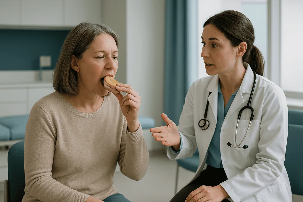
Range of motion is measured with a small ruler between the edges of the upper and lower front teeth, at maximum comfortable opening and then at maximum forced opening, with the examiner noting whether pain appears and where. Sideways movement to each side and forward protrusion of the jaw are measured too. A restricted opening with a hard, abrupt end point suggests something mechanical in the joint; a restricted opening that softens with gentle assistance suggests muscle guarding. The Mayo Clinic lists observing range of motion as a core step in assessing TMJ disorders precisely because it distinguishes these patterns.
Deviation is the next clue. If the chin drifts toward one side during opening and returns to centre at the end, the disc in one joint is likely catching and releasing. If it deviates and stays deviated, that joint may not be translating forward properly. The examiner may place fingertips or a stethoscope just in front of each ear to feel and hear for clicks, pops or the gravelly sound called crepitus, and to note the point in the movement where they occur.
Joint loading tests follow. Pressing over the joint while the mouth is open, or asking you to bite on a cotton roll on one side, shifts pressure onto one joint at a time. Pain appearing in the joint being loaded, and matching your usual pain, points to arthralgia rather than muscle pain. The whole sequence takes a few minutes and involves nothing more invasive than a ruler and two fingers.
What the bite and teeth tell the examiner
Occlusion, the way the upper and lower teeth meet, is one of the most misunderstood parts of orofacial pain diagnosis. For decades an “uneven bite” was blamed for almost every jaw ache, and teeth were ground down or rebuilt to fix it. Current guidance from bodies such as NIDCR and the NHS is far more cautious: bite irregularities are common in people without pain, and irreversible changes to teeth are not recommended as a first response to jaw pain. So the bite check today is less about hunting for the cause and more about gathering context.
The examiner will ask you to close normally and look at how the teeth interlock, whether they meet evenly on both sides, and whether the front teeth show flattened edges or chips that indicate grinding. Wear facets, tiny polished patches where teeth have rubbed, tell a story about night-time clenching that patients often deny because they are asleep when it happens. Scalloped edges on the tongue and a ridge of thickened tissue along the inside of the cheek carry the same message.
Then the teeth themselves are tested as possible pain sources. Tapping each tooth with the handle of a mirror (percussion) identifies an inflamed ligament around a tooth root. A cold stimulus on the tooth surface checks whether the pulp is alive and how it responds; a lingering, exaggerated reaction suggests an inflamed pulp, while no response at all suggests a dead one. Biting on a small device tooth by tooth can reveal a crack that no X-ray shows.
This is where dental and non-dental pain diverge. Genuine tooth pain almost always has a tooth that responds abnormally to at least one of these tests. When every tooth tests normally and the pain persists, the examiner’s attention moves decisively to muscle, joint and nerve, and the patient is spared another filling that would not have helped.
How the muscle examination finds hidden pain generators
Palpation, pressing tissue with the fingers to assess tenderness and texture, is the heart of the muscle examination, and it is more standardised than it looks. The examiner applies a consistent, firm pressure for a few seconds to each site, asks you to rate any discomfort, and crucially asks a second question: “Is that your pain, the one you came in with?” Tenderness alone is common. Tenderness that reproduces the patient’s familiar pain is diagnostic evidence.
The masseter, the thick muscle you can feel bulging at the angle of your jaw when you clench, is examined along its full length from the cheekbone to the jawline, both from outside and, with a gloved finger, from inside the cheek. The temporalis, the fan-shaped muscle over the temple, is checked across its front, middle and back fibres; its tendon, where it attaches inside the mouth behind the upper back teeth, is a frequent and frequently missed source of pain that feels exactly like a bad molar.
Deeper muscles need indirect approaches. The lateral pterygoid, which sits behind the upper jaw and helps open and protrude the mandible, cannot be pressed directly, so the examiner asks you to push your jaw forward or open against gentle resistance and notes whether that provokes pain. Neck and shoulder muscles are included because they refer pain into the face and frequently tighten alongside the jaw muscles.
The concept of referred pain, where pressing one spot produces pain somewhere else, is why this step matters so much. A tender band in the masseter can send pain to an upper molar; a trigger point in the sternocleidomastoid in the neck can send it to the forehead or ear. When pressing a muscle lights up the very tooth or temple the patient has been worrying about, the diagnosis often clicks into place without a single image.
How the nerve examination works and what it is looking for
The nerve examination is quieter than the rest, and patients sometimes wonder why the clinician is brushing cotton wool across their cheek. The purpose is to map the trigeminal nerve’s three branches, which supply the forehead, the mid-face and the jaw respectively, and to detect any area where sensation is reduced, heightened or distorted.
Light touch, a gentle pinprick and sometimes a cool object are applied to matching points on both sides of the face. The examiner is comparing sides: does a stroke that feels normal on the right feel dull, tingly or painfully sharp on the left? Allodynia, pain from a stimulus that should not hurt, such as a light brush or a breeze, is a hallmark of neuropathic pain. Numbness in a defined patch is equally significant, because it means a nerve is not conducting properly and needs an explanation.
Motor function is checked by asking you to clench while the examiner feels both masseters, and by testing the muscles of facial expression supplied by the facial nerve: raising eyebrows, closing eyes tightly, smiling, puffing cheeks. Weakness on one side is a red flag for something beyond ordinary jaw pain. A brief look at eye movements, hearing and the throat covers the other cranial nerves that share the neighbourhood.
For suspected trigeminal neuralgia specifically, the examiner searches for trigger zones, small areas around the mouth, nose or gums where a light touch sets off the characteristic jolt. The NHS describes these attacks as sudden, severe, electric-shock-like pains lasting from a few seconds to about two minutes, often set off by everyday actions such as chewing, talking, brushing teeth or a cold wind. A classic history plus a normal sensory examination between attacks fits typical trigeminal neuralgia; any persistent numbness pushes the clinician toward imaging to look for a cause.
Are there any lab tests used to diagnose trigeminal neuralgia?
Not in the way people hope. There is no blood test, saliva test or nerve conduction study that confirms trigeminal neuralgia. The Mayo Clinic is explicit that the diagnosis rests mainly on the patient’s description of the pain, its type, location and triggers, combined with a neurological examination. The one test that is routinely used is MRI, and its job is not to prove the condition but to look for something else: a blood vessel pressing on the nerve, a tumour, or the plaques of multiple sclerosis, which can produce identical pain.
The same logic applies across orofacial pain. Tests are chosen to answer a specific question raised by the examination, and the table below summarises what each one can and cannot do.
| Test | What it shows well | Typical reason to order | What it cannot show |
|---|---|---|---|
| Dental X-ray / panoramic | Tooth roots, bone loss, abscess, impacted teeth, gross joint bone changes | Suspected dental source; first look at joints | Muscles, disc, nerves, early cracks |
| Cold and percussion tooth tests | Whether a tooth’s pulp is alive and inflamed | Pain that could be dental | Anything outside the tooth |
| CT / cone-beam CT | Fine bone detail of the joint and jaws, sinuses | Suspected arthritis, fracture, sinus disease | Disc position, soft tissue, nerves |
| MRI | Disc, joint fluid, soft tissue, brain, nerve-vessel contact | Suspected disc displacement; any neuropathic pain with abnormal exam | Muscle trigger points, bite problems |
| Blood tests (inflammatory markers) | Systemic inflammation | Older adult with jaw pain on chewing, headache or visual change (possible giant cell arteritis) | TMD, neuralgia, dental pain |
Two habits distinguish careful practice. First, a test is not ordered simply because a patient is worried; a normal MRI in someone whose examination clearly shows muscle pain reassures briefly and explains nothing. Second, when a scan finds an incidental abnormality, the examiner asks whether it matches the clinical picture before acting on it. Disc displacement shows up on MRI in many people who have never had jaw pain.
What is atypical odontalgia, and why does it matter for orofacial pain diagnosis?
Atypical odontalgia is a persistent aching or throbbing pain felt in a tooth or a tooth socket where no dental cause can be found, even after thorough testing. Odontalgia simply means toothache; the “atypical” signals that the tooth itself is not the problem. Many clinicians now group it under the term persistent idiopathic facial pain, or describe it as a persistent dentoalveolar pain disorder, reflecting the view that it is a neuropathic pain, a problem in how the nerves signal, rather than a problem in the tooth.
It matters enormously for diagnosis because it is the condition most likely to lead to a cascade of unnecessary treatment. The pattern is painfully familiar: a tooth aches, tests are borderline, a root canal is performed, the ache continues, the tooth is extracted, and the pain settles into the empty socket as if nothing had happened. Every intervention is done in good faith, and each one fails for the same reason: the generator of the pain was never inside the tooth.
What does the examination show? Typically, the teeth respond normally to cold and percussion, X-rays are unremarkable, and pressing the chewing muscles does not reproduce the pain. Sensory mapping may reveal subtle changes over the area, such as heightened sensitivity to light touch. The pain is usually constant rather than episodic, which separates it from the brief shocks of trigeminal neuralgia, and it does not track a single nerve branch neatly.
Diagnosing atypical odontalgia is largely a matter of exclusion done properly, and it takes courage on both sides. The clinician has to resist the pull of “just one more procedure,” and the patient has to accept that a normal-looking tooth can hurt for reasons that a drill cannot reach. Recognising it early spares teeth and redirects care toward approaches designed for nerve pain, decisions that sit with the treating team.
Who is examined straight away, and who is usually asked to wait and watch
Not every aching jaw needs a full specialist work-up, and not every one can wait. Sorting these groups is itself part of the diagnostic process, and it tends to follow a few consistent principles.
Same-day or urgent assessment is appropriate when facial pain comes with signs that suggest something beyond the jaw and teeth. Persistent numbness or weakness in the face, a jaw that has locked shut or open and will not release, swelling with fever, pain on chewing in someone over about 50 who also has a new headache or changes in vision, or jaw pain that arrives with chest pressure, breathlessness or sweating all fall into this group. The American Heart Association lists jaw pain among heart attack warning signs, and it is treated as such until proven otherwise.
A prompt but routine appointment suits pain that is clearly dental in character: a tooth that reacts to cold, hurts to bite on, or sits beside swollen gum. It also suits neuropathic patterns such as electric-shock pains, because early evaluation shortens the time spent guessing.
The wait-and-watch group is larger than people expect. NIDCR notes that for many people TMD pain is mild and improves on its own without treatment, and both NIDCR and the NHS recommend beginning with conservative, reversible measures such as softer foods, avoiding wide yawning and gum, gentle heat or ice, and awareness of clenching. A clinician who suggests a few weeks of this before further tests is not dismissing you; they are following guidance that reserves imaging and intervention for pain that persists or worsens.
What shifts a person from the watch group to the work-up group is change: pain that spreads, escalates, changes character, or starts to interfere with sleep and eating. A pain diary over that period makes the shift visible and gives the next examination far better material to work with.
What the weeks after an orofacial pain diagnosis usually look like
A diagnosis of TMD, atypical odontalgia or trigeminal neuralgia is rarely the end of the assessment; it is the start of a period of structured observation. The examiner has formed a working hypothesis, and the following weeks test it.
For musculoskeletal pain, the typical first phase is a trial of self-care and reversible measures. Patients are often asked to keep a simple diary noting pain intensity, jaw sounds, clenching episodes and triggers. A follow-up visit repeats the key measurements, opening range, muscle tenderness, joint sounds, so that change can be documented rather than guessed. If the pattern of tenderness has moved or the opening has improved, the muscle-based diagnosis is supported. If the joint still catches or locks, imaging of the disc may be the next step. NIDCR emphasises that treatments in this phase should be reversible and that irreversible ones, such as grinding down teeth or surgery, should not be first-line.
For suspected neuropathic pain, the timeline often includes waiting for an MRI and a review of the result alongside the examination findings. Where a clinician prescribes a medicine that calms over-active nerve signalling, such as an anticonvulsant, its effect is itself used diagnostically: the NHS notes that trigeminal neuralgia pain often responds to this class in a way that ordinary painkillers do not. Any decision about starting, adjusting or continuing such a medicine belongs entirely to the prescribing clinician, who will also monitor for side effects.
Throughout, the diagnosis is held provisionally. Facial pain conditions overlap, and a person can genuinely have both a muscle disorder and a neuropathic component. A good team says so plainly, revisits the examination at each visit, and is willing to revise the label. Patients who understand this find the process far less frustrating than those expecting a single verdict on day one.
What people often get wrong about how orofacial pain is diagnosed
“A normal X-ray means nothing is wrong.” Dental radiographs show teeth and bone; they do not show muscles, the joint disc or nerves. Most orofacial pain that is not dental will have an entirely normal X-ray, and that result is a clue pointing away from the teeth, not proof that the pain is imagined.
“A clicking jaw needs treatment.” Joint sounds are common. NIDCR is clear that clicking or popping without pain or restricted movement is generally not a problem requiring intervention. The examiner records the sound and moves on to what actually hurts.
“My bite must be the cause.” Occlusal irregularities are found in plenty of pain-free mouths. Current guidance does not support permanently altering teeth to treat jaw pain, and a clinician who moves straight to bite adjustment is working from an older model.
“If the pain is in a tooth, the problem is the tooth.” Referred pain from the masseter, temporalis tendon and even the neck can land squarely in a molar. Atypical odontalgia can persist after the tooth is gone. The tooth is guilty only if it fails its own tests.
“There must be a scan or blood test that will settle this.” For trigeminal neuralgia, the Mayo Clinic notes the diagnosis rests on history and neurological examination; MRI is there to exclude other causes. For TMD, imaging is added only when the examination suggests a structural problem.
“Facial pain that has lasted months can’t be serious.” Usually true, but not always. Jaw pain on chewing in an older adult with headache or blurred vision, new numbness, or pain with unexplained weight loss or a lump warrants prompt evaluation regardless of duration.
“The examiner pressed on my face and it hurt, so that proves the diagnosis.” Tenderness alone is common. What counts is whether pressure reproduced your familiar pain. If it produced a different pain, the search is not over.
Questions to ask your care team about your orofacial pain diagnosis
The diagnostic visit moves quickly and it is easy to leave with a label but no understanding of how it was reached. A handful of direct questions turns the appointment into a shared piece of detective work rather than a verdict handed down.
- Which tissue do you think is generating my pain: tooth, joint, muscle or nerve, and what in the examination pointed you there? A confident answer should name a finding, such as “pressing your temporalis tendon reproduced the molar pain.”
- Did any tooth test abnormally, or were all of them normal? This single answer tells you whether more dental treatment is likely to help.
- What did my opening range and jaw movement show, and will you measure them again next time? Documented change is how a working diagnosis is confirmed or revised.
- Do I need imaging now, and if so, what specific question is it meant to answer? A scan ordered to answer a question is far more useful than one ordered to reassure.
- Is there anything in my history or examination that worries you about a more serious cause? You are entitled to hear that red flags were considered and either found or not found.
- Could I have more than one condition at once? Muscle pain and neuropathic pain frequently overlap, and treatment differs.
- What should I track before the next visit, and what change would make you want to see me sooner? This gives you a role and a safety net.
- Which of the options you are suggesting are reversible, and which are not? Guidance from NIDCR favours reversible approaches first for jaw pain; hearing this distinction spoken aloud protects you.
Write the answers down or ask permission to record them. Facial pain has a way of making a person doubt their own account, and a clear record of what the examination found is the best antidote to that uncertainty.
When to call your doctor
Most orofacial pain is not dangerous, and most of it is diagnosed through the calm, methodical examination described above. A small number of presentations, however, should skip the waiting list. Contact your doctor or dentist promptly, or seek emergency care, if any of the following occur.
- Jaw pain with chest pressure, shortness of breath, sweating, nausea or light-headedness. The American Heart Association lists pain in the jaw, neck or back among heart attack warning signs, and notes these are more common presentations in women. Call emergency services rather than a dental office.
- New numbness, tingling that persists, or weakness on one side of the face, a drooping mouth or eyelid, or difficulty speaking or swallowing. Sensory loss means a nerve is not conducting, and it needs a cause.
- Jaw pain or fatigue on chewing in an adult over about 50, especially with a new headache, scalp tenderness, or any change in vision. Giant cell arteritis can threaten sight and is treated as urgent.
- Facial swelling with fever, difficulty opening the mouth, or trouble breathing or swallowing. A spreading dental or throat infection can compromise the airway.
- A jaw that is locked open or closed and will not release.
- Facial pain with unexplained weight loss, night sweats, a growing lump, a non-healing ulcer in the mouth, or bleeding.
- Pain following a blow to the face or jaw, particularly with a change in how the teeth meet or visible deformity.
- Sudden severe headache described as the worst of your life, with or without facial pain.
Outside these red flags, call your care team if pain that had been stable starts to escalate, spread or change character, if it begins to disrupt sleep or eating, if a prescribed medicine produces side effects that concern you, or if you have simply lost confidence that the working diagnosis fits. Every one of these is a reason to be re-examined, and re-examination is how good diagnoses are made. All decisions about testing and treatment rest with your treating team.
Frequently asked questions
Are there any lab tests used to diagnose trigeminal neuralgia?
No blood or laboratory test confirms trigeminal neuralgia. The diagnosis rests on the characteristic history of brief, electric-shock pains along the trigeminal nerve, set off by triggers such as chewing or touch, together with a neurological examination. An MRI scan is commonly ordered, but its purpose is to look for other causes of the same pain, including multiple sclerosis or a blood vessel or tumour pressing on the nerve, rather than to prove the condition itself.
What is atypical odontalgia?
Atypical odontalgia is a persistent, usually constant ache or throb felt in a tooth or an empty socket where thorough dental testing finds no cause. It is regarded as a neuropathic pain, meaning the nerves are signalling abnormally rather than the tooth being diseased. Teeth typically respond normally to cold and tapping and X-rays look unremarkable. Recognising it matters because further dental procedures on the area rarely help and can leave the pain unchanged in the socket.
What are the common causes of facial pain?
The most frequent causes are dental problems such as decay, cracked teeth and gum infection, followed by temporomandibular disorders affecting the jaw joints and chewing muscles. Neuropathic conditions, including trigeminal neuralgia, persistent post-injury nerve pain and burning mouth syndrome, form a third group. Pain can also be referred from sinuses, ears, salivary glands, migraine or the neck. Rarely, jaw or facial pain signals giant cell arteritis or a heart attack, which is why red flags are always screened.
What is the most common orofacial pain disorder?
Excluding toothache, temporomandibular disorder is the most common orofacial pain condition. NIDCR estimates that around 11 to 12 million US adults have pain in the region of the jaw joint, with rates roughly twice as high in women and peaking between ages 35 and 44. TMD is an umbrella term covering muscle pain, joint pain, disc displacement and degenerative change, and the examination is designed to identify which of these is present.
How is a TMJ disorder diagnosis made without a scan?
A clinician watches and measures how far and how straight the jaw opens, feels and listens over each joint for clicks or grinding, presses the chewing muscles and the joint itself, and asks whether each step reproduces your usual pain. Together these findings identify whether the joint, the muscles or both are the source. The Mayo Clinic describes imaging such as X-ray, CT or MRI as an addition when the examination suggests a structural problem, not a starting point.
What does trigeminal neuralgia diagnosis involve at the first appointment?
The clinician takes a detailed history of the pain: its sudden onset, electric-shock quality, duration of a few seconds to about two minutes according to the NHS, its location along one or more branches of the nerve, and its triggers. A neurological examination then checks facial sensation, jaw and facial muscle strength and the other cranial nerves. A typical history with a normal examination between attacks fits the diagnosis; any numbness prompts MRI to look for another cause.
Why does the examiner press on my neck when my pain is in my jaw?
Neck and shoulder muscles share nerve connections with the face and frequently refer pain into the jaw, temple and ear. A tender band in a neck muscle can reproduce pain that feels exactly like a tooth or joint problem. Checking these muscles helps the examiner decide whether the jaw is the true source or the recipient of pain from elsewhere, which changes what is likely to help. It also picks up neck problems that commonly accompany jaw clenching.
Can a normal dental X-ray still mean my tooth is causing the pain?
Occasionally, yes. A cracked tooth or early pulp inflammation may not show on a radiograph, which is why the tooth is also tested by tapping, applying cold and biting on a small device. A tooth that reacts abnormally to one of these tests can be the culprit despite a clean image. If every tooth tests normally and the X-ray is clear, however, the evidence points strongly away from the teeth and toward muscle, joint or nerve.
Is jaw clicking a sign that something is wrong?
Not by itself. Clicking or popping happens when the small disc inside the jaw joint moves slightly out of position and then back as you open. NIDCR notes that joint sounds without pain or limitation of movement are common and generally do not require treatment. The examiner will record the click and the point in movement where it occurs, but will focus on whether the joint is painful when pressed or loaded and whether opening is restricted.
How long does it take to get a definite orofacial pain diagnosis?
Often a working diagnosis is reached at the first examination, but it is usually held provisionally and reviewed over the following weeks. For jaw and muscle pain, guidance from NIDCR and the NHS favours a period of conservative, reversible measures with re-examination, because many cases settle on their own. For suspected nerve pain, the timeline may include waiting for an MRI. Overlapping conditions are common, so the label may be refined as findings change.
References
- TMD (Temporomandibular Disorders) – National Institute of Dental and Craniofacial Research
- Trigeminal neuralgia – NHS
- Temporomandibular disorder (TMD) – NHS
This article is for general information only and is not a substitute for professional medical advice. Please consult a qualified doctor about your individual situation.

More from the Blog
Why Teeth Discolor: How the Cause Decides Whether Teeth Whitening Will Work
Teeth discolor for three broad reasons: pigments from food, drink or tobacco settle on the outer enamel; enamel thins with age so the yellower…
When Dentists Reach for a Laser: Gum Reshaping, Sensitivity and Small Cavity Repair
Dentists use lasers most often for soft-tissue work such as reshaping gum lines, trimming overgrown tissue and cleaning infected gum pockets, and less often…
Why Enamel, Dentin and Pulp Matter: Dental Anatomy Explained Layer by Layer
A tooth has three main living and non-living layers. Enamel is the hard outer shell that resists chewing forces and acid but cannot regrow…

Caring for a Child’s Space Maintainer: Brushing Around the Band and Foods to Avoid
Space maintainer care comes down to three habits: brush twice a day with fluoride toothpaste, angling the bristles to sweep plaque from where the…

What Can You Eat With Metal Braces? Foods That Break Brackets and Easy Swaps
With metal braces you can eat most foods once they are cut small and cooked soft: pasta, rice, eggs, fish, yogurt, soft fruit, steamed…
How Removable Orthodontic Appliances Work: Retainers, Expansion Plates and Functional Devices
Removable orthodontic appliances are custom-made devices patients can take in and out. The main types are retainers (Hawley and clear vacuum-formed) that hold teeth…