

IMRT vs VMAT Radiation Therapy: What the Arc Technique Changes for Your Sessions

Key Takeaways
- VMAT is IMRT delivered while the gantry rotates continuously, with the beam shape, gantry speed, and dose rate all changing at once instead of stopping at fixed angles.
- Published planning comparisons find VMAT typically delivers a session with shorter beam-on time and fewer monitor units, while target coverage and organ sparing are similar to IMRT.
- No large randomized trial has shown VMAT produces better tumor control or fewer serious late effects than IMRT; the proven advantage is speed, not biology.
- The main trade-off of arc delivery is a wider low-dose bath of healthy tissue, which is why some teams still prefer fixed fields for superficial targets or younger patients.
- Side effects depend on the body region treated and the dose to nearby organs, so IMRT and VMAT for the same tumor produce broadly similar side-effect profiles.
- A shorter session does not mean a shorter course; the number of treatments is set by the prescribed dose and its division, not by the delivery technique.
IMRT and VMAT are two ways of delivering the same kind of precisely shaped radiation. IMRT fires from several fixed angles in turn; VMAT sweeps around you in one or more continuous arcs while the beam is shaped on the move. Published comparisons show VMAT usually delivers each session faster, with similar target coverage and organ sparing. Which one you receive is a planning decision made by your radiation oncology team.
The planning appointment is over. Someone has drawn small marks on your skin, a thin mask may be cooling in a box with your name on it, and a physicist you will never meet is about to spend hours turning your CT scan into a treatment plan. Then a phrase drifts past in the corridor: “We’ll do this one as an arc.” You nod. You have no idea what it means.
That is where the imrt vs vmat question usually begins for patients: not in a research paper but in a half-heard sentence. Both are forms of external beam radiation, both shape the dose to hug a tumor, and both are delivered by the same large machine. The difference is choreography, and the choreography changes what your fifteen minutes on the table feel like.
This explainer walks through what each technique does, what the published evidence actually shows about one versus the other, and which questions are worth asking before your first session.
IMRT vs VMAT: the same idea, delivered two ways
Start with the machine. A linear accelerator, usually shortened to linac, is the room-sized device that generates high-energy X-rays for external beam radiation. The part that rotates around you is the gantry. Inside the head of the gantry sits a multileaf collimator: dozens of thin tungsten leaves, each driven by its own tiny motor, that slide in and out to sculpt the beam’s shape in real time.
Intensity-modulated radiation therapy, or IMRT, uses that collimator to vary not only the shape of the beam but how much radiation reaches each part of it. The gantry moves to a fixed angle, stops, delivers a modulated beam, then moves to the next angle and repeats. A typical plan uses several such angles. The MedlinePlus overview of radiation therapy describes this as a way to match the dose closely to the tumor’s three-dimensional outline while limiting exposure to surrounding tissue.
Volumetric modulated arc therapy, or VMAT, takes the same modulation and sets it in motion. Rather than stop-and-shoot, the gantry rotates continuously through an arc while three things change at once: the collimator leaves reshape the aperture, the gantry speed varies, and the dose rate rises and falls. The technique was formally described in a 2008 medical physics paper as IMRT delivered in a single gantry arc, and it has since become a standard option on most modern linacs.
The important thing to hold onto is that VMAT is not a different type of radiation. It is not stronger, and it does not use a different particle. It is a delivery method that grew out of IMRT, engineered to reach comparable dose distributions in less time. Everything else in the imrt vs vmat conversation, from session length to the pattern of low-dose spill into healthy tissue, follows from that one change.
What actually happens during an IMRT or VMAT session
From the table, the two look surprisingly alike. You lie in the position rehearsed at planning, often with a mask, cushion, or molded support to hold you still. Therapists line up the marks on your skin or mask with lasers, step out of the room, and take images on the machine to confirm your position before any treatment dose is delivered. The NHS notes that most of a session is taken up by this careful set-up rather than by the beam itself.
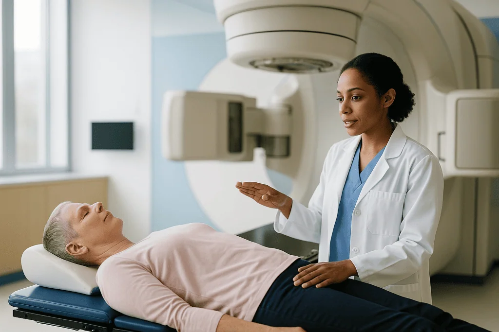
With IMRT, you will hear the gantry move, pause, and buzz. Then it swings to the next angle and does it again. The sound is stop, deliver, reposition, deliver. Depending on the number of angles, the beam-on portion can run several minutes.
With VMAT, the gantry glides around you in a smooth, slow rotation, buzzing continuously, and the multileaf collimator clicks softly as its leaves move. One arc may be enough; complex targets often use two or more, sometimes in opposite directions. A review of VMAT in the British Journal of Radiology reported that beam-on time is typically much shorter than for equivalent IMRT plans, often on the order of a couple of minutes per arc.
Neither technique involves anything you can feel. There is no heat, no pressure, and no sensation of the beam. Some people notice a faint smell or a brief flicker of light, which can happen when radiation passes near the eyes or nose, particularly for head and neck treatment; it is harmless and passes immediately.
The team watches you throughout on cameras and can pause the machine at any moment. If you need to cough, swallow, or move, you raise a hand or speak, and the beam stops. When the session ends, you sit up, the mask or supports come off, and you leave the room in the same state you entered it, with no radiation lingering in your body.
Why the arc makes sessions shorter, and why that matters
The speed difference comes down to wasted motion. In IMRT, every time the gantry stops to change angle, the beam is off. The collimator also has to build each modulated field step by step or by sliding a moving window across the target, which takes time and consumes many monitor units. A monitor unit is simply the machine’s internal counter for how much radiation it has emitted; more monitor units mean the beam is on longer.
VMAT removes the pauses. Because the gantry never stops and the leaves reshape the beam continuously, the same dose distribution can often be painted with fewer monitor units and in a fraction of the beam-on time. The original 2008 description of the technique showed plans deliverable in under two minutes of beam-on time, and the later British Journal of Radiology review confirmed this pattern across multiple tumor sites.
Does a few minutes matter? For the clinic, yes: shorter slots mean more patients treated per day on the same machine. For you, the more meaningful advantages are quieter ones.
- Less time immobilized. Anyone who has worn a head and neck mask, or held their arms above their head for a breast or chest treatment, knows that comfort erodes minute by minute.
- Less opportunity to drift. Small movements during a long session can nudge the target away from where the plan assumed it would be. A faster delivery leaves less room for that.
- Fewer monitor units. Some of the radiation a linac emits leaks through the collimator or scatters within the machine head rather than reaching the target. Fewer monitor units means less of that stray dose.
None of this makes IMRT inadequate. Many people have been treated well with fixed-field IMRT for years. But when two plans meet the same dose goals, most radiation oncology teams will favor the one that gets you off the table sooner, and the arc technique is usually that plan.
Is VMAT better than IMRT? What the evidence actually shows
This is the question people type most often, so it deserves a careful answer rather than a marketing one. The honest summary from the comparative literature is: VMAT is faster and usually at least as good at covering the target and sparing organs; it is not consistently better at the things that matter most to patients, such as long-term tumor control or the rate of serious side effects.
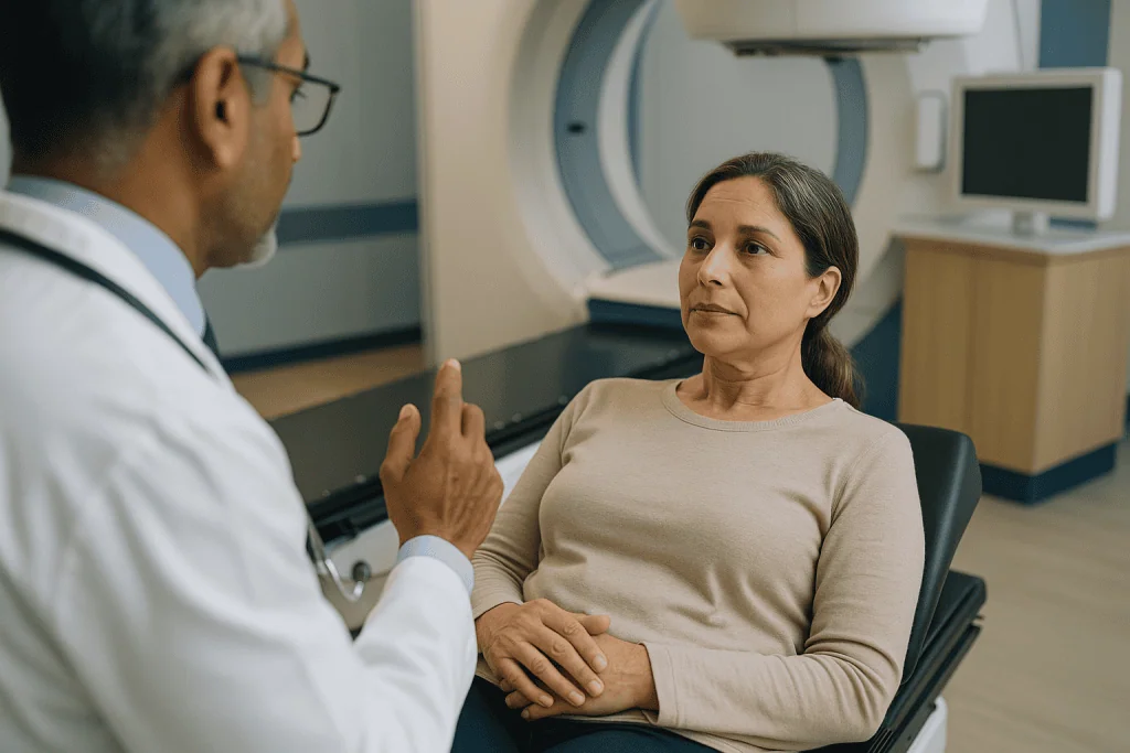
The British Journal of Radiology review that gathered dozens of planning studies found that, across prostate, head and neck, gynecologic, and other sites, VMAT plans generally achieved target coverage equivalent to IMRT with similar or modestly improved sparing of nearby organs at risk. An organ at risk is any healthy structure, such as the spinal cord, rectum, or salivary glands, that planners deliberately try to keep below a dose threshold. The consistent finding was reduced treatment time and reduced monitor units, with dosimetric differences that were often small and site-dependent.
Two caveats matter. First, most of these comparisons are planning studies: the same patient’s scan planned both ways and the numbers compared on a screen. They tell you about dose distributions, not about how people actually fared years later. Head-to-head randomized trials of IMRT versus VMAT with clinical endpoints are scarce, partly because the two are so similar that a trial would need to be enormous to detect a difference.
Second, the quality of a plan depends heavily on the skill and time invested by the planning team, the geometry of the tumor, and the individual anatomy. A carefully optimized IMRT plan can outperform a rushed VMAT plan. The technique is a tool, not a guarantee.
So when someone asks whether VMAT is better, the evidence-based reply is: better for delivery time, comparable for dose quality in most sites studied, and unproven as superior for cure-free outcomes such as recurrence or late toxicity. Your team will choose based on your anatomy, not on which technique sounds newer.
What cancers are treated with VMAT?
Almost any cancer that would be treated with IMRT can be treated with VMAT, because the two produce the same class of dose distribution. The choice tends to be driven by the shape and location of the target rather than the diagnosis on the referral letter.
The National Cancer Institute’s overview of external beam radiation lists IMRT-type techniques as standard options for tumors sitting close to sensitive structures. In practice, the sites where arc delivery is most commonly used include:
- Head and neck cancers, where the target often wraps around the spinal cord and lies beside the salivary glands, and where shorter time in a rigid mask is a real kindness.
- Prostate cancer, where the bladder and rectum press against the target and concave dose shapes help spare them.
- Gynecologic cancers of the cervix and uterus, particularly when pelvic lymph node regions need to be covered while protecting bowel and bladder.
- Brain tumors and skull-base lesions, including single-session or short-course stereotactic treatments, where multiple arcs can concentrate dose sharply.
- Lung and esophageal cancers, with the caveat that breathing motion needs to be managed carefully.
- Anal and rectal cancers, where sparing the skin folds and small bowel matters for quality of life during treatment.
Breast cancer is an interesting exception. Many breast treatments still use simpler tangential fields, sometimes with a modest degree of intensity modulation, because arcing around the chest can spread low dose into the opposite breast and lungs. Some centers use VMAT for complex breast cases involving lymph node regions; others deliberately avoid it. Both approaches can be appropriate.
In short, VMAT is a delivery method rather than a disease-specific treatment. If your team recommends it, the reasoning is geometric: your tumor’s shape and its neighbors make a continuous arc the cleaner way to paint the dose.
Who is usually offered VMAT, and who may be asked to wait or choose differently
Because the two techniques overlap so heavily, there is no strict list of who qualifies for arc therapy. Still, patterns emerge from how planning teams work.
VMAT is often the first choice when the target is irregular, concave, or wrapped around a critical structure, when the treatment involves many sessions and time on the table adds up, or when a patient finds it difficult to lie still for long periods because of pain, breathlessness, anxiety, or claustrophobia in a mask. Shorter beam-on time reduces the burden of every one of those.
There are situations where a team may prefer fixed-field IMRT or even a simpler technique, at least initially:
- When the tumor moves with breathing and the department manages that motion with a gated or breath-hold approach that is easier to coordinate with fixed beams.
- When the target sits at the edge of the body, such as a superficial chest wall or a limb, where arcing through open air adds little and may spread dose unhelpfully.
- When a treatment is being matched to an earlier course of radiation in the same area, and a fixed-field plan makes the junction between old and new dose easier to control.
- When the department’s quality-assurance program for a particular arc technique has not yet been completed for that anatomical site. This is a safety measure, not a sign of a lesser center.
Sometimes a patient is asked to wait, not for VMAT specifically, but for treatment in general: while a dental assessment is completed before head and neck radiation, while a pacemaker’s compatibility is checked, while pregnancy is excluded, or while swelling from recent surgery settles so the planning scan reflects the anatomy that will actually be treated. The Mayo Clinic’s radiation therapy overview describes this preparation phase as an ordinary part of the pathway.
Whatever the recommendation, it comes from the treating team’s assessment of your scans, your other medical conditions, and the equipment available to them. It is reasonable to ask why one technique was chosen, and a good team will be glad to explain.
Difference between IMRT and VMAT at a glance
Words can only do so much when the real distinction is mechanical. The table below lines up the two techniques feature by feature, drawing on the descriptions in the British Journal of Radiology review and the original 2008 description of arc delivery. Where a cell says similar, that reflects the weight of published planning comparisons rather than a guarantee for any individual plan.
| Feature | IMRT (fixed fields) | VMAT (arc) |
|---|---|---|
| Gantry motion during beam-on | Stationary; moves between fields | Continuous rotation |
| Beam shaping | Multileaf collimator, field by field | Multileaf collimator, continuously |
| Dose rate | Usually constant | Varies during the arc |
| Beam-on time per session | Longer, often several minutes | Shorter, typically a couple of minutes per arc |
| Monitor units | Higher | Generally lower |
| Target coverage | High | Similar |
| Sparing of organs at risk | Good | Similar or modestly improved in some sites |
| Volume of tissue receiving low dose | Concentrated along beam paths | Spread more widely around the arc |
| Planning complexity | High | Higher; more variables to optimize |
| Quality assurance | Established | Additional checks for moving components |
| Sensitivity to patient motion | Moderate | Lower exposure time, but interplay with breathing needs care |
Two rows repay a second look. The low-dose row is the genuine trade-off: by spreading beam entry points around a full circle, VMAT tends to bathe a larger volume of healthy tissue in a small dose while sparing the highest doses more effectively. The planning row explains why your first session may be scheduled a little after your planning scan: an arc plan gives the physicist more variables to optimize and more checks to run before the machine is cleared to treat.
What are the disadvantages of VMAT therapy?
Every technique has costs, and it would be dishonest to present arc therapy as a free upgrade. The disadvantages fall into three groups: dose distribution, motion, and complexity.
The dose issue is the low-dose bath. Because the beam enters from a full sweep of angles, a wider ring of healthy tissue receives a small dose than with a handful of fixed fields. For most adults this is not clinically important; the doses involved are far below anything that causes acute symptoms. Where it prompts caution is in younger patients with long life expectancies, in whom radiation-related second cancers decades later are a real, if uncommon, consideration. The British Journal of Radiology review flagged this as an area of ongoing study rather than settled concern.
The motion issue is subtler. VMAT changes gantry position, leaf position, and dose rate simultaneously. If the target also moves with breathing, the interaction between machine motion and tumor motion, called the interplay effect, can in theory blur the intended dose. Teams manage this with breathing control, larger safety margins, or by choosing techniques that are easier to gate. For a target that barely moves, such as a prostate or a brain lesion, this is a non-issue.
The complexity issue lands on the department rather than on you. Arc plans depend on many moving parts behaving exactly as modeled, so they require more rigorous machine quality assurance and patient-specific plan verification before delivery. A department that offers VMAT has invested in that program; it is not something to worry about but something you may notice as a slightly longer gap between planning and starting.
One more limitation is practical. Not every linac can deliver arcs, and not every department has validated the technique for every site. If you are told your treatment will be IMRT rather than VMAT, that is far more likely to reflect a considered match between your anatomy and the available tools than any shortfall in your care.
What is the safest radiation therapy?
This question comes up constantly, and the most useful thing anyone can tell you is that it is asking for the wrong kind of answer. Safety in radiation oncology is not a property of a machine or a technique. It is a property of a whole process: the accuracy of the planning scan, the quality of the contours drawn around the tumor and the organs at risk, the plan optimization, the daily image guidance, the machine’s calibration, and the checks performed by people at each step.
Within that framework, the question becomes: which delivery method gives this particular patient the best balance of target coverage and organ sparing for this particular tumor? For a concave target hugging the spinal cord, a modulated technique, whether IMRT or VMAT, is safer than a simple opposed-field plan because it keeps the cord below its tolerance dose. For a superficial chest wall, a simple tangential plan may be the safer choice because it avoids spraying low dose through the heart and lungs. The NHS radiotherapy guidance makes the same point in plain terms: the team plans treatment to deliver the intended dose to the tumor while limiting exposure to healthy tissue.
Where does the arc technique fit? Shorter beam-on time reduces the chance that you drift during delivery. Fewer monitor units reduce leakage and scatter dose. Both are modest, genuine safety advantages. Set against them is the wider low-dose bath already discussed. Neither factor is large enough to make one technique universally safer than the other.
There is also a broader family of techniques, including proton therapy and stereotactic approaches, that people sometimes have in mind when they ask about safety. Those are separate conversations with their own trade-offs and evidence bases, and whether any of them is relevant to you is a question for your treating team, who can see your scans.
A more answerable question is: what makes this department’s radiation safe? Daily imaging, independent plan checks, and a culture of stopping when something looks wrong are the honest markers, and every accredited radiation oncology service is built around them.
What is VMAT radiotherapy planning like before the first session?
Whichever technique you end up receiving, the weeks before your first treatment follow a similar arc of their own. Understanding the sequence takes some of the mystery out of the wait.
The planning scan, often called simulation, comes first. You lie on a CT scanner in the exact position you will hold for treatment. Supports are molded, a mask may be made from warm thermoplastic mesh that hardens as it cools, and small marks, sometimes permanent tattoo dots the size of a freckle, are placed on your skin. The Mayo Clinic describes this session as the foundation of the entire plan, and it is often the longest single appointment of the course.
Next, out of your sight, the radiation oncologist outlines the target and every organ at risk on each slice of the scan. A dosimetrist or physicist then builds the plan. For VMAT this means choosing the number and range of arcs and letting optimization software search for the combination of leaf motion, gantry speed, and dose rate that best meets the prescribed goals. Multiple iterations are normal. The plan is reviewed, adjusted, and reviewed again.
Before you are treated, the plan is tested on the machine without you present. For arc plans this patient-specific quality assurance step compares what the machine actually delivers to a phantom, a stand-in object with detectors, against what the software predicted. Only when the two agree within tight tolerances is the plan approved.
On your first day, expect extra time. Therapists will take verification images, sometimes a full cone-beam CT on the treatment machine, and compare them against the planning scan. This image guidance, often abbreviated IGRT, is repeated daily or on a schedule your team sets. It is what allows the tight margins that make modulated techniques worthwhile in the first place.
Nothing in this process is a formality. Each step exists because the precision that makes IMRT and VMAT valuable also makes them unforgiving of a target that is not where the plan assumed.
IMRT vs VMAT side effects: what the following weeks usually look like
People often hope that the faster technique will mean fewer side effects. The candid answer is that side effects depend far more on where the radiation goes and how much healthy tissue lies in the way than on whether the gantry paused or kept moving. Because IMRT and VMAT produce similar high-dose distributions, their side-effect profiles are broadly similar for the same target.
Both are typically delivered as a course of daily sessions on weekdays over several weeks, according to the NHS, though shorter courses are used for some tumors and for stereotactic treatments. Each visit, including set-up and imaging, commonly takes somewhere between ten and thirty minutes, with the beam itself accounting for only a few of those.
The early weeks are usually uneventful. Radiation’s effects on healthy tissue build gradually, so most people feel much as they did before treatment for the first stretch. Fatigue tends to creep in around the midpoint and deepen toward the end. Skin in the treated area may redden, dry, or itch. Site-specific effects follow the anatomy: a sore throat and altered taste for head and neck treatment, urinary or bowel irritation for pelvic treatment, and so on. The NHS and MedlinePlus both describe these as expected, temporary, and manageable with support from the team.
After the last session, effects often continue for a week or two before beginning to settle, because tissue reactions lag behind the dose. Most acute side effects ease over the following weeks to a few months. Late effects, meaning changes that appear months or years afterward, are less common with modern modulated techniques than with older methods, precisely because less healthy tissue receives high doses, but they are not eliminated by either IMRT or VMAT.
Where arc delivery may make a difference to your experience is in the sessions themselves. Less time immobilized means less stiffness, less anxiety in a mask, and less bladder discomfort for people asked to hold a full bladder for pelvic treatment. Those are real quality-of-life gains, even if they do not change the biology.
What people often get wrong about IMRT and VMAT
Misunderstandings about these techniques travel fast, partly because the names sound so technical. A few deserve gentle correction.
VMAT is a stronger form of radiation. It is not. The X-rays are the same energy from the same machine. The prescribed dose to the tumor is set by your oncologist based on the diagnosis, not by the delivery technique. VMAT changes how the dose is painted, not how much paint is used.
Faster means rushed. The opposite is closer to the truth. The time saved during delivery is spent beforehand, in planning and quality assurance. Shorter beam-on time is a product of better engineering, not of corners cut.
If I was offered IMRT, I am getting older or inferior treatment. Fixed-field IMRT remains a fully modern technique with decades of outcome data behind it. Teams choose it deliberately for particular geometries and motion strategies. Being offered IMRT says something about your tumor’s shape and position, not about the quality of your care.
VMAT means fewer sessions. The number of sessions is determined by the prescribed dose and how it is divided, which depends on the cancer, its stage, and the treatment goal. A shorter session is not a shorter course.
I will be radioactive afterward. External beam radiation of any kind leaves nothing behind. You can hug children, share a bed, and travel on public transport immediately after each session. The NHS is explicit on this point.
Newer is automatically better. The evidence supports VMAT as faster and dosimetrically comparable; it does not show superior long-term outcomes. That is a fine result for a technique that saves patients time, but it is not the revolution some descriptions imply.
The machine decides everything. Behind every plan is a team of people making judgments about margins, priorities, and trade-offs. Two skilled departments can reasonably choose different techniques for the same patient, and both can be right.
Questions to ask your care team about IMRT vs VMAT
Consultations move quickly, and the moment to ask about delivery technique usually arrives before you have had time to wonder about it. Bringing a short list helps. These questions are phrased so that the answers will tell you something useful, not just reassure you.
- Which technique are you planning to use for me, and what about my anatomy led you to that choice?
- How many arcs or fields will the plan use, and roughly how long will I need to hold still each day?
- What imaging will be done at each session to check my position, and how often?
- Which healthy organs are you working hardest to protect in my plan, and what are the main side effects I should expect from dose to those areas?
- Does my tumor move with breathing, and if so, how will that be managed during delivery?
- Will I need a mask, a full or empty bladder, a specific diet, or any other daily preparation, and can we practice that before the first session?
- How long between my planning scan and my first treatment, and what is happening during that gap?
- If I struggle with lying still, feel claustrophobic, or am in pain on the table, what options are there to make sessions more manageable?
- How will you decide whether the plan needs to be adjusted partway through, for example if I lose weight or swelling changes?
- Who do I contact between sessions if I have a concern, and what should prompt me to call the same day?
You are entitled to ask about the department’s approach to plan checking and machine quality assurance, too. Most teams are proud of that work and happy to describe it in outline. A good radiation oncology service will not be defensive about being asked why one technique was chosen over another. The answer is usually a short, concrete story about your scan, and hearing it tends to make the whole course feel less abstract.
Write the answers down or ask whether a family member can join by phone. Radiation planning involves many people, and the reasoning is easier to remember when it is on paper.
When to call your doctor during or after radiation treatment
Most side effects of IMRT and VMAT are gradual, expected, and discussed with you in advance. Your team will review you regularly, often weekly, during the course. Between those reviews, certain signs should prompt a call the same day rather than waiting for the next appointment. This applies whichever technique you are receiving, because the risks arise from the treated area rather than from the delivery method.
Contact your radiation oncology team or seek urgent care if you notice any of the following, which the NHS and MedlinePlus identify as reasons to seek prompt advice:
- A temperature of 38 degrees Celsius (100.4 degrees Fahrenheit) or higher, shivering, or feeling suddenly unwell, particularly if you are also receiving chemotherapy or other treatments that lower immunity.
- New or worsening breathlessness, chest pain, or coughing up blood, especially with chest or esophageal treatment.
- Difficulty swallowing that is stopping you from taking in fluids, or signs of dehydration such as passing very little urine or feeling faint on standing.
- Skin in the treated area that is broken, weeping, blistered, increasingly painful, or showing spreading redness beyond the treatment field.
- Bleeding from the bowel, bladder, or vagina, or black tarry stools.
- Persistent vomiting or diarrhea that is not settling with the measures your team suggested.
- Severe headache, new confusion, weakness or numbness in a limb, or a seizure, particularly during brain or spinal treatment.
- Sudden swelling, pain, or warmth in one leg, or unexplained shortness of breath, which can indicate a blood clot.
- A rash, hives, or swelling of the face or mouth after any new medicine given alongside treatment.
Your department will give you a contact number for both working hours and out-of-hours advice. Use it. Radiation teams expect these calls and would far rather hear about a problem early than see it at the next scheduled visit. Uncertainty itself is a good enough reason to phone.
Every decision about pausing, adjusting, or continuing your treatment rests with the team that planned it and knows your scans. This article can help you understand the conversation; it cannot replace it.
Frequently asked questions
Is VMAT better than IMRT?
VMAT is faster to deliver and, in most published planning comparisons, produces target coverage and organ sparing similar to IMRT. It has not been shown in large clinical trials to improve tumor control or reduce serious side effects compared with IMRT. Which is better for you depends on your tumor’s shape, its position relative to sensitive organs, and how your department manages motion. Your radiation oncology team makes that call.
What cancers are treated with VMAT?
VMAT is used for many of the same cancers as IMRT: head and neck, prostate, cervical and uterine, brain, lung, esophageal, anal, and rectal cancers among them. It suits targets that curve around critical structures such as the spinal cord or rectum. Some breast treatments still use simpler fixed fields to limit low dose to the lungs and opposite breast. The decision is based on anatomy rather than diagnosis alone.
What is the safest radiation therapy?
There is no single safest technique. Safety comes from the whole process: accurate planning scans, careful outlining of the tumor and organs at risk, plan optimization, daily image guidance, and independent checks. For a given patient, the safest method is the one that best covers the tumor while keeping nearby organs below their tolerance doses, which may be IMRT, VMAT, or a simpler technique. Your team weighs those factors for your specific case.
What are the disadvantages of VMAT therapy?
The main disadvantages are a wider low-dose bath of healthy tissue because the beam enters from many angles, a potential interplay effect when the tumor moves with breathing during a moving arc, and greater planning and quality-assurance complexity for the department. Not every machine can deliver arcs, and not every site is validated everywhere. For most adults with stationary targets, these drawbacks are minor and well managed.
What is the difference between IMRT and VMAT in how a session feels?
Physically you feel nothing with either. The difference is sound and time. With IMRT, the gantry moves to a fixed position, buzzes, stops, and repositions several times. With VMAT, the gantry rotates slowly and continuously around you while the beam is on, and the session is usually over sooner. Set-up and imaging take up most of the appointment in both cases, so the total visit length is often similar.
What is VMAT radiotherapy, in plain terms?
VMAT stands for volumetric modulated arc therapy. It is a way of delivering shaped X-ray radiation in which the treatment machine rotates around you in one or more continuous arcs while tiny metal leaves reshape the beam and the dose rate rises and falls. The goal is to concentrate dose on the tumor and limit it to surrounding organs, using the same physics as IMRT but with faster delivery.
Does VMAT mean fewer treatment sessions?
No. VMAT shortens the time the beam is on during each session, not the number of sessions. The total dose and how it is divided into daily treatments are decided by your oncologist based on the cancer type, stage, and goal of treatment. According to the NHS, most courses run daily on weekdays over several weeks regardless of whether IMRT or VMAT is used.
Are IMRT vs VMAT side effects different?
For the same tumor and prescribed dose, side effects are broadly similar because both techniques produce comparable high-dose regions. Side effects depend on the body area treated: skin changes, fatigue, sore throat for head and neck treatment, or bladder and bowel irritation for pelvic treatment. VMAT’s shorter time on the table can reduce discomfort during sessions, but it does not fundamentally change the tissue reactions that follow.
Why did my team choose IMRT instead of VMAT?
Common reasons include a target that moves with breathing and is easier to manage with fixed beams, a superficial tumor where arcing adds little, the need to match a previous radiation course precisely, or the department’s validation status for a particular site. IMRT is a fully modern technique with decades of outcome data. Ask your team; the explanation is usually a short, concrete story about your scan.
Will I be radioactive after an IMRT or VMAT session?
No. External beam radiation from a linear accelerator passes through you and leaves nothing behind. You can be around children and pregnant people, share meals and beds, and use public transport immediately afterward. This differs from some internal radiation treatments involving implanted or swallowed radioactive sources, which carry temporary precautions. If you are unsure which kind you are receiving, ask your team.
References
This article is for general information only and is not a substitute for professional medical advice. Please consult a qualified doctor about your individual situation.

More from the Blog
Voice and Swallowing After Neck Cancer Treatment: What Rehabilitation Can Restore
Voice changes after neck cancer treatment, such as hoarseness, breathiness, a lower or weaker pitch and difficulty swallowing, are common after surgery, radiation or…
How Ovarian Cancer Treatment Is Planned: Surgery, Chemotherapy and the Order They Come In
Ovarian cancer treatment by stage usually combines surgery to remove as much visible cancer as possible with platinum-based chemotherapy. Early-stage disease confined to the…

Simple, Skin-Sparing or Nipple-Sparing Mastectomy: How the Types Differ and Who Gets Which
The main types of mastectomy differ in how much skin and nipple are kept, not in how much breast tissue is removed. A total…

Sarcoma in Teenagers and Young Adults: How Treatment Planning Considers Growth and School
Sarcoma treatment in teenagers usually combines chemotherapy with surgery, and sometimes radiation, sequenced over many months by a specialist sarcoma team. Because young bodies…

How Do Doctors Judge Whether SBRT Worked? Why the First Follow-Up Scan Waits a Few Months
After stereotactic body radiation therapy (SBRT), the first imaging check usually happens a few months after the final session, because irradiated tumors shrink slowly…

Which Blood and Genetic Tests Guide Lymphocytic Leukemia Treatment Choices?
Treatment choices in chronic lymphocytic leukemia rest on a small set of tests: a complete blood count with flow cytometry to confirm the diagnosis,…