

Minimally Invasive or Four-Gland Exploration Parathyroidectomy: How Is the Approach Chosen?

Key Takeaways
- In roughly 80 percent of primary hyperparathyroidism cases a single benign adenoma is responsible, which is why a focused operation is possible for many people, according to the NIDDK.
- Imaging such as ultrasound, sestamibi and 4D-CT is used to plan where the surgeon will look, not to diagnose the condition; the diagnosis rests on calcium and PTH blood tests.
- A focused parathyroidectomy is usually offered when two imaging studies agree on one gland, while discordant or negative scans generally lead to a four-gland exploration.
- Intraoperative PTH testing works because the hormone clears from blood within minutes, letting surgeons confirm during the operation that no other overactive gland remains.
- Hereditary syndromes such as MEN1, kidney-related secondary hyperparathyroidism and long-term lithium use are associated with multigland disease and usually call for exploration from the start.
- Tingling around the lips or fingertips in the first days after surgery is the typical early sign of low calcium and should prompt a call to the surgical team.
Surgeons choose between a minimally invasive (focused) parathyroidectomy and a four-gland exploration mainly on whether preoperative imaging clearly points to a single overactive gland. When ultrasound, sestamibi or 4D-CT scans agree on one adenoma, a small targeted operation is usually offered; when scans disagree, suggest several glands, or the person has a hereditary or kidney-related cause, exploring all four glands is generally preferred. The treating team makes the final call.
The envelope from the surgeon’s office holds two scan reports and one sentence that keeps catching the eye: “localization concordant for a left inferior adenoma.” Underneath, in a different pen, someone has written “candidate for focused approach.” You read it three times. Focused, as opposed to what, exactly?
That question sits at the heart of the minimally invasive parathyroidectomy vs exploration decision, and it is one of the few surgical choices where a scan read on a Tuesday can change the shape of the incision on Friday. Both operations treat the same problem, an overactive parathyroid gland pushing calcium too high. They differ in how much of the neck the surgeon looks at before deciding the job is done.
This article walks through what each approach involves, what tips a team one way or the other, and what the days afterward usually look like. It will not tell you which operation you should have; that conversation belongs to you and your surgeon.
What does a parathyroidectomy actually do?
Most people carry four parathyroid glands, each about the size of a grain of rice, tucked behind the thyroid at the front of the neck. Their only job is to release parathyroid hormone (PTH), the chemical signal that tells bones to release calcium, kidneys to hold onto it, and the gut to absorb more of it. In a healthy body, PTH rises when blood calcium dips and switches off when calcium is back in range.
Primary hyperparathyroidism is the condition in which one or more glands keep producing PTH regardless of how much calcium is already circulating. According to the National Institute of Diabetes and Digestive and Kidney Diseases, in roughly 80 percent of cases a single benign growth, called an adenoma, is responsible. Less often, two or more glands are enlarged (hyperplasia), and very rarely the cause is a parathyroid cancer.
A parathyroidectomy is simply the removal of the gland or glands that have stopped listening to calcium. Once the overactive tissue is out, the remaining normal glands take over, PTH falls, and blood calcium drifts back toward normal. The operation does not rebuild bone overnight or dissolve existing kidney stones, but it removes the engine driving both problems.
Two broad strategies exist. A four-gland exploration, sometimes called a bilateral neck exploration, means the surgeon inspects every parathyroid gland on both sides before removing the abnormal ones. A minimally invasive or focused parathyroidectomy means the surgeon goes directly to the gland that imaging flagged, removes it, confirms with a blood test that hormone levels have dropped, and stops. The rest of this article is about how a team decides which story your neck is telling.
Minimally invasive parathyroidectomy vs exploration: the core difference
The difference is less about incision size than about certainty. Both operations are done through a cut low on the front of the neck, and both are usually finished within a couple of hours. What separates them is how the surgeon answers one question: have I found everything that is overactive?
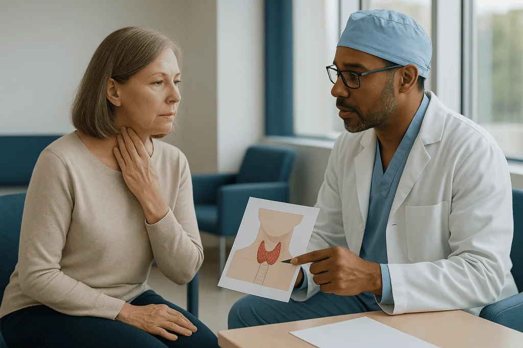
In a four-gland exploration, the answer comes from direct inspection. The surgeon looks at each gland, judges its size and appearance, and removes those that are enlarged. For decades this was the only approach, and it remains the reference against which newer techniques are measured. It handles the unexpected well: if a second abnormal gland is hiding on the far side of the neck, the surgeon sees it.
In a focused parathyroidectomy, the answer comes from a combination of preoperative imaging and an intraoperative blood test. The surgeon trusts the scans to point to the right spot, removes the suspect gland through a smaller opening, and then measures PTH in the operating room. Because PTH clears from the blood within minutes, a sharp fall tells the team the remaining glands are behaving normally. If the hormone does not drop as expected, the surgeon can widen the operation into a full exploration on the spot.
The Mayo Clinic describes minimally invasive parathyroidectomy as appropriate when a single gland is clearly identified beforehand, and bilateral exploration as the approach when imaging is inconclusive or more than one gland is suspected. Neither is “better” in the abstract. One is better suited to a neck with a single, well-mapped adenoma; the other is better suited to a neck where the map is unclear or where the disease is known to involve several glands.
Why the sestamibi scan for parathyroid disease decides so much
Here is a point that surprises many people: imaging is not used to diagnose hyperparathyroidism. The diagnosis comes from blood tests showing high calcium alongside PTH that is high or inappropriately normal. Scans are ordered only after the decision to operate has been made, and their purpose is to plan the operation, not to justify it.
Three tools dominate. Neck ultrasound is quick, painless and radiation-free, and in experienced hands it can often spot an enlarged gland sitting behind the thyroid. A sestamibi scan uses a small amount of a radioactive tracer that overactive parathyroid tissue absorbs and holds longer than surrounding tissue; a camera then shows where the tracer lingers. Four-dimensional CT (4D-CT) is a CT scan timed to how contrast dye washes in and out of tissue, which helps distinguish a parathyroid adenoma from a thyroid nodule or lymph node.
When two different studies independently point to the same gland, surgeons describe the imaging as concordant. That agreement is the single strongest argument for a focused approach. The Cleveland Clinic notes that a minimally invasive operation is typically planned when imaging shows one abnormal gland.
When the studies disagree, when neither finds anything, or when one shows two possible targets, the imaging is called discordant or negative. This does not mean surgery is off the table; it means the surgeon will probably need to look at all four glands rather than trust a single picture. People sometimes feel that a negative scan means they “failed” a test. In reality, a negative scan tells the surgeon nothing about whether the disease exists, only that a focused shortcut may not be safe to take.
Who needs parathyroid surgery, and who is usually asked to wait?
Not everyone with high calcium is rushed toward an operation. Guidance summarized by the NIDDK and the NHS points to surgery when hyperparathyroidism is causing measurable harm or is likely to: kidney stones, reduced kidney function, osteoporosis or fragility fractures, calcium that is well above the normal range, or symptoms such as persistent fatigue, bone pain or difficulty concentrating that cannot be explained another way. Younger age at diagnosis also pushes toward surgery, since a person has more decades over which untreated disease can erode bone and kidney health.

People who are typically asked to wait, or offered monitoring instead, tend to have mild, stable calcium elevation, normal bone density, normal kidney tests and no stones or symptoms. Monitoring usually means periodic blood tests, bone density scans and kidney checks, with surgery revisited if anything changes. The NHS notes that regular monitoring is a reasonable path for people with mild disease and no complications.
Whether a person qualifies for surgery and whether they qualify for a focused operation are two separate questions. The first depends on the biology of the disease and the person’s overall health. The second depends almost entirely on imaging and on the suspected cause. Someone with clear symptoms, kidney stones and a single concordant adenoma on two scans is a textbook candidate for a focused approach. Someone with equally clear indications but scans that light up nowhere will likely be offered an exploration instead.
Other factors matter at the margin. Prior neck surgery, a large thyroid goiter, or anatomy that makes imaging unreliable can tilt a surgeon toward exploration even when one scan looks convincing. These are judgment calls, and reasonable surgeons can land in different places on the same case.
When is four-gland exploration the clearer choice?
Certain situations make a focused operation a poor bet before anyone looks at a scan, because the underlying biology almost guarantees that more than one gland is involved.
Hereditary syndromes are the clearest example. Multiple endocrine neoplasia type 1 (MEN1) and related inherited conditions cause several parathyroid glands to enlarge over time. Removing one adenoma in such a person treats a fraction of the problem and leaves the rest to resurface. Surgeons therefore plan to inspect all four glands, and often remove most of the parathyroid tissue while preserving a small remnant.
Secondary hyperparathyroidism, in which glands enlarge in response to chronic kidney disease or long-standing vitamin D deficiency, behaves similarly: all glands tend to be driven, so all need assessment. Long-term treatment with lithium, a mood stabilizer, is associated with multigland disease as well. In each case the question is not “which gland?” but “how much of the total tissue needs to come out?”
Discordant or negative imaging is the other large category, covered above. Even when a single scan suggests one target, a surgeon may choose exploration if the suspected gland is in an unusual location, if the person has had previous neck surgery that distorts the anatomy, or if intraoperative hormone testing is not available at that center.
There is also a learned-humility argument. Mayo Clinic and Cleveland Clinic patient guides both acknowledge that a focused operation can be converted to a full exploration partway through if the hormone level does not fall. Surgeons who know they may need to widen the operation sometimes prefer to plan for it from the outset rather than discover it mid-procedure. That preference is legitimate, and it is one of the reasons two surgeons can recommend different approaches for similar scans.
What is intraoperative PTH monitoring and why does it matter?
The focused operation only became practical when laboratories learned to measure PTH quickly enough to return a result while the patient was still asleep. Intraoperative PTH monitoring is the practice of drawing blood before the gland is removed and again a short while after, then comparing the two numbers in the operating room.
The logic rests on PTH’s short life in the bloodstream. Once the overactive gland is out, hormone already in circulation breaks down within minutes. If the remaining glands are normal, they will have been suppressed by months of high calcium and will not immediately start producing much. The result is a steep fall: a drop to roughly half the starting level within about ten minutes is the benchmark most surgical teams use to conclude that no other hyperactive tissue remains.
If the hormone level stays high or falls only modestly, the surgeon has useful information before closing the incision: something else is still producing PTH. That is the moment a focused operation turns into an exploration. Patients should understand this possibility in advance, because it means a small planned incision can occasionally become a somewhat larger one, and the consent conversation should cover it.
The test is not perfect. Occasionally the hormone falls appropriately even though a second small abnormal gland is present and simply not active enough to register, which is one reason a small proportion of focused operations are followed by a return of high calcium later. Equally, hormone can linger in some people for reasons unrelated to leftover disease. Surgeons interpret the number alongside what they can see and feel, not in isolation. Ask whether your center uses intraoperative PTH; if it does not, a focused approach relies more heavily on imaging alone.
Focused parathyroidectomy and exploration side by side
The table below summarizes how the two approaches differ in the situations where patients most often ask about them. It is a general guide drawn from patient information published by the Mayo Clinic, Cleveland Clinic and MedlinePlus, not a scoring system, and individual surgeons weigh these factors differently.
| Feature | Minimally invasive (focused) parathyroidectomy | Four-gland (bilateral) exploration |
|---|---|---|
| Typical candidate | Single adenoma identified on concordant imaging | Negative or discordant imaging; suspected multigland disease; hereditary syndromes; secondary hyperparathyroidism |
| Incision | Small, often placed to one side of the neck | Longer central incision allowing access to both sides |
| Glands examined | Usually one | All four, with abnormal ones removed |
| Confirmation that disease is treated | Intraoperative PTH drop, plus imaging | Direct inspection of every gland |
| Anesthesia | General, or occasionally regional with sedation | Almost always general |
| Chance of conversion | Can be widened to full exploration if PTH does not fall | Not applicable |
| Hospital stay | Same-day discharge or one night is common | Same-day or one night is common; overnight observation slightly more usual |
| Risk to other glands and nerves | Lower exposure of unaffected side | Both sides handled, so slightly more tissue at risk |
Two patterns stand out. First, the focused approach trades direct visual certainty for indirect biochemical certainty; it works because PTH testing is reliable, not because surgeons have become less thorough. Second, the exploration’s main advantage is completeness, which matters most precisely in the cases where imaging is least helpful. Seen this way, the two operations are complementary tools for different maps of the neck, rather than old and new versions of the same thing.
What actually happens on the day of surgery?
Whichever approach is planned, the morning follows a familiar pattern. You arrive having fasted as instructed, meet the anesthesia team, and have a final conversation with the surgeon that often includes a marker drawn on the neck to indicate the side imaging flagged. If intraoperative PTH is being used, a baseline blood sample is taken before or just after anesthesia begins.
For a focused operation, the surgeon makes a small incision, moves the thin strap muscles aside, and gently lifts the thyroid lobe to reach the gland behind it. An adenoma is usually darker and firmer than normal parathyroid tissue, and it is removed in one piece. A second blood sample is drawn, and the team waits for the laboratory result while keeping the incision open. A satisfactory fall ends the operation; an inadequate one prompts a look at the other glands.
For a four-gland exploration, the incision sits in the midline and the surgeon works systematically through both sides, identifying each gland in turn. Enlarged glands are removed. If all four are enlarged, as in hereditary or secondary disease, the surgeon typically removes three and part of the fourth, or removes all four and transplants a small fragment into a muscle in the forearm or neck where it can continue making hormone. Removed tissue is sent to pathology for confirmation.
Throughout both operations the surgeon takes care around the recurrent laryngeal nerves, the pair of nerves that run beside the parathyroids and control the vocal cords. Many centers use nerve monitoring to help protect them. The incision is closed with fine sutures or glue, and MedlinePlus notes that most people are awake and talking within an hour or two. A nurse checks your voice, your ability to swallow, and signs of a tingling sensation around the mouth or fingertips that can signal falling calcium.
Parathyroid surgery recovery time: what the first weeks look like
Recovery is usually brisker than people expect from an operation on the neck, but it is not uniform, and the main variable is calcium rather than the incision.
In the first day or two, a sore throat from the breathing tube, a stiff neck and mild swelling around the incision are ordinary. MedlinePlus describes same-day discharge or a single overnight stay as typical, with the overnight option used more often after exploration or when calcium needs watching. Many surgeons prescribe calcium and sometimes vitamin D for a short period, because the previously suppressed normal glands can take days to weeks to wake fully. How much, for how long, and whether to taper are decisions for the surgical team based on your blood results; this article deliberately does not describe regimens.
The tingling or numbness around the lips and fingertips that signals low calcium (hypocalcemia) most often appears in the first few days. Teams tell patients what to watch for and what to do, and it is worth writing those instructions down before leaving the hospital.
By the end of the first week, most people have returned to desk work and light activity. The Mayo Clinic’s patient guidance notes that heavy lifting and strenuous exercise are generally paused for a short period while the incision heals, and that a follow-up blood test is scheduled to confirm calcium and PTH have settled. A review visit typically falls a few weeks after surgery.
Longer-term, bone density is rechecked after a year or so, since recovery of bone mass continues over many months. Some symptoms such as fatigue and low mood improve noticeably within weeks for some people; others notice little change, which usually means those symptoms had other causes. Honest expectation-setting before surgery prevents disappointment afterward.
What are the risks, and do they differ between approaches?
Parathyroid surgery is generally considered low-risk among neck operations, and patient resources from the NHS, Mayo Clinic and Cleveland Clinic describe a similar short list of complications for both approaches. The differences between focused and exploratory surgery are differences of degree, not kind.
Temporary low calcium is the most common issue and is covered above; it is more likely after exploration, especially when several glands are removed, because the remaining tissue has been handled more extensively. Permanent low calcium requiring lifelong supplementation is uncommon but is a recognized risk, again weighted toward multigland operations.
Voice change results from irritation or injury to a recurrent laryngeal nerve. Temporary hoarseness or voice fatigue after surgery is relatively common and usually resolves over weeks. Permanent weakness of a vocal cord is rare. In principle a focused operation exposes only one nerve, while exploration exposes both, so the theoretical nerve risk is slightly higher with exploration; in practice, both are small.
Bleeding into the neck after surgery is rare but is the complication that most needs rapid attention, because swelling can compress the airway. This is one reason for the overnight observation some teams prefer.
Persistent or recurrent hyperparathyroidism is the risk most specific to the focused approach. If a second abnormal gland was missed because imaging and hormone testing both pointed to a single target, calcium can remain high or rise again later, and a second operation may be discussed. Exploration reduces this risk by design but does not eliminate it, since glands can sit in unusual places such as within the chest.
Infection, a visible scar, and the usual risks of general anesthesia apply to both. None of these figures should be read as personal probabilities; your surgeon can tell you how they apply to your anatomy and health.
What people often get wrong about choosing the approach
“A smaller incision means a better operation.” The incision is a byproduct of the plan, not its goal. A focused approach is chosen because the disease is believed to sit in one place, and the small incision follows from that. Choosing it in the face of uncertain imaging simply to keep the scar short raises the chance of leaving disease behind.
“A negative scan means I don’t have hyperparathyroidism.” The diagnosis is made from blood tests, full stop. Scans localize; they do not diagnose. The NIDDK is explicit that imaging is used to plan surgery, not to decide whether the condition exists. People with convincing biochemistry and unhelpful scans are still candidates for surgery; they are candidates for exploration.
“Exploration is the outdated operation.” Bilateral exploration remains the standard against which focused surgery is judged, and for hereditary and multigland disease it is the primary approach rather than a fallback. Many high-volume surgeons continue to perform it routinely.
“The surgeon will know in advance exactly what they will do.” Often, yes. But the intraoperative PTH test exists precisely because plans sometimes need to change. A focused operation that becomes an exploration is not a failure; it is the system working as designed.
“Once the gland is out, calcium is fixed for life.” For most people with a single adenoma, treatment is durable. Recurrence is possible, particularly in hereditary disease, and lifelong occasional calcium checks are reasonable.
“Medicines can do the same job.” Drugs can lower calcium or protect bone, but they do not remove the overactive tissue. They have a role for people who cannot or prefer not to have surgery, which the next section covers, but they are not an equivalent alternative for everyone.
Are there alternatives to surgery for hyperparathyroidism?
Surgery is the only treatment that removes the source of excess hormone, and the NHS and NIDDK both describe it as the main treatment for primary hyperparathyroidism. For people who are not candidates for an operation, or who choose to defer it, the alternatives aim at the consequences rather than the cause.
Monitoring is the simplest. For mild, asymptomatic disease with normal bones and kidneys, periodic blood tests, bone density scans and kidney assessments can be continued for years, with surgery revisited if anything deteriorates. Staying well hydrated and keeping dietary calcium at a normal level rather than restricting it are commonly advised, since very low calcium intake can paradoxically drive PTH higher; the NIH Office of Dietary Supplements discusses typical calcium intake ranges for adults.
Two medicine classes come up in conversation. Calcimimetics are drugs that make the parathyroid glands more sensitive to calcium, so they release less hormone; they lower blood calcium but have not been shown to improve bone density, and they are generally reserved for people who cannot have surgery or whose disease is secondary to kidney failure. Bisphosphonates are bone-protecting drugs that slow bone breakdown; they can help preserve density in people with hyperparathyroidism-related bone loss, though they do not lower PTH or calcium meaningfully.
Vitamin D is often checked and, if low, corrected before surgery, because deficiency can exaggerate PTH levels and complicate recovery. All of these decisions, including whether any medicine is appropriate and how it should be used, rest with the prescribing clinician.
Newer approaches such as ablating an adenoma with heat or alcohol under ultrasound guidance have been reported, mainly in people unable to have surgery. The evidence remains limited to small series, and they are not an established substitute for either operative approach.
Questions to ask your care team before deciding
A good consultation leaves you knowing not only which operation is planned but why. The questions below are ones surgeons generally welcome, because they lead to clearer consent and fewer surprises.
- Which imaging studies did I have, and did they agree on a single gland? If not, what did each one show?
- Based on those results, are you planning a focused operation or a four-gland exploration, and what would make you change that plan during surgery?
- Do you use intraoperative PTH monitoring? If not, how will you confirm the disease has been treated before closing?
- Is there anything in my history, such as kidney disease, lithium use or a family pattern of endocrine tumors, that makes multigland disease more likely?
- Roughly how many parathyroid operations does your team perform, and how do you handle a gland that cannot be found?
- What is your approach to calcium and vitamin D after surgery, and how will I know whether my levels are safe at home?
- What symptoms would you want me to call about in the first week, and who answers that call out of hours?
- What follow-up blood tests and bone density checks do you recommend, and over what period?
- If my calcium stays high or rises again, what would the next steps be?
- Is monitoring without surgery a reasonable option for me, and what would prompt you to recommend surgery later?
Write the answers down or bring someone who can. Surgeons describe anatomy quickly and fluently, and it is easy to nod along and forget the details by the car park. If the recommended approach differs from what you expected after reading about focused surgery, ask what specifically about your scans or history drove the choice. The reasoning is usually concrete, and hearing it tends to settle the question.
When to call your doctor after parathyroid surgery
Most recoveries are uneventful, but a few signs warrant a same-day call to the surgical team, and a smaller set warrant emergency care without waiting.
Call your care team promptly if you notice tingling or numbness around the lips, in the fingertips or toes, or muscle cramps and twitching, particularly in the first several days. These are the classic early signs of low calcium described by MedlinePlus and the NHS, and the team will usually want a blood test and may adjust your post-operative plan. Also call for a fever, increasing redness, warmth or discharge at the incision, a voice that is weak or hoarse and not improving after the first few days, or difficulty swallowing that is getting worse rather than better.
Seek emergency care immediately if you develop rapidly increasing swelling or tightness in the neck, difficulty breathing, a sensation that the throat is closing, or severe muscle spasms affecting the hands, face or throat. Neck swelling after surgery can indicate bleeding that compresses the airway, and severe spasms can indicate calcium that has fallen to a dangerous level. Both need urgent assessment.
Before surgery, people with hyperparathyroidism should also know the warning signs of very high calcium: confusion, severe nausea or vomiting, intense thirst with frequent urination, marked drowsiness, or an irregular heartbeat. The Mayo Clinic lists these among the features of a hypercalcemic crisis, which is uncommon but requires emergency treatment.
None of this is intended as a checklist for self-diagnosis. If something feels wrong and you are unsure which category it falls into, calling the team is always the right choice; surgical units expect these calls and would far rather hear from you than not. Your discharge paperwork should include a number that is answered around the clock.
Frequently asked questions
What is a focused parathyroidectomy?
A focused parathyroidectomy is a targeted operation in which the surgeon removes only the single parathyroid gland that preoperative scans identified as overactive, usually through a small incision. A blood test for parathyroid hormone taken during surgery confirms the hormone level has fallen, indicating no other gland is overactive. It is offered when imaging clearly points to one adenoma. The term is used interchangeably with minimally invasive parathyroidectomy.
Who needs parathyroid surgery rather than monitoring?
Surgery is generally advised when primary hyperparathyroidism is causing or likely to cause harm: kidney stones, reduced kidney function, osteoporosis or fractures, calcium well above normal, or persistent symptoms. Younger age at diagnosis also favors surgery. People with mild, stable calcium elevation, normal bones and kidneys and no symptoms are often offered monitoring with periodic blood and bone tests instead. The treating team weighs these factors for each person.
How accurate is a sestamibi scan for parathyroid adenomas?
A sestamibi scan detects many single adenomas but can miss small glands, multiple enlarged glands, or tissue in unusual locations, and thyroid nodules can occasionally mimic a parathyroid signal. Accuracy varies by center and by the person’s anatomy, which is why surgeons often combine it with ultrasound or 4D-CT and look for agreement. A negative scan does not rule out hyperparathyroidism; it simply means a focused operation may not be appropriate.
What is the usual parathyroid surgery recovery time?
Most people go home the same day or after one night and return to desk work and light activity within about a week, according to patient guidance from MedlinePlus and the Mayo Clinic. Strenuous exercise and heavy lifting are typically paused for a short period while the incision heals. Calcium levels are checked in the following days and weeks, and bone density is reassessed after roughly a year. Individual timelines vary.
Can a minimally invasive parathyroidectomy turn into a full exploration during surgery?
Yes. If the intraoperative parathyroid hormone level does not fall as expected after the suspected gland is removed, the surgeon can extend the incision and examine the remaining glands in the same operation. This possibility is a built-in safety feature of the focused approach rather than a complication, and it should be covered in the consent discussion so that a slightly larger incision is not a surprise afterward.
Why would a surgeon choose four-gland exploration if my scan shows one adenoma?
Several reasons are common: the scan may be only one study without a second confirming it, your history may suggest multigland disease (for example a hereditary syndrome, kidney disease or long-term lithium use), prior neck surgery may have altered the anatomy, or the center may not use intraoperative hormone testing. Some surgeons also prefer exploration based on experience. Ask what specifically drove the recommendation in your case.
What happens to the other parathyroid glands after one is removed?
The remaining normal glands, which have been suppressed by months or years of high calcium, gradually resume producing parathyroid hormone. This can take days to weeks, which is why temporary low calcium is common after surgery and why teams monitor blood levels and may advise short-term supplementation. In most people with a single adenoma, the remaining glands eventually maintain normal calcium on their own.
Is hoarseness after parathyroid surgery permanent?
Usually not. Temporary hoarseness or voice fatigue is relatively common and typically results from irritation of the nerves beside the parathyroid glands or from the breathing tube; it generally improves over days to weeks. Permanent weakness of a vocal cord is a recognized but rare complication. A voice that is weak or hoarse and not improving after the first few days should be reported to the surgical team.
Does parathyroid surgery help bone density?
Removing the source of excess parathyroid hormone stops the ongoing loss of calcium from bone, and bone density tends to improve over the following one to two years in people who had hyperparathyroidism-related bone loss. The degree of recovery varies with age, baseline density and other factors. Bone density is typically rechecked about a year after surgery so the team can see how your bones are responding.
Are there medicines that can replace parathyroid surgery?
No medicine removes the overactive tissue. Calcimimetics can lower blood calcium by making the glands more sensitive to it, and bisphosphonates can protect bone, but neither treats the underlying cause. They are generally considered for people who cannot have surgery or whose disease is secondary to kidney failure. Whether any medicine is appropriate, and how it should be used, is a decision for the prescribing clinician.
References
- MedlinePlus: Parathyroid gland removal
- NHS: Hyperparathyroidism
- NIDDK (NIH): Primary Hyperparathyroidism
This article is for general information only and is not a substitute for professional medical advice. Please consult a qualified doctor about your individual situation.

More from the Blog

Exercise With Type 1 Diabetes: How Activity Affects Glucose and How Plans Are Adjusted Safely
People with type 1 diabetes can exercise safely, and most guidelines encourage regular aerobic and strength activity. The challenge is that injected insulin cannot…
When Does Hashimoto Thyroiditis Need Treatment, and When Is Monitoring Enough?
Hashimoto thyroiditis itself is not treated; the underactive thyroid it can cause is. Treatment with thyroid hormone replacement is usually recommended when blood tests…

What Does Radiosurgery Change for Pituitary Tumors Compared With Open Surgery?
Radiosurgery for a pituitary tumor replaces cutting with precisely aimed radiation, so there is no incision, usually no hospital stay, and no direct removal…
Why Can Voice Changes and Low Calcium Follow Thyroidectomy, and When Do They Need a Call?
Voice changes and low calcium can follow thyroidectomy because the thyroid sits against the nerves that move the vocal cords and beside the four…

Recovering From Diabetic Ketoacidosis: From Intensive Care to Going Home and the Weeks After
DKA recovery time typically runs in two phases. In the hospital, intravenous fluids, insulin and electrolyte replacement usually clear the acidosis within roughly 24…

Life After Cushing Syndrome Treatment: Blood Pressure, Blood Sugar and Bone Health Follow-Up
Life after Cushing syndrome treatment usually means months of gradual recovery rather than an overnight change. Cortisol levels often fall abruptly after surgery, so…