

Preparing for an Eye Examination: Glasses, Old Reports, Medication Lists and a Driver
Key Takeaways
- The glasses you actually wear are read on a lensometer and compared with the new measurement, which shows whether a change is in your eyes or in your lenses.
- Dilating drops take roughly 15 to 30 minutes to work and leave near vision blurry and eyes light-sensitive for several hours, so a driver and sunglasses are part of preparation.
- Glaucoma, diabetic retinopathy and early macular degeneration usually cause no symptoms, which is why the National Eye Institute recommends dilated exams every one to two years for people at higher risk.
- Corticosteroids, hydroxychloroquine, amiodarone and alpha-blockers each have documented eye effects, so a complete medication list changes how findings are interpreted.
- Redness-relief drops used before a visit constrict surface vessels and hide exactly what the clinician needs to see; arrive with the eye as it is.
- Mayo Clinic guidance suggests healthy adults in their 20s and 30s can go 5 to 10 years between exams, while adults 65 and over are usually seen every 1 to 2 years.
For an eye exam, bring the glasses and contact lenses you actually use, any previous prescriptions or eye reports, a complete list of your medicines and supplements with the reason you take each one, your identification and insurance details, a pair of sunglasses, and a driver if your pupils may be dilated, because dilation blurs near vision and increases light sensitivity for several hours.
The night before her appointment, a retired teacher lines things up on the kitchen table: reading glasses with one arm taped, a pharmacy printout, a folded letter from an eye doctor she saw in another state years ago. Then she pauses over the car keys. Someone mentioned drops. Will she be able to drive home?
That small kitchen-table moment is the whole question of what to bring to an eye exam. Most of the visit is done by the clinician, but a surprising amount of its accuracy depends on what you carry through the door. The glasses you wear every day tell a story the chart cannot. The medicine list explains a dry cornea or a pressure reading. The old report turns a single snapshot into a trend.
This guide walks through each item, why it matters, what actually happens in the room, and how to plan the ride home.
What to bring to an eye exam: the short list that actually matters
An eye exam is one of the few medical visits where the physical objects you bring change the quality of the result. The clinician can measure your eyes without any of them, but each item removes a guess.
- The glasses you actually wear, including reading glasses, computer glasses and sunglasses with a prescription. Not the pair in the drawer; the pair on your face.
- Contact lenses in your eyes or in their case, plus the box or blister foil that shows brand, power and curvature.
- Previous prescriptions and eye reports, even old or partial ones. A photo on your phone counts.
- A full medication list: prescription drugs, over-the-counter products, eye drops, vitamins and herbal supplements, with the reason you take each.
- Your health history in brief: diagnoses such as diabetes or high blood pressure, past eye surgery or injury, and eye conditions in close relatives.
- A list of your questions and a note of any symptom, however minor, with roughly when it started.
- Identification and insurance or referral paperwork, as the office requests.
- Sunglasses for the trip home.
- A driver if there is any chance your pupils will be dilated, which the Mayo Clinic notes can leave vision blurry and light-sensitive for several hours.
None of this is obscure, yet the items people most often forget are the everyday glasses (“I only wear them for driving”) and the supplements (“they aren’t real medicine”). Both matter, for reasons the next sections explain. If you are unsure whether the visit will include dilation, call ahead and ask; it is a normal question and the office will have a clear answer for the type of appointment you have booked.
Why your current glasses matter more than the prescription on paper
A written prescription describes what a clinician intended. Your glasses describe what you have been living with, and the two are not always the same. Lenses can be made slightly off target, frames can be bent so the optical center no longer sits in front of the pupil, and coatings can craze or scratch until the world looks softly foggy. A person who says “my vision has gotten worse” may simply have glasses that no longer match their eyes, or glasses that never quite matched.
The office will usually place your glasses on a lensometer, an instrument that reads the power ground into a lens. Comparing that reading with the new measurement answers a practical question: is the change in your eyes, or in the glasses? A small shift is common and expected with age. A large or lopsided shift, or a big difference between what the lenses say and what your file says, is a clue worth following.
Bring every pair you use in a normal week. Reading glasses reveal your near-vision needs. Computer glasses show the working distance you spend hours at. Prescription sunglasses tell the clinician whether your outdoor vision is being corrected the same way as your indoor vision. Even a pair you have abandoned can be informative: if you stopped wearing them because they “never felt right,” that is a data point about how your eyes tolerate change.
Wear the glasses into the room rather than handing them over in a case. Part of the exam is watching how you see with your usual correction, then measuring how much better you could see. That comparison is the heart of a refraction, the test that determines your lens power, and it works best when the starting point is the real thing.
Contact lens wearers: bring the box, and ask when to take them out
Contact lenses sit directly on the cornea, the clear front window of the eye, and they gently reshape it while they are worn. That reshaping is temporary, but it is enough to nudge a refraction and to alter how the corneal surface looks under magnification. Clinicians therefore often ask patients to leave lenses out for a period before a full exam, especially when a new glasses prescription or a contact lens fitting is planned.
How long varies with the lens type. Soft lenses rebound faster than rigid gas-permeable lenses, and overnight or orthokeratology lenses (rigid lenses worn during sleep to flatten the cornea) change the surface for longer. Rather than guessing, ask the office when you book. A typical instruction is to arrive wearing glasses and to carry your lenses in a clean case with fresh solution, so the clinician can assess how they fit and move on the eye later in the visit if needed.
Bring the packaging. The blister foil or box lists the brand, base curve, diameter and power, details that are hard to guess by looking at a lens and are essential when a fitting is being updated. If you buy lenses in bulk and have thrown the box away, take a photo of the label next time, or ask the pharmacy or previous provider for the parameters.
Be honest about habits. Sleeping in lenses not designed for it, topping up old solution, showering or swimming in lenses, and stretching a two-week lens to two months are all common and all raise the risk of infection and inflammation. The clinician is not there to scold; they are looking at a cornea and trying to explain what they see. Your candor makes their explanation accurate and keeps the advice relevant to how you actually wear your lenses.
Old eye reports and prescriptions: how history sharpens the exam
A single eye exam is a photograph. Two or more, laid side by side, become a film, and a film shows direction. Most of the conditions that quietly threaten sight, including glaucoma (damage to the optic nerve, often linked to eye pressure) and macular degeneration (wear in the central retina, the light-sensing layer at the back of the eye), are diagnosed and monitored by change over time rather than by a single reading.
Consider eye pressure. A measurement that is a little high on one day might be a normal variation, a stiff cornea, or the start of something. If you carry a report from five years ago showing the same figure, the picture calms. If the old figure was much lower, the clinician now has a trend worth investigating. The same logic applies to the size of the optic nerve cup, the thickness of the retina on a scan, and the power of your lenses.
What counts as a useful report? A printed prescription with the date. A discharge letter after surgery, especially cataract or laser refractive surgery, since the implanted lens or reshaped cornea changes how measurements are interpreted for life. Imaging printouts, retinal photographs or scans, if you were given copies. Even a pediatric school-screening card from decades ago can help when a lazy eye is suspected.
Do not worry if what you have is incomplete or from another country. Partial data is better than none, and the clinician can often request the rest with your permission. If you have had records from several places, a short handwritten timeline (“glasses since age 12, contacts in my twenties, one eye infection in my thirties”) is worth its weight. It takes two minutes to write and saves the exam from starting at zero.
Your medication list: which medicines can change what the doctor sees
The eye is a small organ with a large blood supply and delicate tissues, so it registers the effects of medicines taken for entirely unrelated reasons. A complete list, including over-the-counter products and supplements, lets the clinician interpret findings correctly and decide whether any monitoring is needed. This is information, not a prompt to change anything; decisions about your medicines stay with the doctor who prescribes them.
A few classes illustrate why the list matters. Corticosteroids, whether taken as tablets, inhaled, injected or applied as eye drops, can raise pressure inside the eye in some people and are associated with a type of cataract, a clouding of the eye’s natural lens. Hydroxychloroquine, used for autoimmune conditions, can affect the retina with long-term use, which is why baseline and follow-up retinal examinations are typically recommended in guidelines. Amiodarone, a heart rhythm medicine, commonly deposits harmlessly in the cornea but occasionally affects the optic nerve. Alpha-blockers used for prostate symptoms can make the iris floppy during cataract surgery, a fact surgeons want to know well in advance.
Everyday medicines matter too. Antihistamines, some antidepressants, bladder medicines and decongestants dry the eye’s surface and can slightly widen the pupil, which changes both comfort and measurements. Isotretinoin for acne is well known for causing dryness. Topiramate, a seizure and migraine medicine, has a rare link with a sudden rise in eye pressure.
Bring the list in whatever form is easiest: a pharmacy printout, a photo of the bottles, or the medication page from your health app. Include the reason for each medicine, because the condition being treated is often as relevant as the drug. Note anything you have stopped in the last year. If a medicine has a known eye effect, your clinician will explain what they are looking for and how often it is usually checked, and will coordinate with your prescriber rather than asking you to act on your own.
Health history to have ready: diabetes, blood pressure and family eyes
The back of the eye is the one place in the body where a clinician can look directly at small blood vessels and a piece of the nervous system without cutting anything. That is why the questions at the start of an eye exam reach well beyond your eyes.
Diabetes sits at the top of the list. High blood sugar damages the tiny vessels of the retina, causing diabetic retinopathy, and MedlinePlus and the National Eye Institute both stress that this often has no symptoms until it is advanced, which is why people with diabetes are advised to have a dilated exam at least yearly. Bring your diagnosis date, your most recent blood sugar control figure if you know it, and any prior retinal photographs. High blood pressure and high cholesterol also leave signatures on retinal vessels. Autoimmune diseases, thyroid conditions, migraines and sleep apnea all have eye connections worth mentioning.
Family history is the second pillar. Glaucoma runs in families, and the National Eye Institute lists a family history of glaucoma, diabetes, and being aged 60 or older, or Black and aged 40 or older, among the reasons for a dilated exam every one to two years. Macular degeneration, high nearsightedness and some retinal conditions also cluster in relatives. You do not need a full genealogy; a sentence about parents, siblings and grandparents is enough.
Round out the picture with your own eye history: injuries, infections, surgery, lazy eye, a patch as a child, laser vision correction. Note your job and hobbies, because a welder, a truck driver, a night-shift nurse and a violinist have different visual demands and different risks. Finally, think about symptoms honestly. Flashes, floaters, halos, double vision, eye pain and headaches with reading are all worth mentioning, along with when they began. Write them down before the visit; memory has a way of going blank in an exam chair.
Do I need a driver for an eye exam? What dilation actually does
Dilation means widening the pupil with eye drops so the clinician can see a much larger area of the retina and optic nerve. The pupil is the aperture of the eye; a narrow one shows the examiner a keyhole view, a wide one opens the door. The National Eye Institute describes the dilated exam as the only way to check for many eye diseases early, when they are most manageable.
The drops work in one of two ways: some relax the muscle that normally constricts the pupil, others stimulate the muscle that widens it. Many clinics use a combination. The drops sting briefly, take roughly 15 to 30 minutes to work according to the Mayo Clinic, and their effect does not switch off when the exam ends.
Two things follow. First, a wide pupil admits far more light, so bright rooms and sunshine feel harsh and glaring. Second, some drops also relax the focusing muscle inside the eye, so reading, phone screens and dashboards go blurry while distance vision may seem almost normal. The Mayo Clinic notes these effects usually last several hours, and warns that driving may be unsafe during that time.
Whether you need a driver therefore depends on whether dilation is planned. A quick glasses check might not include it; a comprehensive exam, a diabetic eye check or any visit for flashes, floaters or a new symptom very likely will. Ask when you book. If dilation is possible, plan as if it will happen: arrange a ride, bring sunglasses, and avoid scheduling fine visual work or a long drive afterward. Some offices offer to postpone dilation to a second visit if you arrive alone, but that means another appointment and a delayed look at the retina. The simpler path is a companion, a taxi or public transport, and a slow afternoon.
What actually happens during a comprehensive eye exam
Knowing the sequence takes the mystery out of the room. A comprehensive exam typically runs 30 to 90 minutes, longer if dilation is included, and the Mayo Clinic and MedlinePlus describe a broadly similar set of steps.
It begins with conversation: symptoms, history, medicines, the items covered above. Then visual acuity, the sharpness test in which you read letters on a chart from a set distance, first with your current glasses and then without. Acuity is recorded as a fraction such as 20/20, meaning you see at 20 feet what a person with typical vision sees at 20 feet.
Refraction follows. An autorefractor, a machine that shines light into the eye and measures how it focuses, gives a starting estimate. The clinician then refines it with a phoropter, the large device with dials that flips lenses in front of your eyes while you say which of two views is clearer. There is no wrong answer; “they look the same” is useful information.
Next come tests of how the eyes work together: tracking a moving target, covering one eye and then the other to check alignment, and a peripheral vision check in which you signal when you notice a light or a hand at the edge of your view. Eye pressure is measured either with a puff of air or with a gentle probe after a numbing drop; both take seconds.
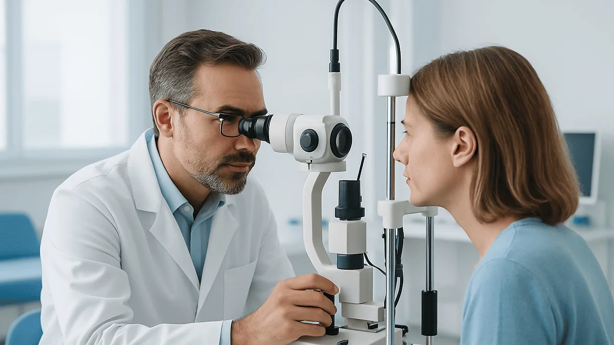
The slit lamp, a microscope with a bright thin beam, lets the clinician inspect the eyelids, cornea, iris and lens in fine detail. If dilation is planned, the drops go in here and you wait. Once the pupils are wide, the clinician uses a handheld light or a lens to look at the retina, the optic nerve and the blood vessels, and may take a photograph or a scan for the record. The visit ends with a plain explanation of findings and a plan, which may be as simple as a new prescription and a date for the next check.
Who is usually asked to come sooner, and who can usually wait
How often you should sit in that chair depends less on age alone than on risk, and the common guidance from major sources is consistent in shape if not in every number. The table below summarizes typical recommendations for adults without symptoms, drawn from the Mayo Clinic’s eye exam overview and the National Eye Institute’s dilated exam guidance. It is a starting point, not a rule; your clinician sets your interval.
| Situation | Typical suggested interval | Why |
|---|---|---|
| Adults in their 20s and 30s, no symptoms, no risk factors | Every 5–10 years (Mayo Clinic) | Low rates of disease; prescription changes are usually gradual |
| Ages 40–54 | Every 2–4 years (Mayo Clinic) | Near-focusing declines; glaucoma and early lens changes become more common |
| Ages 55–64 | Every 1–3 years (Mayo Clinic) | Rising risk of cataract, glaucoma and macular change |
| Age 65 and over | Every 1–2 years (Mayo Clinic) | Highest prevalence of age-related eye disease |
| Diabetes, family history of glaucoma, age 60+, or Black and age 40+ | Dilated exam every 1–2 years (National Eye Institute) | Higher risk of silent retinal and optic nerve disease |
| Glasses or contact lens wearers, any age | Usually 1–2 years (Mayo Clinic) | Prescription accuracy and contact lens safety |
Who is asked to come sooner than the table suggests? Anyone with new symptoms, anyone taking a medicine that requires eye monitoring, people with a prior retinal problem, and children with a family history of strabismus (misaligned eyes) or amblyopia (lazy eye). The NHS recommends most adults have an eye test every two years and more often if advised.
Who can usually wait? A healthy young adult with a stable prescription and no symptoms is generally not asked to return every year. Waiting is a decision, not a default, and it should be made with the clinician who last examined your eyes rather than assumed because nothing feels wrong.
What not to do before an eye exam
Preparation is mostly about not muddying the water. A handful of common habits can quietly distort the measurements or the clinician’s view.
Do not arrive with tired, strained eyes if you can help it. A long stretch of close screen work immediately before the visit can leave the focusing muscle temporarily locked on near objects and the eye surface dry, both of which can nudge a refraction. Give your eyes an easy hour beforehand where possible.
Do not wear eye makeup heavily on the day. Mascara flakes and liner along the lash line show up under the slit lamp, can smear into the tear film and complicate the assessment of eyelid glands and dry eye. Clean lids make the exam clearer and quicker.
Do not put in contact lenses if the office has asked you to leave them out, and do not use redness-relieving drops to “tidy up” a pink eye before the visit. Those drops constrict surface blood vessels and hide exactly what the clinician needs to see. If your eye is red, let it be red.
Do not stop or skip a prescribed medicine, including eye drops for glaucoma, because you have an appointment. Pressure readings are meant to reflect your treated state, and interrupting treatment can be harmful. If you are unsure about timing a drop around the visit, ask the office in advance; only your prescriber should change a regimen.
Do not turn up on a rushed schedule. Dilation adds waiting time, and a hurried refraction is a worse refraction. Block out the appointment plus a generous margin, and leave the car at home if drops are likely.
Finally, do not rehearse the letter chart or try to “pass.” The point is not to score well; it is to find out how your eyes actually see, so that whatever help you need is the right help.
Can I drink the night before, and is morning or afternoon better?
Two questions come up constantly and neither has a dramatic answer.
On alcohol: no eye-care guideline forbids a drink the evening before a routine exam, and a single glass with dinner is unlikely to shift a prescription meaningfully. Heavy drinking is a different matter. Alcohol dehydrates the body, and a dehydrated eye surface is drier and less smooth, which can blur vision and make the tear film look poorer than usual. A hangover brings light sensitivity, redness and difficulty concentrating, all of which make the “which is clearer, one or two” part of the exam harder to answer reliably. Alcohol also transiently affects eye movements and coordination. If you have overdone it, the exam will still be valid for most purposes, but tell the clinician; it may explain a red eye or an unusually dry surface, and it lets them judge whether a borderline finding deserves a recheck on a better day.
On timing: there is no guideline preference for morning over afternoon, and for most people the time of day does not change the result in a way that matters. A few practical points can tip the choice. If your eyes feel driest and most tired late in the day, an afternoon visit may make dry-eye symptoms easier to demonstrate but may also make the refraction feel harder. If you wear contact lenses and have been asked to leave them out beforehand, a morning slot lets you do that overnight. If dilation is planned, remember that the effects last several hours according to the Mayo Clinic, so a late-afternoon appointment can mean blurry near vision well into the evening, while a mid-morning one gives the day room to recover.
The best time, in short, is the one when you are rested, unhurried, and have a way home.
The rest of the day: what the hours after the exam usually look like
Walking out of an eye exam without dilation is uneventful; you may have a new prescription in hand and nothing else to manage. Walking out after dilation is a small event, and it helps to know the shape of it.
The first thing most people notice is light. The world outside the clinic door looks bleached and glaring, and the sky can be uncomfortably bright even on an overcast day. This is the wide pupil doing exactly what it was asked to do. Sunglasses help immediately, and many offices hand out disposable dark shields if you have forgotten your own.
The second is near blur. Reading a text message, a menu or a receipt becomes difficult because the focusing muscle has been relaxed along with the pupil. Distance vision often feels close to normal, which is why some people are tempted to drive. Resist that. Glare recovery is slower and judging distance is less precise than it feels, and the Mayo Clinic advises against driving until the effects wear off.
Duration varies with the drops used and with your own eyes. Lighter irises tend to stay dilated longer than darker ones. The Mayo Clinic describes the effect as lasting several hours; some people are fully back to normal by evening, others notice a slightly wide pupil the next morning, especially if a stronger drop was used for a child or for a detailed retinal check. Both are within the expected range.
Plan the afternoon accordingly: no fine detail work, no long screen sessions, generous breaks, and hydration. A headache from squinting is common and settles as the pupils recover. Mild stinging or a gritty feeling from the drops fades within an hour or two. What should not happen is pain, sudden loss of vision, or worsening rather than easing symptoms; those are covered in the final section, and they warrant a call rather than patience.
What people often get wrong about eye exams
“My vision is fine, so my eyes are fine.” This is the most consequential misunderstanding. Glaucoma, diabetic retinopathy and early macular degeneration typically cause no symptoms in their early stages, which is precisely why the National Eye Institute recommends dilated exams for people at risk even when they see well. Sharp acuity says nothing about eye pressure or the state of the retinal blood vessels.
“An online vision test is the same thing.” Screen-based tests estimate acuity and sometimes refraction. They cannot measure eye pressure, examine the retina, detect cataract or check how the eyes align. They are a partial substitute for one part of the visit and no substitute for the rest.
“Dilation is optional and just for older people.” Dilation is what turns a glasses check into a health check. It is routinely recommended for children with suspected eye problems, for anyone with diabetes, and for new symptoms at any age.
“If I get the letter chart wrong, I’ll end up with the wrong glasses.” The refraction is built from many cross-checked measurements and your answers are one input among several. Hesitation and “they look the same” are expected and useful.
“Supplements don’t count as medicines.” High-dose vitamin A, niacin, some herbal products and blood-thinning supplements all have documented eye or bleeding effects. List them.
“Reading in dim light or sitting close to screens damages the eyes.” These habits cause fatigue and dryness, not structural damage. The clinician will be more interested in how many hours you spend at near tasks than in your lighting.
“A stronger prescription means my eyes are getting worse.” Lens power describes focusing, not health. Many people need more help with age while their eyes remain perfectly healthy, and the reverse is also true.
Questions to ask your care team
A good exam ends with understanding, and a short list of questions turns a rushed handover into a conversation. Bring these written down; you will not remember them with a wide pupil and a bright light in your face.
- Did you see anything today that needs monitoring, and what specifically are you watching?
- What was my eye pressure, and how does it compare with previous readings if you have them?
- Are there any early signs of cataract, glaucoma or macular change, and what would make you more concerned at the next visit?
- Does anything in my medication list need eye monitoring, and how often is that usually done?
- How has my prescription changed since the last one, and is the amount of change what you would expect?
- Do I need different glasses for different tasks, such as screens or driving at night?
- If I wear contact lenses, is my current type and wearing schedule appropriate for how my eyes look today?
- When should I come back, and what would make me come back sooner?
- Which symptoms should prompt an urgent call rather than waiting for the next appointment?
- Can I have a copy of today’s findings and any images, so I can bring them next time or to another clinician?
Ask, too, about anything that puzzled you during the exam. If the clinician mentioned a term such as “cup-to-disc ratio” or “astigmatism” without explanation, request the one-sentence version. Most are simple once translated: astigmatism, for example, just means the front of the eye is curved more steeply in one direction than the other, like a spoon rather than a ball, and it is corrected with a lens shaped to match.
Write the answers down or ask for them in the summary. The point of preparing so carefully is to leave with a clearer picture, and the picture belongs to you.
When to call your doctor
Most eye exams, including dilated ones, end with nothing more than a few hours of blur and glare. A small number of symptoms, whether they appear after a visit or on an ordinary day, should not wait for a routine appointment. MedlinePlus, the Mayo Clinic and the NHS list the following as reasons to seek care urgently:
- Sudden loss of vision in one or both eyes, even if it is partial or lasts only minutes.
- A sudden shower of new floaters, flashes of light, or a curtain or shadow moving across your vision, which can signal a retinal tear or detachment.
- Severe eye pain, especially with a red eye, headache, nausea and blurred vision or halos around lights, which can indicate a sudden rise in eye pressure.
- Sudden double vision, a drooping eyelid, or an eye that will not move normally.
- An injury to the eye, a chemical splash, or something embedded in the eye.
- A painful red eye in a contact lens wearer, particularly with light sensitivity or discharge.
- Vision loss accompanied by weakness, numbness, slurred speech or confusion, which needs emergency stroke assessment.
After dilation specifically, call the office if blur and light sensitivity are worsening rather than easing after several hours, if you develop eye pain or a headache that does not settle, or if you feel unwell in a way you cannot explain. These reactions are uncommon but they are the reason the office gives you a number to call.
For anything less dramatic, such as a prescription that feels slightly off after a week of wearing new glasses, a persistent gritty feeling, or a question you forgot to ask, a routine call is the right route. Adaptation to a new prescription commonly takes days, and small adjustments are normal.
Every judgment about what a finding means, what to monitor and when to return rests with the clinician who examined you. This article prepares you to make the most of that conversation; it does not replace it.
Frequently asked questions
Do I need to bring anything for an eye exam?
Yes: bring your current glasses and contact lenses with their packaging, any old prescriptions or eye reports, a complete list of medicines and supplements, your health and family eye history, identification and insurance details, sunglasses, and a driver if dilation is likely. Each item removes a guess from the exam and makes the result more accurate and more comparable with future visits.
What is a good eye exam checklist the night before?
Lay out every pair of glasses you use, your lens box or foil, a pharmacy printout of your medicines, any old reports, and a short note of symptoms and questions. Confirm whether dilation is planned and arrange a ride if it is. Charge your phone so you can photograph the prescription, and set aside sunglasses. Aim for a normal night’s sleep and light screen use in the morning.
Is there anything I shouldn't do before an eye exam?
Avoid heavy eye makeup, redness-relieving drops, long stretches of close screen work immediately beforehand, and wearing contact lenses if the office has asked you to leave them out. Do not stop or skip prescribed medicines, including glaucoma drops, on the day of the exam; pressure readings are meant to reflect your treated state, and only your prescriber should change a regimen.
Can I drink the night before an eye exam?
A single drink with dinner is unlikely to affect a routine exam. Heavy drinking dehydrates the eye surface, causes redness and light sensitivity, and makes it harder to judge which lens is clearer, so it can reduce the reliability of the refraction. If you have had a heavy night, keep the appointment but mention it, so the clinician can interpret a dry or red eye correctly.
Is it better to have an eye exam in the morning or afternoon?
No guideline prefers one over the other, and for most people the time of day does not change the result meaningfully. Choose a time when you are rested and unhurried. If dilation is planned, remember the effects last several hours, so an earlier appointment gives the day room to recover. Contact lens wearers asked to leave lenses out may find a morning slot easier.
Do I need a driver for an eye exam?
You need a driver only if your pupils will be dilated, which is common in comprehensive exams, diabetic eye checks and visits for new symptoms. Dilation blurs near vision and increases glare for several hours, and the Mayo Clinic advises against driving during that time. Ask when you book; if dilation is possible, plan for a companion, taxi or public transport.
How long does an eye exam usually take?
A comprehensive exam typically runs 30 to 90 minutes, longer when dilation is included because the drops take roughly 15 to 30 minutes to work before the retina can be examined. A simple glasses check without dilation is shorter. Allow a generous margin in your schedule, since a hurried refraction is a less accurate one.
How to prepare for an eye exam if I wear contact lenses?
Ask the office how long to leave your lenses out beforehand, since the answer depends on whether they are soft, rigid or overnight lenses. Arrive in glasses, carry your lenses in a clean case with fresh solution, and bring the box or foil showing brand, power and curvature. Be candid about how you actually wear and clean them.
Should I bring my medication list even if none of my medicines are for my eyes?
Yes. Many medicines taken for other conditions affect the eyes: corticosteroids can raise eye pressure, hydroxychloroquine can affect the retina, and antihistamines dry the surface. Include over-the-counter products and supplements, and note the reason for each medicine. The clinician uses this to interpret findings and to coordinate any monitoring with your prescriber; it is not a signal to change anything yourself.
What if I can't find my old eye prescriptions or reports?
Bring whatever you have, even a photo or a partial record, and write a brief timeline of your eye history from memory. The exam is fully valid without old documents; they simply make trends easier to see. With your permission the office can often request records from previous providers, and you can ask for a copy of today’s findings to start a folder for next time.
References
This article is for general information only and is not a substitute for professional medical advice. Please consult a qualified doctor about your individual situation.

More from the Blog

Esotropia, Exotropia and Beyond: How Strabismus Disorders Differ and How Care Is Planned
Strabismus is a misalignment of the eyes, and the main types of strabismus are named for the direction of the turn: esotropia (inward), exotropia…
Laser, Injections or Vitrectomy: How Retina Specialists Match the Treatment to the Problem
Retinal treatment options fall into three main groups: laser, which seals tears or treats leaking blood vessels; injections into the eye, which block the…

Observation, Laser, Injections or Surgery: How Diabetic Retinopathy Treatment Is Chosen
Diabetic retinopathy treatment options are chosen by stage and by what is threatening sight. Early disease without swelling of the central retina is usually…

What Happens at a Contact Lens Fitting: Tear Film Checks, Trial Lenses and Wear Rules
A contact lens fitting appointment is a separate visit, or an add-on to an eye exam, in which an eye care professional measures the…

When an Intraocular Lens Needs Repositioning or Exchange: What Happens Next
An intraocular lens exchange is an operation to remove an artificial lens implanted during earlier cataract surgery and replace it with a new one.…

ILASIK Step by Step: The Femtosecond Flap, the Excimer Laser and the Minutes in Between
ILASIK is an all-laser, wavefront-guided form of LASIK. The typical procedure steps are: detailed corneal and wavefront mapping, numbing drops and an eyelid holder,…