

Scalp Reconstruction Techniques: Skin Grafts, Local Flaps and Tissue Expansion Explained
Key Takeaways
- Scalp skin barely stretches because of the galea, a tough fibrous sheet beneath the fat layer, so wounds only a few centimeters wide on the crown often cannot be stitched closed directly.
- Only techniques that move living scalp with its own blood supply, such as local flaps and tissue expansion, can restore hair; a skin graft from the thigh stays permanently hairless.
- MedlinePlus notes that a healthy skin graft begins to develop new blood vessels connecting it to surrounding tissue within about 36 hours, which is why early stillness and a pressure dressing matter so much.
- Bare skull bone without its periosteum cannot support a skin graft, so wounds that expose bone are steered toward flaps or staged approaches.
- Tissue expansion requires two operations and repeated clinic visits over weeks to months, and produces a visible bulge during the growing phase that many people find the hardest part.
- Hair along scalp incisions commonly falls out temporarily from surgical shock and typically begins regrowing within a few months, so the appearance at the first dressing change is not the final result.
Scalp reconstruction techniques are surgical methods for closing a wound on the scalp that cannot simply be stitched shut, most often after skin cancer removal, trauma, burns or infection. The main options are skin grafts, which transplant a thin layer of skin; local flaps, which rotate nearby scalp into the gap; and tissue expansion, which slowly stretches healthy scalp to grow extra hair-bearing skin. Surgeons choose based on wound size, depth and the health of surrounding tissue.
The stitches came out on a Tuesday. The dermatologist had been clear and kind: the basal cell carcinoma was gone, the margins were clean, and now there was a coin-sized hole on the crown that no comb could hide. The next appointment was with a reconstructive surgeon, and the referral letter used a word that sounded larger than the wound itself: reconstruction.
People in this position usually arrive with the same two worries. Will it heal? And will I look like myself afterward? The answers depend on a set of scalp reconstruction techniques that surgeons have refined for decades, each with a different logic, timeline and trade-off. Some borrow skin from elsewhere on the body. Some shift scalp sideways. One asks the body to grow extra skin on purpose.
None of them is a beauty treatment, and none deserves the mystery that surrounds them. What follows is a plain walk through what each involves, who tends to be offered which, what the weeks afterward look like, and the questions worth bringing into the consultation room.
Why is the scalp harder to repair than skin elsewhere?
Ask a surgeon what makes the scalp difficult and they will point to two things: it barely stretches, and it sits directly on bone. Skin on the forearm or abdomen can be pinched into a generous fold. Scalp skin over the crown moves only a few millimeters before it stops, because beneath the skin and fat lies a tough fibrous sheet called the galea, a tendon-like layer that connects the forehead muscles to the muscles at the back of the head. That sheet is what gives the scalp its strength and also its stubbornness.
The layers matter for planning. From the outside in, the scalp has skin, a dense fat layer packed with blood vessels, the galea, a loose gliding layer, and then the periosteum, the thin membrane that nourishes the skull bone. A wound that stops above the galea behaves very differently from one that exposes bone. Bone without its periosteum cannot support a skin graft on its own, because a graft needs a living, blood-rich bed to survive.
Blood supply is the scalp’s great advantage. Several named arteries run upward from the face and neck and link with each other across the top of the head, which is why scalp cuts bleed so dramatically and also why scalp flaps tend to survive well when designed around those vessels. Hair adds a further layer of complexity. Hair follicles sit in that fat layer, so any repair that removes or thins this layer changes how hair will grow afterward.
Put those facts together and the challenge is clear: close a gap in a tight, hair-bearing, bone-backed surface without leaving bald patches, distorting the hairline or the eyebrows, or starving the new tissue of blood. Every technique described below is a different answer to that single problem.
How scalp reconstruction techniques work: the reconstructive ladder
Surgeons often describe their options as rungs on a ladder, starting with the simplest and climbing only as far as the wound demands. The image is useful for patients too, because it explains why two people with scalp cancer can leave with entirely different plans.
On the lowest rung is healing by secondary intention, which means letting a shallow wound close on its own with dressings and time. Above that sits primary closure, where the edges are brought together and stitched. The scalp’s tightness means this only works for small wounds, and pulling too hard risks the edges dying or the scar spreading wide.
The next rung is the skin graft. A graft is a piece of skin lifted completely free of its own blood supply and laid onto the wound, where it must pick up new vessels from below. It is quick and covers large areas, but it depends on the wound bed being healthy and cannot carry hair.
Higher still is the local flap. A flap is tissue moved into the wound while remaining attached to its original blood supply. On the scalp, flaps are cut from neighboring skin and rotated, advanced or transposed into the defect, bringing hair and full-thickness cover with them.
Tissue expansion occupies its own rung. Rather than borrowing skin, it grows it: a soft silicone balloon is placed under healthy scalp and gradually inflated over weeks to months so that the stretched skin can later be advanced across the gap.
At the top are free flaps, where tissue from a distant site is detached with its artery and vein and reconnected to vessels in the neck or temple under a microscope. These are reserved for the largest or most complex wounds.
The ladder is not a rule that lower is always better. Surgeons frequently skip rungs, choosing a flap over a graft for a small crown defect because the result will match the surrounding hair. The point is that each technique solves a specific problem of size, depth and location.
What happens before surgery: mapping the wound
Reconstruction begins with measurement, not incisions. The surgeon assesses three things about the defect: how wide it is, how deep it goes, and what surrounds it.
Width is measured in centimeters, but its meaning changes with location. A gap near the ear or over the temple, where the scalp is looser, may close directly. The same gap on the vertex, the flat top of the head where the galea is tightest, may need a flap. Depth is checked by identifying which layer forms the wound floor. Healthy fat or intact periosteum will accept a graft. Bare bone, bone that has been treated with radiation, or bone that has been burred or partly removed during cancer surgery will not, and a flap or a staged plan becomes necessary.
Surroundings matter as much as the hole itself. Prior radiotherapy leaves scalp that is thin, poorly supplied with blood and slow to heal, which pushes surgeons toward bringing in fresh tissue from outside the treated zone. Old scars from previous surgery can block the path of a planned flap. Smoking, diabetes and long-term steroid use all reduce the small-vessel blood flow that grafts and flaps depend on, and surgeons will ask about them directly.
When the wound follows skin cancer removal, timing depends on the pathology. If the tumor was removed with Mohs surgery, a technique in which thin layers are removed and examined under a microscope until no cancer cells remain, the margins are already confirmed and reconstruction can often be planned promptly. If a standard excision is awaiting a pathology report, many teams wait for the result before committing to a flap, because re-excision through a freshly rotated flap is far harder than through an open wound.
Imaging is not routine for skin-level defects, but a CT scan may be requested when bone involvement or a deeper tumor is suspected. Photographs are usually taken so the team can plan flap geometry and so you have a record of the starting point.
Scalp skin graft: how a piece of skin learns to live somewhere new
A skin graft is the reconstructive equivalent of transplanting a seedling. The surgeon takes skin from a donor site, commonly the thigh, and lays it on the prepared wound. From that moment the graft has no blood supply of its own. It survives its first day or two by absorbing fluid from the wound bed, and, according to MedlinePlus, a healthy graft begins to develop new blood vessels connecting it to the surrounding tissue within roughly 36 hours. If that connection fails, part or all of the graft dies and turns dark.
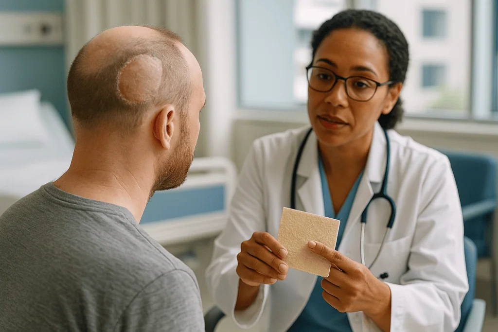
Grafts come in two thicknesses. A split-thickness graft takes the epidermis and only part of the dermis, the deeper skin layer that gives skin its strength. It is thin, takes readily, and the donor site heals on its own because deeper skin cells remain to regrow. A full-thickness graft takes the entire dermis, gives a thicker and better-colored result, but demands a richer wound bed and leaves a donor wound that must be stitched.
On the scalp, split-thickness grafts are the more common choice because they are forgiving and can cover very large areas after extensive cancer resection or burns. The trade-off is honesty about appearance: grafted scalp is smooth, hairless, often paler or shinier than surrounding skin, and slightly sunken because it lacks the fat layer beneath. For a wound on the crown of someone with dense hair, that is a real cosmetic cost. For someone with little hair, or a wound in a hidden area, it may be a very reasonable one.
After surgery the graft is protected with a bolster dressing that presses it gently against the bed and prevents shearing. MedlinePlus advises keeping the grafted area still and avoiding stretching or pressure during the early healing window, which on the scalp means no hats that rub, no lying directly on the site, and no vigorous hair washing until the team says so.
Scalp flap surgery: borrowing hair-bearing skin from next door
Where a graft is a seedling, a flap is a branch bent across a gap while still attached to the tree. Local scalp flaps are cut from healthy scalp adjacent to the wound and moved into it without ever losing their blood supply. Because the flap brings its own fat layer and hair follicles, the repaired area can grow hair that matches its neighbors, which is the main reason surgeons favor flaps for visible, hair-bearing defects.
Several geometries are in common use. A rotation flap is a large semicircular section pivoted around a fixed point into the wound. An advancement flap slides straight forward. A transposition flap jumps over a bridge of intact skin. Bilobed and rhomboid flaps are smaller, angular designs suited to moderate defects. When the wound is too large for a single flap, surgeons may use two or three flaps meeting in the middle, sometimes described as a pinwheel pattern.
Flap design on the scalp is largely a question of size. Because scalp skin barely stretches, a flap often needs to be several times larger than the hole it fills so that the tissue arrives without tension. Surgeons frequently score the underside of the galea with shallow parallel cuts, a step called galeal scoring, to gain a little extra reach.
Moving a flap leaves a second wound at the donor site. If it is small, it is closed directly. If it is too large, the surgeon may place a skin graft there instead, choosing a spot that sits lower on the head or under longer hair where a smooth patch is less noticeable. This is a common and deliberate compromise: a graft goes in the hidden place, a flap goes in the visible one.
Flaps also carry hair in a fixed direction. A rotated flap can make hair grow at an angle to its surroundings, and a poorly oriented one can pull the hairline or an eyebrow upward. Good planning minimizes this, but it cannot always be eliminated, and it is a fair question to raise during consultation.
Tissue expansion scalp surgery: growing new skin on purpose
Tissue expansion borrows a trick the body already knows. Skin over a growing pregnancy or a slowly enlarging swelling does not simply thin; it multiplies its cells and genuinely increases in area. Surgeons harness that response with a silicone balloon, called a tissue expander, placed in a pocket beneath healthy scalp next to the area that needs covering.
The procedure is staged. In the first operation the empty expander is inserted under the galea and the incision is closed. After the scalp has healed, the patient returns to the clinic at intervals for the expander to be gradually filled with sterile saline through a small port, either built into the device or placed under the skin nearby. Each filling stretches the overlying scalp a little, and the skin responds by growing. The process unfolds over weeks to months, with the pace set by the surgeon according to how the skin tolerates the stretch. During this phase the expander is visible as a smooth, rounded bulge, which many people find the hardest part of the experience.
When enough extra scalp has grown, a second operation removes the expander, excises the scar, graft or bald area, and advances the expanded, hair-bearing skin across the gap. Because the new skin comes with its own follicles, expansion is one of the few ways to replace a large hairless area with hair that matches in color, texture and growth pattern.
Its strengths make it a common choice for children with large congenital scalp lesions, for burn scars and for revising old grafts once cancer surveillance has settled. Its demands are equally clear: two operations, repeated clinic visits, a period of living with a visible bulge, and a small but real risk that the expander becomes infected or works its way through the skin, which usually means removing it and starting over later. Expansion is rarely used when a tumor is still under active treatment, because the timeline conflicts with cancer care.
Free flaps and dermal substitutes: options for the largest wounds
Some defects outrun every local option. A wound that spans most of the scalp after removal of an aggressive tumor, exposes a large area of bone, or sits in heavily irradiated tissue may need skin brought in from far away with its own blood vessels reconnected on arrival. This is a free flap, and it is microsurgery: an artery and vein in the flap are sewn to vessels in the neck or in front of the ear using stitches finer than a human hair.
The most familiar donor is the latissimus dorsi, the broad flat muscle of the back, taken with or without its overlying skin. Another common choice is a thin flap from the outer thigh. These flaps deliver a thick, well-supplied blanket that can cover exposed bone, tolerate later radiotherapy, and survive where a graft would fail. The costs are longer surgery, a hospital stay with close flap monitoring, a donor-site scar on the back or thigh, and a repaired scalp that is smooth and hairless, sometimes noticeably bulky at first.
A separate development is the dermal substitute, a sheet of collagen-based scaffold laid onto a wound to encourage the body to build a new dermis layer before a thin skin graft is applied on top. It is used mainly when bone is exposed and a flap is not appropriate, for example in frail patients. The evidence base is mostly case series and small studies rather than large randomized trials, so it is best described as an established option in the right setting rather than a proven equal to flap coverage. The Cleveland Clinic and other centers describe these products as part of a staged approach that adds several weeks before the graft can be placed.
Most people facing scalp reconstruction will never need this rung of the ladder. It exists because some wounds are genuinely large, and knowing it is there can be reassuring rather than alarming.
Comparing scalp reconstruction options at a glance
The table below summarizes how the main techniques differ. It is a map, not a prescription: the same wound can reasonably be treated in more than one way, and the treating team weighs factors that no table can capture, such as your other medical conditions, whether further cancer treatment is expected, and what matters most to you about the result.
| Technique | What it is | Typically suited to | Hair in the repaired area | Main trade-offs |
|---|---|---|---|---|
| Primary closure | Edges stitched together | Small wounds, looser scalp regions | Yes, existing hair | Tension can widen scar or cause edge breakdown |
| Split-thickness skin graft | Thin skin from thigh laid on wound | Large, shallow wounds with healthy bed or periosteum | No | Smooth, pale, slightly sunken patch; donor site discomfort |
| Local flap | Adjacent scalp rotated or advanced with its blood supply | Small to moderate visible defects | Yes, may change direction | Long incisions; donor site may need a graft; hairline shift possible |
| Tissue expansion | Balloon grows extra scalp over weeks to months | Large hairless areas, scars, congenital lesions, graft revision | Yes, closely matched | Two operations; visible bulge; repeated visits; infection or exposure risk |
| Free flap | Distant tissue reconnected by microsurgery | Very large, deep or irradiated wounds, exposed bone | No | Long surgery; hospital stay; donor scar; bulkiness |
| Dermal substitute plus graft | Collagen scaffold then thin graft | Exposed bone when flap not appropriate | No | Staged over weeks; evidence mostly from small studies |
Two patterns are worth noticing. Hair follows blood supply: only techniques that move living scalp with its vessels intact can restore hair. And complexity follows depth: the deeper the wound and the poorer the surrounding tissue, the further up the ladder the plan tends to climb. A surgeon who explains where your wound sits on both axes has usually explained most of the reasoning behind the recommendation.
Who is scalp reconstruction usually for, and who is asked to wait?
The most common route to scalp reconstruction runs through skin cancer. The scalp receives years of direct sun, particularly in people with thinning hair, and basal cell carcinoma, squamous cell carcinoma and melanoma all occur there. The NHS notes that surgery to remove the cancer is the main treatment for most non-melanoma skin cancers, and when the removed area is too large to stitch closed, reconstruction follows. Trauma, burns, scalp infections that destroy tissue, and rare congenital conditions in which a child is born with an area of missing scalp make up most of the remainder. Some people seek revision of old grafts or scars years after the original event.
Being a good candidate is less about age than about tissue and timing. Surgeons look for a wound with a clean, healthy bed, confirmed clear margins if cancer was involved, and surrounding scalp with good blood flow. Older adults are frequently excellent candidates for flaps precisely because their scalp is looser and their expectations are practical.
Several groups are commonly asked to wait or to choose a simpler option first. If pathology is still pending, teams often hold off on flaps until margins are confirmed. If radiotherapy is planned for the same area, the sequence of reconstruction and radiation is negotiated between the surgeon and the oncologist, because irradiated flaps behave differently from irradiated grafts. Active smokers are often asked to stop for a period before elective flap or expansion surgery, since nicotine constricts the small vessels that keep flaps alive. Poorly controlled diabetes, active infection at the site, and blood-thinning medicines that cannot be safely paused are all reasons for delay, and the decision about any medicine change rests with the prescribing clinician, never with the patient acting alone.
Tissue expansion has its own gatekeeping. It suits people who can attend repeated visits, tolerate a visible bulge for months, and are not in the middle of active cancer treatment. For a child, that also means a family able to manage the schedule.
What the first days and weeks after scalp reconstruction usually look like
The first week is about protection. Grafts are covered with a pressure dressing that stays undisturbed until the team removes it, typically at the first follow-up visit. Flaps are usually closed with sutures or staples and may have a small drain for a day or two. Swelling and bruising commonly track down into the forehead and around the eyes because gravity pulls fluid from the scalp, which alarms people who were not warned; it is expected and settles. Numbness across the top of the head is nearly universal after scalp surgery, since the sensory nerves run upward through the tissue that has been moved, and sensation returns slowly and sometimes incompletely.
Sleeping with the head raised on extra pillows reduces swelling. Hair washing is usually paused until the dressing is off and then restarted gently with the team’s guidance. MedlinePlus’s self-care guidance for skin flaps and grafts stresses keeping the site clean and dry, avoiding stretching or pressure, and not picking at scabs or crusts, which can lift a healing graft edge.
Sutures and staples on the scalp are usually removed at a follow-up visit within the first two weeks, though the timing is set by the surgeon based on how the wound looks. Split-thickness graft donor sites on the thigh are often the sorest part of recovery; they weep for several days and heal beneath a dressing over the following weeks.
Most people return to desk-based work within a week or two of a graft or local flap, and later after a free flap, which typically involves several days in hospital for monitoring. Heavy lifting, bending and straining raise blood pressure in the head and are usually restricted for a few weeks to protect the repair. Tissue expansion has a different rhythm: recovery from each operation resembles a flap, but the months of filling visits in between are the defining feature. Timelines vary with wound size and personal health, and the treating team’s instructions override any general range.
Hair, scars and appearance: an honest account of the result
The question people ask most quietly is what it will look like. The honest answer is that scalp reconstruction aims for a durable, safe closure first and a natural appearance second, and the two do not always align perfectly.
Grafted areas are smooth and hairless for life. Skin from the thigh carries no scalp follicles and never will. The patch usually matures over months from pink or purple to a paler shade than surrounding skin, and it sits slightly below the level of the scalp because the fat layer is missing. Sunscreen or a hat matters here, because grafted skin lacks pigment protection and burns easily.
Flaps look different at first than they will later. In the early weeks the rotated tissue is swollen, the incision lines are red, and hair along the cut edges may fall out temporarily. This shock loss occurs because follicles near an incision pause their growth cycle, and Cleveland Clinic and other sources describe hair regrowth around scalp incisions typically beginning within a few months. Hair that grew under a flap may point in a new direction, which can be noticeable with short styles and less so with longer ones. Incision lines on the scalp tend to heal well because they are hidden by hair, but a scar always exists, and scars on the scalp can be wider than elsewhere because of tension.
Tissue expansion produces the closest match, since the advanced skin is genuine neighboring scalp. Even so, expanded scalp can be slightly thinner with hair spaced a little more widely than before, and the final scar line runs along the advanced edge.
Later refinement is common. Scar revision, small adjustments to hairline position, or hair transplantation into a grafted area are all options discussed after the primary reconstruction has fully matured and, in cancer cases, after surveillance has established stability. None of this is a promise about outcome, but knowing that refinement is possible often takes pressure off the first result.
Risks and complications in plain language
Every reconstruction carries risks, and a good consultation names them without drama. The most technique-specific risk is failure of the repair to survive. A graft can fail to take if blood collects beneath it, if it shifts before vessels grow in, if the bed is poor, or if infection sets in. Partial loss is more common than total loss and is often managed with dressings while the surviving portion heals in. A flap can lose blood flow at its far edge, the part furthest from the vessels that feed it, producing a strip of darkened skin that may scab and heal or may need trimming. Free flaps carry the added possibility that the microsurgical connection clots in the first days, which is why they are watched closely in hospital.
Bleeding beneath the repair, called a hematoma, is a particular concern on the scalp because of its rich blood supply. It shows as rapidly increasing swelling and tightness and sometimes requires return to the operating room. Infection presents with spreading redness, warmth, increasing pain or discharge and is treated according to the team’s assessment.
Tissue expansion adds device-related risks: the expander can become infected, can push through thinned skin, or can leak. Any of these usually means removing it and allowing the scalp to recover before deciding whether to try again.
Longer-term issues include numbness that does not fully return, itching or tenderness in scars, hairline or eyebrow distortion, and dissatisfaction with the appearance of a graft. Wound breakdown is more likely in irradiated scalp, in smokers, and in people with diabetes or conditions that impair small-vessel circulation.
Alternatives always exist, and they include doing less. For a frail person with a shallow wound, healing by secondary intention with regular dressings can be a legitimate choice. The role of the treating team is to lay out each option’s risks alongside its benefits so that the decision is shared rather than assumed.
What people often get wrong about scalp reconstruction
Myths gather around any surgery that involves the head, and a few deserve correcting because they change decisions.
The first is that a skin graft will grow hair once it settles. It will not. Hair follicles live in the scalp’s fat layer, and a graft from the thigh carries thigh skin. A smooth patch is permanent unless a later procedure moves hair-bearing tissue or transplants follicles into it.
The second is that flaps are riskier than grafts. On the scalp the opposite is often true. A well-designed local flap carries its own blood supply and tolerates a less-than-perfect wound bed, while a graft depends entirely on the tissue beneath it. The flap’s risks lie in its length of incision and its effect on hairline, not in survival.
Third, people assume tissue expansion is a cosmetic luxury. Its origins and its most common uses are reconstructive: children born with large scalp lesions, burn survivors, and adults replacing failed grafts. It is chosen because it restores hair-bearing scalp, not because it is quicker or easier. It is neither.
Fourth, a common worry is that the skull is exposed or damaged during these operations. In most skin-level reconstructions the bone is never touched. When bone is involved, the surgeon says so explicitly, because it changes the plan.
Fifth, many believe the result seen at the first dressing change is the final result. Early grafts look mottled, flaps look swollen and lopsided, and hair near incisions falls out. Scar maturation on the head, as elsewhere, takes many months, and appearance improves substantially over that time.
Finally, there is a belief that any reconstruction after skin cancer means the cancer is gone for good. Reconstruction closes the wound; it does not replace follow-up. The NHS notes that people who have had one non-melanoma skin cancer have a higher chance of developing another, which is exactly why surveillance continues after the scalp has healed.
Questions to ask your care team before scalp reconstruction
Consultations move quickly, and the most useful questions are often the ones people think of on the drive home. Writing a list beforehand helps, and there is no question too basic to ask.
- Which layer does my wound reach, and is any bone exposed or affected?
- Which of the scalp reconstruction techniques are you recommending for me, and what made you choose it over the others?
- Will the repaired area grow hair, and if so, will the hair direction or hairline change?
- Where will the incisions be, and how long will they be?
- If I need a skin graft, where will it come from, and what will that donor site feel like while it heals?
- Are my cancer margins confirmed, and could I need further surgery through this repair?
- Is radiotherapy planned, and how does that affect the timing and type of reconstruction?
- What should I do about my regular medicines, and who decides whether any are paused?
- How long will the dressing stay on, and when can I wash my hair?
- What would make you want to see me urgently before my scheduled follow-up?
- If the graft or flap does not fully survive, what is the usual next step?
- What refinements might be possible later, and how long would you wait before considering them?
Two questions deserve a place at the top for anyone considering tissue expansion: how many clinic visits are anticipated over the expansion period, and what the expander will look like at its largest. Seeing photographs of the expanded stage in advance prevents a genuine shock later.
It also helps to ask who will see you at each stage. Reconstruction after skin cancer often involves a dermatologist, a reconstructive surgeon, and sometimes an oncologist, and knowing which team member to call for which problem avoids delay when something does not look right. Bringing a family member or friend to take notes is a small step that consistently pays off.
When to call your doctor after scalp reconstruction
Most recoveries are uneventful, but the scalp’s blood supply means problems that do occur can develop quickly. Contact the team promptly, or seek urgent care as instructed on your discharge paperwork, if any of the following appear.
- Bleeding that soaks through the dressing or does not stop with steady, gentle pressure applied for several minutes.
- Rapidly increasing swelling, tightness or a firm lump beneath the repair, which can signal blood collecting under a graft or flap.
- A graft or the edge of a flap turning dark purple, gray or black, or feeling cold compared with surrounding skin.
- Spreading redness beyond the incision line, increasing warmth, worsening pain after the first few days, or pus or foul-smelling discharge, all of which suggest infection.
- Fever, chills or feeling generally unwell.
- A wound edge that opens or a graft that lifts or slides, especially after a knock or a fall.
- Clear fluid draining persistently from a wound over the skull, which needs assessment.
- For tissue expanders: sudden deflation, the device becoming visible through thinning skin, or redness and pain over the expander or its port.
- Severe headache, confusion, drowsiness, new weakness, or a seizure, which are rare but require emergency evaluation.
Numbness, mild itching, some bruising around the eyes and a small amount of clear or pale pink ooze in the first days are expected and do not usually need a call, but no one is ever wrong to ask. If you cannot reach the surgical team and the sign is on the list above, emergency services are the right next step.
The pattern to watch is change. A repair that looked settled and then becomes more swollen, more painful or more discolored is telling you something, and the team would far rather assess a false alarm than a late complication.
Frequently asked questions
What is scalp reconstruction after skin cancer?
Scalp reconstruction after skin cancer is surgery to close the wound left when a tumor is removed and the gap is too large or too deep to stitch shut. Options include skin grafts, local flaps that rotate nearby scalp into the defect, tissue expansion, and for very large wounds, free flaps from elsewhere on the body. The choice depends on wound size, depth, confirmed margins and whether radiotherapy is planned.
Will a scalp skin graft grow hair?
No. A skin graft taken from the thigh or another donor site carries no scalp hair follicles and will remain smooth and hairless permanently. It also tends to look paler and slightly sunken compared with surrounding scalp. If a hair-bearing result matters, surgeons consider a local flap or tissue expansion instead, or discuss later hair transplantation into the grafted area once healing and any cancer surveillance have settled.
How does scalp flap surgery differ from a skin graft?
A flap is scalp tissue moved into the wound while still attached to its original blood supply, so it arrives alive with its fat layer and hair follicles. A graft is skin fully detached from its source and laid on the wound, where it must grow new vessels from the bed beneath. Flaps preserve hair and tolerate poorer wound beds; grafts cover larger areas quickly but cannot carry hair.
What is tissue expansion scalp surgery and how long does it take?
Tissue expansion places a silicone balloon under healthy scalp and gradually fills it with saline over a series of clinic visits, stretching the skin so that it grows extra hair-bearing tissue. A second operation then removes the expander and advances the new scalp across the defect. The process unfolds over weeks to months, with the pace set by the surgeon based on how the skin responds.
Is scalp reconstruction painful?
Discomfort is usually moderate and most noticeable in the first few days. Many people find a split-thickness graft donor site on the thigh sorer than the scalp itself. The scalp is often numb after surgery because sensory nerves pass through the moved tissue, and that numbness can last months. Pain that worsens after the first days, rather than easing, should be reported because it can signal infection or bleeding.
How long does recovery from scalp reconstruction take?
Recovery varies with technique and wound size. Dressings over grafts typically stay in place until the first follow-up, sutures or staples are usually removed within the first couple of weeks, and desk-based work often resumes within one to two weeks after a graft or local flap. Free flaps involve a hospital stay for monitoring. Scars and grafts continue maturing for many months, and the team’s instructions override general ranges.
Can the skull be exposed during scalp reconstruction?
In most skin-level reconstructions the skull is never touched. When cancer or trauma has exposed bone, the surgeon will say so, because bare bone without its periosteum cannot support a skin graft. Such wounds are directed toward flaps that bring their own blood supply, or toward staged approaches using a dermal substitute before grafting. Exposed bone changes the plan, and teams explain this explicitly.
Why did hair fall out around my scalp incision?
Temporary hair loss along incision lines is common and results from surgical stress on follicles near the cut, which pauses their growth cycle. This shock loss is not usually permanent. Regrowth around scalp incisions typically begins within a few months. Hair that grew on a rotated flap may point in a new direction afterward. Persistent bald strips beyond that period are worth raising with the surgical team at follow-up.
Who should not have scalp reconstruction right away?
Teams often delay flaps or expansion when cancer margins are still unconfirmed, when radiotherapy timing has not been agreed with the oncologist, when there is active infection at the site, or when someone smokes heavily, since nicotine narrows the small vessels flaps depend on. Poorly controlled diabetes and blood-thinning medicines that cannot safely be paused are further reasons to wait. Any medicine decision belongs with the prescribing clinician.
Can a scalp reconstruction be improved later?
Often, yes. Once the initial repair has fully matured, which takes many months, options such as scar revision, small hairline adjustments, or hair transplantation into a grafted area can be discussed. In cancer cases, teams usually wait until surveillance shows the area is stable. Whether refinement is appropriate, and which kind, depends on the individual result and is a decision made together with the treating team.
References
- MedlinePlus Medical Encyclopedia: Skin graft
- MedlinePlus: Skin flaps and grafts – self-care
- Cleveland Clinic: Skin Graft
- NHS: Non-melanoma skin cancer – Treatment
This article is for general information only and is not a substitute for professional medical advice. Please consult a qualified doctor about your individual situation.

More from the Blog
How to Wash Long Hair After a Hair Transplant Without Shaving: Protecting the Grafts
Washing hair after an unshaven hair transplant follows the same cautious timeline as any transplant: most surgical teams allow a first gentle rinse within…

U-FUE vs Standard FUE: How Unshaven Extraction Changes Session Length and Graft Handling
U-FUE (unshaven follicular unit extraction) removes individual grafts while surrounding hair stays long, whereas regular FUE usually shaves the donor area first. Because long…
Sapphire Hair Transplant Myths: Does the Blade Alone Decide Density and Natural Direction?
No. In a sapphire hair transplant, the sapphire blade only makes the tiny recipient slits; it does not decide density or direction. Density depends…
Questions to Ask Before Robotic Hair Surgery: Who Places the Grafts and How Results Are Checked
Before robotic hair surgery, ask who performs each stage: the robot usually harvests follicular units, but a surgeon or trained technicians still make recipient…
Why the Cause of Hair Loss Sets the Plan: Tests Before Non-Surgical Hair Restoration
Before non-surgical hair restoration, clinicians usually confirm the cause first: a history and scalp examination, a gentle pull test, magnified scalp imaging (trichoscopy), and…
Who Is a Candidate for Laser Hair Therapy? Early Thinning vs Long-Standing Bald Areas
A laser hair therapy candidate is usually an adult with early to moderate hereditary (pattern) thinning, where follicles have shrunk but are still alive,…