

SI Joint Pain: The Often-Missed Cause of Low Back and Buttock Pain

Key Takeaways
- Mainstream estimates attribute 15 to 30 percent of chronic low back pain to the sacroiliac joints, making it a common diagnosis that is simply named less often.
- The SI joint moves only two to four degrees and is held by some of the body's strongest ligaments, so it cannot be 'popped back into place' by a stretch or manipulation.
- Three or more positive provocation tests out of five, confirmed by an image-guided anaesthetic injection, is the most reliable way to identify the joint as the pain source; scans alone are not.
- Mechanical SI pain is worse with stairs, standing on one leg and rolling in bed, whereas inflammatory sacroiliitis brings prolonged morning stiffness and night pain that improve with movement.
- Exercise programmes targeting the gluteus maximus, gluteus medius and deep abdominal muscles have the broadest trial support of any treatment, with benefits appearing over weeks rather than days.
- Pain that travels below the knee with numbness points to a lumbar nerve root, and pain in the groin that flares when crossing the legs points to the hip, not the SI joint.
SI joint pain comes from the sacroiliac joints, the two joints that connect the base of the spine to the pelvis. It usually causes one-sided low back or buttock pain that worsens with standing, stairs or rolling over in bed. Mainstream estimates attribute 15 to 30 percent of chronic low back pain to these joints. Diagnosis relies on examination and, when needed, a diagnostic injection; most people improve with targeted exercise and time.
As of September 2026, the phrase “SI joint pain” is climbing search charts for a reason that has little to do with new science. A wave of short videos promises that a single twist, a chiropractic “pop” or a thirty-second stretch will “put the pelvis back in place” and end months of buttock pain. Millions have watched. Many have tried it in their kitchens. Some felt better for an hour. Most were left asking the same question they started with.
Here is the quieter story behind the trend. The sacroiliac joints, where the triangular bone at the base of the spine meets the two wings of the pelvis, are a genuinely under-recognised source of low back pain. They sit low, they hide behind the disc and the hip, and they rarely show anything dramatic on a scan. That is why someone can carry a diagnosis of “nonspecific back pain” for years while the real culprit is two inches to the side of the spine.
This piece sorts the viral claims from what examination, injection studies and randomised trials actually support.
Why SI joint pain is so often missed
The sacroiliac joint, or SI joint, is where the sacrum (the wedge-shaped bone at the bottom of the spine) meets the ilium (the broad upper bone of the pelvis) on each side. You have two of them. Each is a strong, irregular joint wrapped in some of the thickest ligaments in the body, and each moves only a few degrees. Its job is to pass load from the upper body into the legs, roughly the way a keystone transfers weight in an arch.
Three things make it easy to overlook. First, its pain pattern overlaps with the lumbar disc, the hip and the piriformis muscle, so the same ache can be blamed on any of them. Second, routine X-rays and most MRI scans look at the lumbar spine and often stop just above the joint, or show age-related change that proves nothing about where the pain is coming from. Third, there is no single blood test or image that confirms a painful SI joint; the diagnosis is built from history, examination and sometimes a numbing injection.
Cleveland Clinic and other mainstream sources estimate that 15 to 30 percent of chronic low back pain originates in the SI joints. That is not a rare condition. It is a common one wearing an unfamiliar name. Johns Hopkins and Mayo Clinic describe a typical picture: pain concentrated over the dimple at the top of the buttock, usually on one side, that flares with prolonged standing, climbing stairs, getting out of a car or rolling over in bed.
Knowing that the joint exists is half the battle. The other half is recognising how it behaves differently from the disc or the hip, which is where the rest of this article goes.
What changed recently
The honest answer is that the anatomy has not changed; the attention has. Several developments explain why the topic feels new.
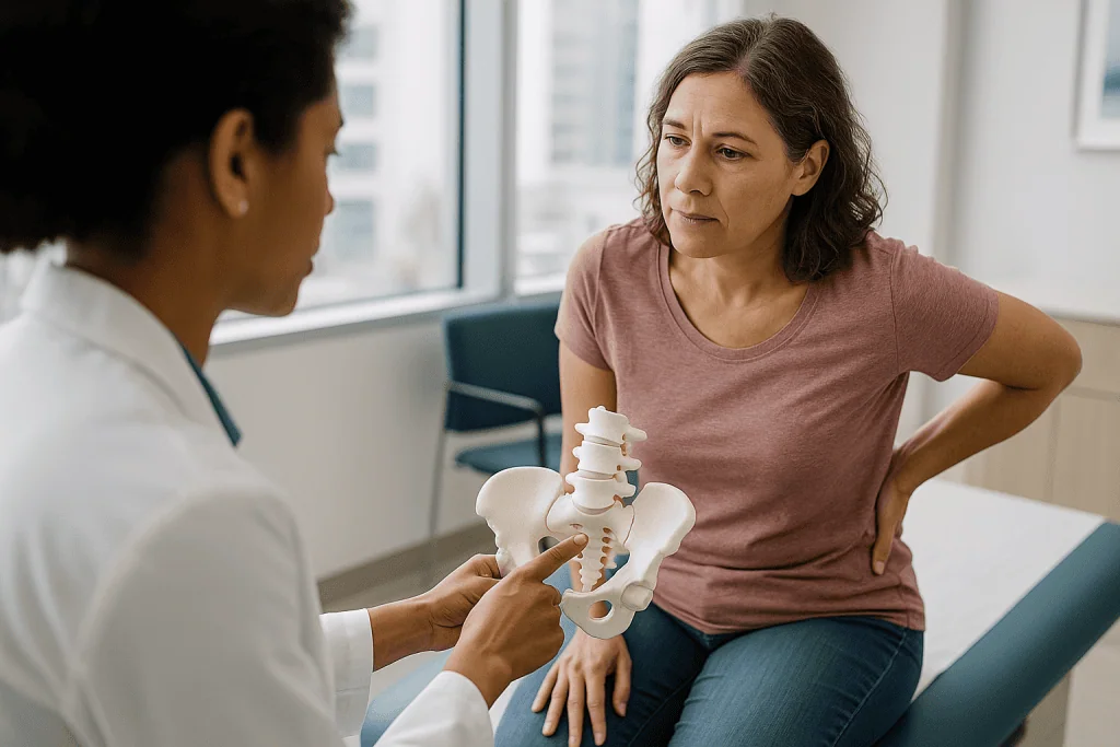
The first is diagnostic. Since 2009, when international rheumatology groups introduced the concept of “non-radiographic axial spondyloarthritis,” MRI of the sacroiliac joints has been used to detect inflammation years before it shows on a plain X-ray. Inflammatory sacroiliitis (inflammation of the SI joint, often as part of ankylosing spondylitis) is therefore being identified earlier and in younger adults, and MedlinePlus now lists sacroiliac inflammation as a hallmark early feature of ankylosing spondylitis.
The second is procedural. Minimally invasive SI joint fusion devices, placed through small incisions to stabilise the joint, have been in clinical use in the United States for well over a decade, and the body of published trials comparing them with non-surgical care has grown through the 2020s. Mayo Clinic’s current treatment guidance lists joint fusion as a last-resort option after injections and rehabilitation have been tried, which is a shift from a time when the operation was rarely mentioned to patients at all.
The third is cultural. In 2025 and 2026, self-adjustment and “pelvic reset” videos spread widely, often filmed in homes rather than clinics. They are the main reason search volume spiked. They are also the main source of misunderstanding, because they present a complex, ligament-stabilised joint as something that slips out and clicks back.
None of this alters the first-line approach described by NHS, Mayo Clinic and Cleveland Clinic: stay active, use targeted strengthening, and reserve injections and procedures for pain that persists despite good rehabilitation. What has changed is how early the joint gets considered, and how many people now know to ask about it.
Sacroiliitis symptoms vs mechanical sacroiliac joint dysfunction
Two different problems share one joint, and they call for different conversations with a clinician.
Mechanical sacroiliac joint dysfunction means the joint is painful because of how it is loaded or how it moves: too little motion, too much motion, or uneven stress from one side to the other. It is the version most people searching “si joint pain” have. The pain is usually one-sided, sits low in the back or upper buttock, and can run down the back of the thigh, rarely below the knee. It is worse with standing on one leg, stairs, long drives and getting up from a chair. It is often better lying flat. Mornings are not dramatically stiffer than evenings.
Sacroiliitis, in the strict sense, means inflammation inside the joint. Mayo Clinic describes it as a feature of inflammatory conditions such as ankylosing spondylitis, psoriatic arthritis and inflammatory bowel disease, and occasionally of infection. Its signature is different: pain and stiffness that are worst in the early morning or after sitting still, that ease with movement rather than rest, that may wake you in the second half of the night, and that frequently alternate between buttocks. It tends to start before age 45 and can be accompanied by eye inflammation, psoriasis or bowel symptoms.
Why the distinction matters is simple. Mechanical pain is managed primarily with movement, load management and, if needed, injections. Inflammatory sacroiliitis is managed by a rheumatologist, usually with anti-inflammatory medicines such as naproxen or ibuprofen first, and in persistent cases with biologic medicines such as adalimumab, which block the inflammatory signal TNF. Every medicine decision belongs to the prescribing clinician, but the pattern of your pain is something you can describe accurately, and it steers which door you walk through.
What causes sacroiliac joint dysfunction?
The SI joint rarely fails on its own. It usually becomes painful because something changed the way force travels through it.
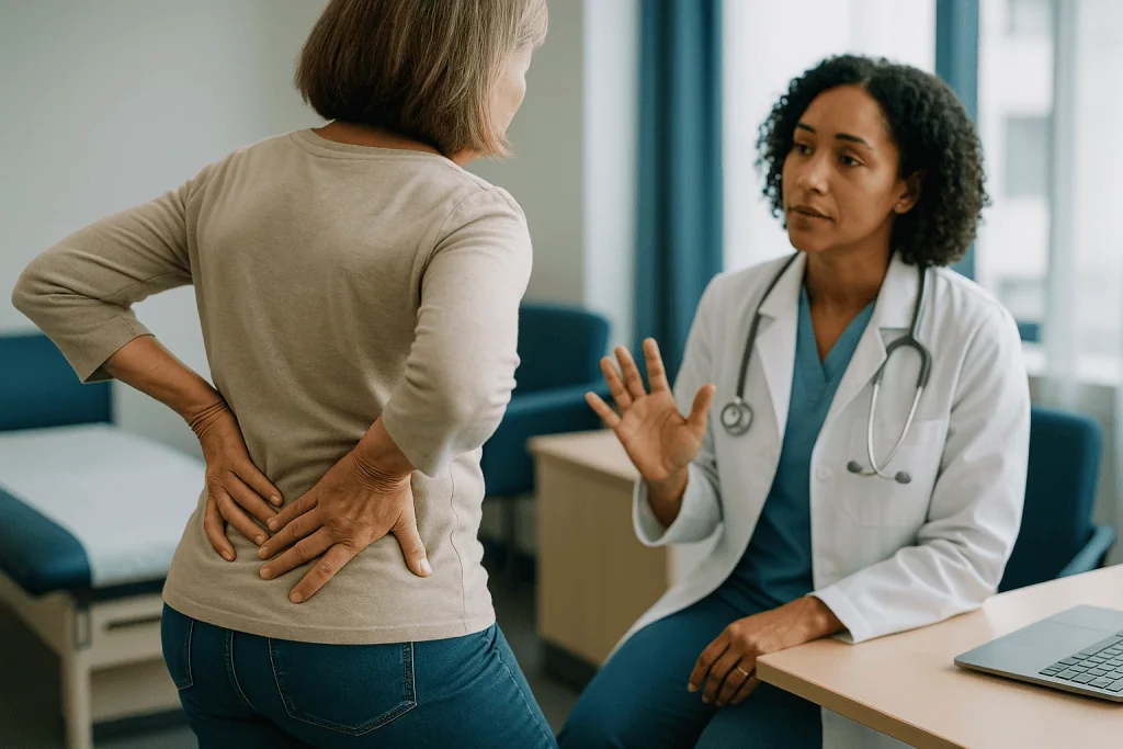
- Pregnancy and the postpartum year. The hormone relaxin loosens pelvic ligaments so the birth canal can widen. Combined with weight gain and an altered walking pattern, this makes the SI joints one of the most common sources of pelvic girdle pain in pregnancy, and the laxity can persist for months afterwards.
- A leg-length difference or a stiff hip. If one leg is functionally shorter, or one hip cannot rotate fully, the pelvis tilts and one SI joint takes more load with every step.
- A fall or sudden jolt. Landing hard on one buttock, a missed step or a rear-end collision can sprain the joint’s ligaments.
- Previous lumbar fusion. When vertebrae above are fused, the segments that still move take more stress. The SI joint is next in line, and Cleveland Clinic lists prior spinal fusion as a recognised risk factor.
- Osteoarthritis. Like any joint, the SI joint can lose cartilage with age. The catch is that worn-looking joints on X-ray are common in pain-free people, so the image alone does not make the diagnosis.
- Repetitive one-sided loading. Golf, racket sports, carrying a toddler on the same hip, or standing for hours with weight shifted to one leg.
- Inflammatory arthritis or infection. Discussed above, and the reason a clinician will ask about morning stiffness, skin, eyes and bowel symptoms.
In many people, no single trigger stands out. What they share is a mismatch between the load the joint receives and the stability the surrounding muscles provide. That is also why the gluteal and deep abdominal muscles sit at the centre of every credible rehabilitation plan.
SI joint pain so bad you can't walk: what could be causing it?
This exact question appears in search thousands of times a month, and it deserves a straight answer rather than reassurance.
A mechanically irritated SI joint can be surprisingly disabling. Because the joint is involved in every stride, an acute flare can make each step feel like a stab at the top of the buttock. Protective muscle spasm in the gluteals and lower back then stiffens the whole region, so standing up from a chair becomes a two-handed project. In this situation the problem is often a sprain of the joint’s ligaments after a fall, a long drive, an awkward lift or a sudden increase in activity. It is painful, it is frightening, and it usually settles over days to a few weeks with gentle movement, short periods of rest and, if a clinician agrees, an over-the-counter anti-inflammatory.
Late pregnancy and the weeks after delivery are another common setting for severe, walk-limiting SI pain, driven by ligament laxity. Obstetric and physiotherapy teams see this often and have specific strategies for it.
Severe inability to bear weight can also point away from the SI joint entirely. A hip stress fracture, a sacral insufficiency fracture (a crack in the sacrum that occurs in thin or osteoporotic bone, sometimes after no obvious injury), or a lumbar disc pressing on a nerve root can all masquerade as “SI joint pain.” Infection of the joint is rare but produces fever and unrelenting pain even at rest.
The practical rule is this: if you genuinely cannot walk, if the pain followed a fall, if you are over 65 or have osteoporosis, if you have fever, or if there is numbness, weakness or any change in bladder or bowel control, this is not a wait-and-see situation. Those signs are listed again in the “When to see a doctor” section below, because they override every self-care tip in this article.
How is si joint pain diagnosed?
There is no single test, which is exactly why the diagnosis gets missed. Clinicians build it from three layers.
History. Pain below the belt line and off to one side, pointing with one finger to the dimple over the joint, worse with stairs and transitions, better lying flat. Pain that does not travel below the knee. A plausible trigger such as pregnancy, a fall or a recent spinal fusion.
Provocation tests. These are examination manoeuvres that stress the joint in specific directions while the examiner watches for reproduction of your familiar pain. The commonly used set includes the thigh thrust, compression, distraction, Gaenslen’s and FABER tests. Research summarised by Cleveland Clinic and others suggests that no single test is reliable alone, but three or more positive tests out of five raise the likelihood of SI joint origin considerably. One positive test means little.
Diagnostic injection. The closest thing to a reference standard is an image-guided injection of local anaesthetic into the joint. If the pain drops by 75 percent or more for the duration of the anaesthetic, the joint is very likely the source. Many specialists use two injections on separate days to reduce false positives.
Imaging plays a supporting role. X-rays can show arthritis or fracture; MRI is valuable when inflammatory sacroiliitis is suspected because it reveals bone-marrow swelling around the joint before the X-ray changes. Blood tests such as inflammatory markers and the HLA-B27 gene are ordered when the pattern suggests spondyloarthritis.
The key limitation is that degenerative change on a scan is common in people without pain. A scan showing “SI joint arthritis” does not prove the joint hurts, and a normal scan does not prove it doesn’t. The examination and the injection carry more weight than the picture.
SI joint, disc, hip or piriformis: how the look-alikes compare
Four conditions account for most one-sided low back and buttock pain. Their patterns overlap, which is why a careful examiner tests all four rather than guessing from the location alone. The table summarises the features that most often separate them. None is absolute, and two problems can coexist.
| Feature | SI joint | Lumbar disc / sciatica | Hip joint | Piriformis syndrome |
|---|---|---|---|---|
| Where it hurts most | Top of buttock, over the joint dimple | Low back, often central, radiating down leg | Groin, outer thigh, sometimes knee | Deep in the buttock |
| Below the knee? | Rarely | Often, to foot | Rarely | Sometimes |
| Numbness or weakness | No | Common | No | Occasional tingling |
| Worse with | Stairs, standing on one leg, rolling in bed | Sitting, bending, coughing | Walking, putting on shoes and socks | Prolonged sitting, hip rotation |
| Typical onset | Pregnancy, fall, post-fusion, gradual | Lifting, bending, gradual | Gradual, age-related or athletic | Overuse, long sitting |
| Key exam finding | 3 or more positive provocation tests | Positive straight-leg raise, nerve signs | Painful, limited hip rotation | Pain on resisted hip rotation |
| Most useful confirmation | Diagnostic injection | MRI matching symptoms | X-ray, hip injection | Diagnosis of exclusion |
Two patterns are worth memorising. Pain that reaches the foot with numbness points toward the nerve root, not the SI joint. Pain in the groin that flares when you try to cross your legs points toward the hip. The SI joint stays stubbornly local: buttock, back of the thigh at most, and never the calf.
What the evidence actually says about si joint pain treatment
Grading the evidence matters more here than in most back-pain topics, because the gap between what is marketed and what is proven is wide.
Exercise and physical therapy: moderate evidence, strongest for function. Randomised trials of pelvic stabilisation exercise, mostly in pregnancy-related and postpartum pelvic girdle pain, show meaningful reductions in pain and disability compared with general advice. For non-pregnancy SI pain the trials are smaller, but the direction is consistent and the approach is endorsed by NHS, Mayo Clinic and Cleveland Clinic. No trial has shown that one specific stretch outperforms a general strengthening programme.
Manual therapy and manipulation: low to moderate evidence, short-term. Several randomised trials show that manipulation can reduce pain for days to weeks. Benefits beyond that are not consistently demonstrated, and no study supports the idea that a manipulation “realigns” a displaced joint.
Corticosteroid injection: moderate evidence for short-term relief. Image-guided steroid injections reduce pain for weeks to a few months in many patients, more reliably in inflammatory sacroiliitis than in mechanical pain. Relief is temporary, and repeated injections carry diminishing returns and cumulative risks.
Radiofrequency ablation: low to moderate evidence. This procedure uses heat to interrupt the small nerves carrying pain from the joint. Randomised trials, a handful in total, show greater pain relief than sham at three to six months in carefully selected patients who responded to diagnostic blocks. Certainty is limited by small numbers and variable technique.
Minimally invasive fusion: moderate evidence in selected patients, with caveats. Randomised trials comparing fusion with non-surgical management report larger improvements in pain and function at one to two years. Most were industry-funded, patients were highly selected, and long-term and comparative data remain limited.
Belts, cushions, braces: expert opinion and small studies. Helpful for symptom control in some people; not disease-modifying.
The pattern is clear: the least invasive options have the broadest support, and each step up the ladder requires stricter patient selection.
SI joint pain exercises: what actually helps and why
The goal of exercise is not to “stretch the joint open.” It is to give the joint a stronger muscular corset so the ligaments stop being asked to do all the work. Three muscle groups matter most.
The gluteus maximus and medius. These are the large and side buttock muscles. The maximus crosses the back of the SI joint and tensions the ligaments that lock it; the medius keeps the pelvis level when you stand on one leg. Weakness in either lets the pelvis drop and shear the joint with each step. Bridges, side-lying leg lifts (clamshells), and step-ups build them.
The deep abdominal and pelvic floor muscles. The transversus abdominis is the corset-like muscle that wraps the lower trunk. Gentle “drawing in” of the lower abdomen, then holding that tension while moving a leg, trains it to fire before load arrives.
The hip rotators and hamstrings. Tightness here tilts the pelvis. Figure-four stretches and hamstring stretches address it, done without forcing.
A sensible starting sequence, performed within comfortable limits:
- Glute bridge: lie on your back, knees bent, lift hips until the body forms a line from shoulders to knees.
- Clamshell: side-lying with knees bent, open the top knee while keeping the feet together and the pelvis still.
- Bird-dog: on hands and knees, extend the opposite arm and leg without letting the lower back arch.
- Single-leg stand: hold for 20–30 seconds beside a counter for safety.
- Knee-to-chest and figure-four stretches: hold gently, no bouncing.
Expect weeks, not days. Pain that is worse during an exercise and settles within an hour afterwards is usually acceptable; pain that is sharper the next morning means the load was too high. A physical therapist can tailor the programme and check that you are not substituting back muscles for glutes, the most common error.
Does losing weight help, and is cycling good for the SI joint?
Two of the most-searched questions, and both have nuanced answers.
Weight. Mechanically, every additional kilogram of body weight increases the load passing through the SI joints with each step, and observational studies consistently find higher rates of low back pain in people with higher body weight. For the SI joint specifically, direct trial evidence that weight loss reduces pain is lacking; the inference comes from biomechanics and from broader back-pain data. What the evidence does support, more strongly, is that the activity involved in healthy weight management, especially walking and strength training, improves back pain independently of the number on the scale. So the honest framing is this: if weight loss is something you and your clinician are pursuing for health reasons, your SI joints will likely benefit; but you do not need to wait for weight change to start the exercises that help most.
Cycling. Cycling is generally kind to the SI joint. It is low-impact, it loads both sides symmetrically, and the sitting position takes the ligaments off stretch. For many people with SI pain it is the most comfortable form of cardiovascular exercise. There are two caveats. A saddle set too high forces the pelvis to rock side to side with each pedal stroke, which irritates the joint; lowering the seat so the knee keeps a slight bend at the bottom of the stroke fixes this. And an aggressively forward, racing posture can aggravate pain in some people, in which case a more upright setup or a stationary recumbent bike works better. If pain increases during or after rides despite these adjustments, that is useful information to bring to a physical therapist rather than a reason to push through.
SI joint pain relief at home: cushions, belts and sleeping positions
Home measures do not fix the underlying problem, but they can make the weeks of rehabilitation far more tolerable.
Seat cushions. People ask for the “best” cushion, and the honest answer is that no brand has been tested in a trial. What matters is the shape. A wedge cushion that tilts the pelvis slightly forward reduces slumping, which otherwise loads the back of the joint. A cushion with a cut-out under the tailbone helps if sitting pressure on the sacrum is the trigger. Firm foam holds its shape; very soft foam lets the pelvis sink and rotate. For long drives, the most effective change is often not a cushion at all but a break every 30–45 minutes to stand and walk.
SI belts. A pelvic belt is a wide, firm strap worn low around the hips, just above the hip bones, that compresses the SI joints. Small studies and consistent clinical experience suggest it reduces pain during walking and standing, particularly in pregnancy and the postpartum period. It is a short-term aid for flares and activity, not something to wear all day for months, because long-term reliance can let the stabilising muscles weaken.
Sleeping. Side sleeping with a pillow between the knees keeps the top leg from dragging the pelvis forward. Back sleepers often do better with a pillow under the knees. Rolling over hurts because the joint shears; keeping knees together and moving as one unit reduces that.
Heat and cold. Heat relaxes protective muscle spasm; cold dulls an acute flare. Neither changes outcomes, both are safe for most people in 15–20 minute applications with skin protection.
Medicines. Over-the-counter anti-inflammatories such as ibuprofen or naproxen can ease a flare. Whether they are appropriate for you, given your stomach, kidneys, heart and other medicines, is a question for a pharmacist or doctor, not a package label.
Injections, radiofrequency ablation and fusion: what the procedures do
When several months of well-executed rehabilitation have not moved the needle, clinicians climb a ladder of procedures, each with a clear purpose.
Steroid injection. A corticosteroid (a potent anti-inflammatory medicine) mixed with local anaesthetic is placed in the joint under X-ray or ultrasound guidance. Beyond relief, it confirms the diagnosis if the pain reliably disappears. Benefits typically last weeks to a few months. Risks are low but real: infection, temporary blood-sugar rise in people with diabetes, and, with repetition, weakening of local tissue. Mayo Clinic notes that injections are usually limited to a few per year for these reasons.
Radiofrequency ablation. The SI joint is supplied by small nerve branches that run along the back of the sacrum. Ablation uses a heated probe to interrupt those branches so pain signals do not reach the brain. Candidates are usually people who had clear, temporary relief from two diagnostic nerve blocks. Relief, when it occurs, lasts six to twelve months on average because the nerves regrow; the procedure can be repeated.
Minimally invasive fusion. Through a small incision in the buttock, implants are placed across the joint to stop its motion and encourage bone to grow across it. It is reserved for people with pain confirmed by diagnostic injection, who have failed non-surgical care, and in whom other causes have been excluded. Trial evidence shows meaningful improvement in selected patients, but the operation is permanent, carries surgical risks, and transfers load to neighbouring joints.
What none of these procedures do is replace the strengthening work. Even people who proceed to fusion do better when the gluteal and trunk muscles are conditioned beforehand. The decision about whether and when to step up the ladder belongs with the treating clinician, informed by how precisely the joint has been confirmed as the source.
Common myths about SI joint pain
The viral claims that drove this topic to the top of search deserve point-by-point correction.
“My SI joint is out of place and needs to be popped back in.” The joint is held by ligaments strong enough to withstand the forces of running and childbirth. It moves two to four degrees. Studies using imaging before and after manipulation have not shown measurable repositioning. The click you hear is gas releasing from a joint capsule, and the short-term relief is from changes in muscle tone and pain signalling, not from realignment. That relief is real; the explanation is not.
“One stretch will fix it.” No randomised trial supports a single stretch. Programmes that help combine strengthening of the gluteals and trunk with gradual return to activity over weeks.
“If it hurts to move, I should rest until it stops.” NHS guidance on back pain is unambiguous: prolonged rest slows recovery. Modify activity, keep walking, avoid the specific movements that spike pain, and build back up.
“The scan shows arthritis, so that’s the cause.” Degenerative change in the SI joints is common in pain-free adults, especially after 50. Imaging findings must match the examination to mean anything.
“SI joint pain always means ankylosing spondylitis.” Most SI joint pain is mechanical. Inflammatory sacroiliitis has a distinct pattern of morning stiffness, night pain and improvement with activity, and is confirmed with MRI and blood tests, not assumed.
“A belt or cushion will solve it.” Both ease symptoms. Neither strengthens a muscle or changes the joint. They are scaffolding while the real work happens.
“Fusion is the modern answer.” It is an option for a small, carefully confirmed group after everything else has been tried. Mainstream guidance places it last for good reason.
When to see a doctor about SI joint pain
Most SI joint pain is uncomfortable rather than dangerous, and most people can start with activity modification and the exercises above. Certain features change that calculation and should prompt a prompt appointment, and in some cases urgent care.
Seek urgent medical attention if you have:
- New numbness around the groin or inner thighs, difficulty passing urine, or loss of bladder or bowel control. These can indicate pressure on the nerves at the base of the spine and are an emergency.
- Fever, chills or feeling unwell together with severe back or buttock pain, which can signal infection in the joint or spine.
- Inability to bear weight after a fall or accident, especially if you are over 65, have osteoporosis or take long-term steroids, because a fracture of the hip or sacrum is possible.
- Progressive weakness in a leg or foot.
Book a routine appointment if:
- Pain has lasted more than four to six weeks despite staying active and modifying what aggravates it.
- Pain wakes you regularly in the second half of the night, is markedly stiffer in the morning for more than 30 minutes, and improves with activity, particularly if you are under 45 or have psoriasis, inflammatory bowel disease or a history of eye inflammation. This pattern warrants assessment for inflammatory sacroiliitis.
- You have a history of cancer, unexplained weight loss, or pain that is constant and unrelated to position.
- You are pregnant or postpartum and pain is limiting walking or sleep; specialised physiotherapy is available and effective.
- You are considering any injection, ablation or surgical option, or you want to use anti-inflammatory medicines regularly.
Every decision about medicines, injections and procedures belongs with your treating clinician, who can weigh your examination, your other health conditions and your goals. The role of an article like this is to help you describe your pain precisely enough that the SI joint gets considered, rather than overlooked for another year.
Frequently asked questions
What does SI joint pain feel like?
It usually feels like a deep, one-sided ache or sharp catch at the top of the buttock, right over the dimple beside the base of the spine. It flares with stairs, standing up from a chair, getting out of a car and rolling over in bed, and often eases lying flat. Pain may spread to the back of the thigh but rarely passes the knee, and it does not cause numbness or weakness.
What are the symptoms of sacroiliitis compared with ordinary SI joint strain?
Sacroiliitis, meaning inflammation inside the joint, typically causes stiffness lasting more than 30 minutes in the morning, pain that wakes you in the second half of the night, buttock pain that alternates sides, and improvement with activity rather than rest. Mechanical strain behaves the opposite way: worse with loading, better with rest. The inflammatory pattern, especially before age 45, warrants assessment for conditions such as ankylosing spondylitis.
I have sacroiliac joint pain so bad I can't walk. What could be causing it?
An acute ligament sprain of the joint, often after a fall, long drive or sudden lift, can be severe enough to limit walking for days, as can late-pregnancy ligament laxity. Severe inability to bear weight can also signal a hip or sacral fracture, a disc pressing on a nerve, or rarely infection. If you cannot walk, had a fall, have fever, or notice numbness or bladder changes, seek medical care promptly.
What is the best seat cushion for SI joint pain?
No specific cushion has been tested in a clinical trial, so shape matters more than brand. A firm wedge that tilts the pelvis slightly forward reduces slumping, which loads the back of the joint, and a tailbone cut-out helps if direct pressure on the sacrum triggers pain. Very soft cushions let the pelvis sink and rotate. For long sitting, standing and walking every 30 to 45 minutes helps more than any cushion.
Does losing weight help sacroiliac joint dysfunction?
Probably, though direct trial evidence for the SI joint specifically is lacking. Higher body weight increases the load through the joints with every step, and observational studies link it to more low back pain. The activity involved in healthy weight management, especially walking and strength work, improves back pain on its own. Start the strengthening exercises now rather than waiting for weight to change, and discuss weight goals with your clinician.
Is cycling good for the SI joint?
Generally yes. Cycling is low-impact, loads both sides evenly and takes the pelvic ligaments off stretch, so many people with SI pain find it the most comfortable cardiovascular exercise. Set the saddle so the knee keeps a slight bend at the bottom of the stroke; a seat that is too high makes the pelvis rock and irritates the joint. If pain rises during or after rides despite adjustments, review it with a physical therapist.
Which SI joint pain exercises should I start with?
Start with glute bridges, clamshells, bird-dogs and single-leg standing, which strengthen the buttock and deep trunk muscles that stabilise the joint, plus gentle knee-to-chest and figure-four stretches. Work within comfortable limits; pain that settles within an hour afterwards is acceptable, pain that is worse the next morning means too much load. Expect improvement over weeks, and ask a physical therapist to check your form.
Can a chiropractor or physical therapist put the SI joint back in place?
No, because it is not out of place. The joint moves only a few degrees and is held by very strong ligaments; imaging studies have not shown repositioning after manipulation. Manipulation can still reduce pain for days to weeks by changing muscle tone and pain signalling, which randomised trials support in the short term. Lasting improvement comes from strengthening and graded activity, not from a one-time adjustment.
How long does SI joint pain take to get better?
An acute flare or sprain often settles over two to six weeks with modified activity. Pregnancy-related SI pain commonly improves in the months after delivery, especially with pelvic stabilisation exercise. Longstanding mechanical pain typically needs eight to twelve weeks of consistent strengthening before clear change. Pain persisting beyond six weeks despite good self-care, or with night pain and morning stiffness, should be assessed by a clinician.
What is the most effective si joint pain relief when exercise alone is not enough?
After a proper rehabilitation trial, image-guided corticosteroid injection has moderate evidence for relief lasting weeks to months and also confirms the diagnosis. Radiofrequency ablation, which interrupts the small nerves carrying pain from the joint, helps selected patients for six to twelve months. Minimally invasive fusion is a last resort for confirmed cases. Which step is appropriate, and when, is a decision for your treating clinician.
References
This article is for general information only and is not a substitute for professional medical advice. Please consult a qualified doctor about your individual situation.

More from the Blog
Heel Pain: Plantar Fasciitis, Achilles or Something Else: How to Tell
Heel pain is most often plantar fasciitis, which causes a sharp pain under the heel with the first steps of the morning. Pain at…

Anterior Hip Replacement: How the Front Approach Differs, and for Whom
Anterior hip replacement reaches the hip joint through the front of the thigh, working between muscles rather than detaching them. Research suggests modestly less…
Posture Correctors: Do They Work, or Weaken the Muscles That Should Hold You Up?
A posture corrector can work as a short-term reminder that gently pulls the shoulders back, and small studies show people slouch less while wearing…
Is My Cast Too Tight? Numbness, Color Changes and Fracture Warning Signs to Report
A cast may be too tight if the pain keeps climbing despite elevation and prescribed pain relief, if fingers or toes turn pale, blue…

When a Broken Shoulder Is Replaced Rather Than Repaired: Shoulder Prosthesis After Fracture
A shoulder prosthesis for fracture is an artificial ball, or ball and socket, implanted when the top of the upper arm bone has shattered…

Aesthetic Foot Surgery Results: What Toe Shortening and Foot Narrowing Realistically Change
Aesthetic foot surgery results are usually modest and structural: toe shortening removes a small segment of bone so a long toe sits level with…