

Spinal Compression Fractures Explained: When Kyphoplasty or Vertebroplasty Is Considered

Key Takeaways
- Osteoporosis is the most common cause of spinal compression fractures, which is why a bone density scan and bone-health plan are part of treatment even when no procedure is ever discussed.
- MedlinePlus gives a typical healing range of about 8 to 10 weeks for compression fractures managed with rest, bracing and pain relief, with acute pain usually easing well before the bone is fully united.
- Kyphoplasty differs from vertebroplasty mainly by inflating a balloon to create a cavity before injecting thicker cement at lower pressure, which is associated with less cement leakage in studies.
- Two sham-controlled trials found vertebroplasty no better than a placebo procedure for many osteoporotic fractures, while later trials restricted to recent, severe fractures reported benefit, so the evidence remains genuinely divided.
- A cement-filled vertebra is stiffer than its osteoporotic neighbors, and new fractures at adjacent levels are a recognized concern that treatment of the underlying bone disease is meant to address.
- New leg weakness, numbness, or loss of bladder or bowel control after a compression fracture is a medical emergency, because osteoporotic fractures very rarely affect the nerves and these signs point to something else.
Most spinal compression fractures are treated without surgery: short-term pain control, gradual return to movement, sometimes a brace, and treatment of the underlying osteoporosis. Kyphoplasty or vertebroplasty, in which bone cement is injected into the collapsed vertebra, is usually considered only when severe pain persists despite several weeks of conservative care, when imaging confirms the fracture is unhealed, and when the treating team judges the likely benefit outweighs the risks.
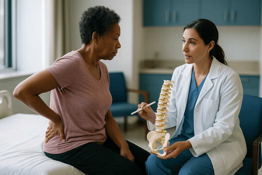
It is rarely a dramatic fall. More often it is a woman in her seventies who bent to lift a bag of potting soil, felt something give in the middle of her back, and assumed she had pulled a muscle. Two weeks later she is still sleeping in a recliner because lying flat is unbearable, and an X-ray shows that one of the bones in her spine has quietly collapsed like a stepped-on cardboard box.
What follows is a conversation many families are not prepared for. A doctor explains that spinal compression fracture treatment usually begins with time, pain relief and careful movement, and that a procedure involving bone cement exists but is not for everyone. The words kyphoplasty and vertebroplasty enter the household vocabulary, along with a reasonable question: if the bone is broken, why wait?
This explainer walks through what the fracture is, why most heal without a needle, what the cement procedures actually do, and where the evidence is honestly unsettled. The decisions belong to you and your treating team; the aim here is to make that conversation clearer.
What is a spinal compression fracture, and why does the bone give way?
The spine is a stack of roughly thirty-three bones called vertebrae, each shaped a little like a drum with a bony arch behind it. The drum, known as the vertebral body, carries most of the weight. A compression fracture happens when that body cracks and loses height, most often at the front, so the vertebra ends up shaped like a wedge rather than a block.
In a young, healthy spine this takes real force, such as a car crash or a fall from height. In an older spine it can take almost nothing. Osteoporosis, a condition in which bone becomes porous and fragile, is the usual culprit. The internal scaffolding of the vertebra thins until a cough, a misstep off a curb or the act of sitting down heavily is enough to buckle it. MedlinePlus lists osteoporosis as the most common cause, with trauma and tumors that weaken bone accounting for most of the rest.
The fractures cluster in the lower thoracic and upper lumbar spine, the region roughly behind the bottom of the ribcage, where the relatively rigid upper back meets the more mobile lower back and mechanical stress concentrates. Many people have one and never know it; the collapse is gradual and the pain is mistaken for arthritis or a strain. Others feel a sharp, sudden pain that is worse standing and eases lying down.
Over time, several wedged vertebrae stack into a forward curve of the upper back. That curve is what the word kyphosis describes, and it explains why kyphoplasty carries the name it does. Understanding this mechanism matters, because it frames the central truth of treatment: the fracture is a symptom of the bone, and fixing one vertebra does nothing for the others unless the bone itself is addressed.
Spinal compression fracture treatment: what conservative care actually involves
Conservative sounds passive. In practice it is a structured plan with several moving parts, and for most people it is the whole treatment.

Pain control comes first, because pain that keeps someone in bed causes its own harm. Clinicians typically start with simple pain relievers and, for severe acute pain, may add stronger options for a short, defined period. Which medicines, at what strength and for how long is a decision for the prescribing clinician, weighed against age, kidney function and other conditions. The goal is not to erase pain but to make walking, washing and sleeping possible while the bone knits.
Movement is the second pillar, and it surprises people. Prolonged bed rest weakens muscle and bone further and raises the risk of blood clots and pneumonia. MedlinePlus advises rest only briefly, then a gradual return to activity, often guided by a physical therapist who teaches how to rise from a chair, roll out of bed and lift without bending the spine forward.
Bracing is the third element, and the most debated. A rigid or semi-rigid back brace can limit forward bending and remind the wearer to keep upright posture. Some clinicians prescribe one for a few weeks; others avoid them because they can weaken trunk muscles and are uncomfortable in hot weather. The evidence does not clearly favor either approach, so this often comes down to the individual and the fracture location.
Running alongside all of this is the question nobody should skip: why did the bone break? A bone density scan, blood tests for vitamin D and calcium, and a review of medicines such as long-term corticosteroids are standard parts of spinal compression fracture treatment even when no procedure is ever discussed.
How long does a compression fracture take to heal?
Bone heals on its own schedule, and for a compression fracture that schedule is measured in weeks, not days. MedlinePlus gives a typical range of about 8 to 10 weeks for most compression fractures treated with rest, bracing and pain relief, with a longer road to full activity when surgery has been involved.
That figure needs some interpretation. The sharp, acute pain often improves well before the bone is fully healed, frequently within the first two to four weeks, as inflammation settles and the fractured surfaces begin to bind. Full radiographic healing, in which the bone looks solid again on imaging, can lag behind how someone feels. Conversely, some people report lingering aching for months after the fracture has healed, because the muscles and ligaments around a newly wedged vertebra have to adapt to a changed shape.
Several factors stretch the timeline. Very low bone density heals more slowly. Smoking impairs bone repair. Long-term steroid use, uncontrolled diabetes and poor nutrition all slow the process. Age itself matters less than these underlying factors, though older adults tend to carry more of them.
A fracture that has not healed by roughly three months may be described as a nonunion, meaning the bone fragments have failed to join. Some collapse further over this period, a process called progressive vertebral collapse. Both situations are among the reasons a treating team may start discussing kyphoplasty or vertebroplasty, and they are why follow-up imaging at intervals is standard rather than optional.
The honest answer to the healing question, then, is a range rather than a date. Most people are substantially better in two to three months. A minority are not, and that minority is where the rest of this article turns.
What actually happens during kyphoplasty and vertebroplasty
Both procedures belong to a family called vertebral augmentation, a term that simply means reinforcing a fractured vertebra from the inside. They are performed by interventional radiologists, spine surgeons or pain specialists, usually with the patient lying face down on a table equipped with live X-ray guidance.
After the skin over the back is cleaned and numbed, the operator passes a hollow needle through a small incision, angled toward the collapsed vertebra. Continuous X-ray images confirm the needle tip sits inside the bone and well away from the spinal canal and the nerves that exit on either side. This positioning step is the heart of the procedure and where most of the time is spent.
In vertebroplasty, a bone cement made from a medical acrylic is mixed to a toothpaste consistency and injected slowly through the needle. It flows into the honeycomb spaces of the fractured bone and hardens within minutes, stabilizing the cracked fragments the way epoxy stiffens a split table leg. The vertebra is not restored to its original height; it is fixed in its current shape.
Kyphoplasty adds a step. Before cement, a small balloon is threaded into the vertebra and inflated. This compacts the surrounding bone and creates a cavity, and it may push the collapsed end plate partly back toward its original position. The balloon is withdrawn and the cavity is filled with cement, which is thicker and injected under lower pressure than in vertebroplasty.
Cleveland Clinic describes the whole procedure as typically taking about an hour, longer if more than one vertebra is treated, and notes many people go home the same day. It is usually done under sedation with local anesthetic, though general anesthesia is sometimes chosen. Afterward the incision is covered with a small dressing; there are no stitches to remove.
Kyphoplasty vs vertebroplasty: what is the difference and does it matter?
Patients often assume one is the modern version of the other. In reality they are close relatives with a few real differences and several theoretical ones.
| Feature | Vertebroplasty | Kyphoplasty |
|---|---|---|
| Balloon used | No | Yes, inflated before cement |
| Aim for height restoration | Stabilize only | Stabilize and may partly restore height |
| Cement consistency and pressure | Thinner, higher pressure | Thicker, lower pressure into a pre-formed cavity |
| Cement leakage in studies | Reported more often | Reported less often; most leaks in both are silent |
| Anesthesia commonly used | Local with sedation | Local with sedation or general |
| Typical setting | Outpatient | Outpatient or overnight |
The height question is the most marketed and the least settled. Balloon inflation can lift a collapsed end plate in the moments before cement is placed, particularly in a fresh fracture. Whether that gain persists once the person stands and loads the spine, and whether a few millimeters of height translate into less pain or better posture years later, is not clearly established.
Pain relief appears broadly similar between the two in the comparative research that exists, and both share the same central uncertainty about how they compare with good conservative care, which the evidence section below addresses. Cement leakage, meaning cement escaping the vertebra into surrounding tissue, is reported somewhat less often with kyphoplasty, likely because the cavity lets the operator use thicker cement at lower pressure. Most leaks cause no symptoms.
In practice, the choice frequently reflects the operator’s training, the fracture’s age and shape, and local availability more than a firm evidence hierarchy. Asking why a particular technique is being proposed for your fracture is a fair question and one a good team will welcome.
Who is usually offered a cement procedure, and who is asked to wait
There is no single scoring system that decides this. Teams weigh pain, function, imaging and the person in front of them, but some patterns recur across guidelines and clinical practice.
A procedure is more likely to be discussed when severe, function-limiting pain persists after a fair trial of conservative treatment, commonly several weeks; when imaging, often an MRI, shows the fracture is still unhealed, with swelling inside the bone that suggests it remains active; when the pain clearly comes from the fractured level rather than from arthritis or muscle strain nearby; and when the person cannot tolerate the pain relief or the immobility that conservative care requires, for instance because bed rest is causing pressure injuries, pneumonia risk or a steep functional decline. Fractures caused by cancer weakening the bone form a separate category where stabilization is often considered earlier.
People are usually asked to wait, or are steered away altogether, when the fracture is only days old and improving on its own, since many will settle without intervention; when the bone has already healed, because cement adds nothing to a united fracture; when there is an active infection anywhere in the body or at the intended skin site; when a bleeding disorder or anticoagulant treatment cannot be safely managed around the procedure; when the fracture has pushed bone into the spinal canal or is causing nerve symptoms, which may call for a different operation; or when the vertebra has collapsed so completely that there is no room for a needle and cement.
Frailty sits in the middle. Some very frail people are poor candidates because of anesthesia risk; others are candidates precisely because lying immobile for two months is more dangerous for them than a procedure. Only the treating team, with the full picture, can make that call.
What the evidence really says about kyphoplasty and vertebroplasty
For roughly a decade, vertebroplasty was widely adopted on the strength of before-and-after observations and studies that compared cement with usual care. People walked out of the procedure room feeling better, and that felt like proof.
Then two randomized trials published in the New England Journal of Medicine compared vertebroplasty with a sham procedure, in which participants received the needle placement and local anesthetic but no cement, so neither they nor their assessors knew who had been treated. Pain and disability improved in both groups, and the differences between real and sham were not statistically significant at one month or beyond. Those results reshaped the field and led several guideline bodies to recommend against routine vertebroplasty for osteoporotic fractures.
The story did not end there. Critics noted the trials enrolled many people with fractures that were weeks to months old and included some with only moderate pain. Later sham-controlled trials that restricted enrollment to fractures under about six weeks old with severe pain reported a benefit for vertebroplasty, while another trial with a somewhat broader population again found none. Systematic reviews continue to disagree, largely because they weigh these trials differently.
Kyphoplasty has been compared with conservative care in randomized trials showing earlier pain and function improvement, but it has not been tested against a sham procedure in the same way, so the placebo question remains open.
Where does that leave a patient? With a fair summary: cement procedures are safe in experienced hands, they are not clearly better than sham for older or less painful fractures, they may help selected people with recent, severe, unhealed fractures, and part of the relief people report probably reflects natural healing and the expectation of benefit. That is not nothing, but it is not the certainty the early enthusiasm implied.
What are the risks of vertebral augmentation?
Serious complications are uncommon, which is one of the reasons these procedures are offered to older adults who might not tolerate a larger operation. Uncommon is not the same as absent, and the risks fall into a few groups.
Cement leakage is the most frequent event. Cement can seep through cracks in the vertebra into the disc space, the veins around the spine or, rarely, toward the spinal canal. Most leaks are seen only on imaging and cause no symptoms. When cement reaches the canal or a nerve exit, it can cause new pain, numbness or weakness, and occasionally requires an urgent operation to remove it. Cement that enters veins can travel to the lungs; small fragments usually cause no symptoms, but larger emboli are a recognized rare hazard.
Infection at the skin or within the bone is rare but serious, and is why active infection elsewhere postpones the procedure. Bleeding is generally minor. Allergic reactions to cement components or contrast dye are uncommon. Anesthesia carries its own risks, particularly for people with heart or lung disease.
Adjacent-level fracture deserves its own mention. A vertebra filled with hardened cement is stiffer than its osteoporotic neighbors, and some studies report an increased rate of new fractures immediately above or below a treated level. Whether cement causes these fractures or simply marks people whose bones were going to break anyway is debated; osteoporotic spines fracture in clusters regardless. Either way, it is a reason the underlying bone disease must be treated whatever happens to the individual vertebra.
Finally, there is the possibility of no benefit. The procedure may go perfectly and the pain may not change, either because the pain came from somewhere else or because the fracture was already on its way to healing. Your team should be candid that this outcome is possible.
What the days and weeks after kyphoplasty or vertebroplasty usually look like
Recovery is generally brisk compared with open spine surgery, though the word quick sets up expectations that do not always hold.
Immediately afterward, people typically lie flat for a short period while the cement finishes hardening, then are helped to sit and stand. Cleveland Clinic notes that many go home the same day; some stay overnight, particularly if general anesthesia was used or if other health conditions warrant observation. The needle site is usually sore for a few days, a bruise-like ache that responds to ice and simple pain relief.
Some people notice reduced back pain within a day or two, which is often described as the procedure’s main appeal. Others improve gradually over one to two weeks. A small number feel no different, and a few feel temporarily worse before settling. Because the acute pain of a fracture often eases on its own around the same time, it is genuinely hard to know, in any individual, how much of the improvement the cement produced.
Activity advice is usually to walk from the first day and to avoid heavy lifting, twisting and deep forward bending for a few weeks while the surrounding bone and soft tissue adjust. Driving typically resumes once sedation has cleared and someone can turn comfortably to check mirrors. Physical therapy focused on trunk strength and posture is commonly recommended, and the instructions for protecting the spine during daily tasks are the same ones taught in conservative care.
Follow-up usually includes a clinic visit and, at some point, imaging to check cement position and to look for new fractures at neighboring levels. The other essential follow-up is with whoever manages bone health, because a cement-filled vertebra sitting in an untreated osteoporotic spine is a repair, not a solution. Timelines vary with age and fitness, and your team’s specific instructions override anything general written here.
Treating the fracture is not the same as treating the bone
A compression fracture in someone over fifty is a diagnosis of osteoporosis in all but name, and guidelines treat it that way. The NIH describes osteoporosis as a disease that weakens bones until they break from minor stresses, and notes that having one fracture sharply raises the risk of the next. Whatever happens to the individual vertebra, the bone-health conversation is the part of spinal compression fracture treatment with the longest reach.
Assessment usually starts with a bone density scan, a low-dose X-ray of the hip and spine that produces a T-score comparing your bone density with that of a healthy young adult; Mayo Clinic and NIH define osteoporosis as a T-score of minus 2.5 or lower. Blood tests check vitamin D, calcium, kidney and thyroid function and, in men particularly, hormone levels, because secondary causes are common and correctable.
Medicine to strengthen bone is then discussed. The main classes work in one of two ways. Antiresorptive medicines, which include the bisphosphonates and a monoclonal antibody, slow the cells that break bone down, letting bone-building cells catch up; their measurable effect on density develops over months to years. Anabolic medicines, which are synthetic versions of parathyroid hormone or related molecules, directly stimulate bone formation and are generally reserved for people at very high fracture risk. Which class, in which form, for how long, and with what monitoring is a decision for the prescribing clinician, weighed against kidney function, dental health, swallowing ability and personal preference. Never start, stop or change these medicines on your own account.
Nutrition matters too. NIH’s Office of Dietary Supplements sets recommended intakes for calcium and vitamin D that rise with age, and notes that food sources are preferred with supplements filling genuine gaps. Weight-bearing exercise, stopping smoking and limiting alcohol round out the plan. None of these is glamorous. All of them affect the next vertebra.
What people often get wrong about spinal compression fracture treatment
Misunderstandings cluster around this condition, partly because the word fracture sounds like an emergency and partly because the cement procedures were once promoted more confidently than the evidence supported.
The first myth is that a broken bone must be fixed. Most compression fractures are stable, meaning the fragments are not going anywhere, and they heal as reliably as a broken wrist in a cast. Time is a treatment, not the absence of one.
The second is the opposite: that if a doctor recommends waiting, the fracture is being ignored. Watchful conservative care includes scheduled imaging precisely to catch the fractures that fail to heal or continue to collapse, and to escalate when that happens.
Third, people assume kyphoplasty straightens the back. It may partly restore the height of one vertebra at the moment of the procedure. It does not reverse an established curve built from several old, healed wedges, and it does not lift a stooped posture caused by muscle weakness.
Fourth, there is a belief that cement makes the spine strong again. It stabilizes one bone. The others remain as fragile as before, and the treated segment may be stiffer than its neighbors. Bone-strengthening treatment, not cement, changes future fracture risk.
Fifth, many believe bed rest protects a healing spine. Beyond the first few days, it weakens muscle and bone, raises clot risk and slows recovery. Guided movement is protective.
Sixth, people fear that a compression fracture means paralysis is imminent. Osteoporotic compression fractures very rarely damage the spinal cord, because the collapse is at the front of the vertebra, away from the canal. Nerve symptoms are a red flag to report, not an expected outcome.
Finally, some assume a normal X-ray rules the fracture out. Fresh fractures can be subtle, and MRI is often needed to tell an old, healed wedge from a new, painful one.
Questions to ask your care team before deciding
A good consultation about a cement procedure should feel like a discussion of trade-offs, not a sales pitch or a dismissal. Taking a written list helps, and so does bringing someone to listen. These are the questions that tend to matter most.
- Is my fracture confirmed as recent and unhealed on imaging, and is it the source of my pain rather than the arthritis or muscle strain nearby?
- How long have I actually been on conservative treatment, and what would a full trial look like before we decide it has failed?
- If I do nothing further, what do you expect to happen over the next two to three months, and how will we know if the fracture is collapsing further?
- Why are you proposing kyphoplasty rather than vertebroplasty, or the reverse, for this particular fracture?
- What are the specific risks in my case, given my other medical conditions, my medicines and my bone density?
- How many levels would be treated, and what anesthesia do you plan to use?
- What happens if the procedure does not relieve my pain?
- What is the risk of a new fracture at a neighboring level, and how are we going to reduce it?
- Who is managing my osteoporosis, and when will that treatment start regardless of whether I have the procedure?
- What should I be able to do at one week, one month and three months, and what should prompt me to call?
- Is there anything in my case that makes the evidence for or against a procedure more or less applicable to me?
Notice that several of these questions are about the bone, not the needle. The answer to the last one is the most revealing. A team that acknowledges uncertainty and explains how your situation fits the research is usually the team to trust with the decision, which remains theirs and yours together.
Living well with a healed fracture and preventing the next one
The fracture heals. The person carries on with a spine that is slightly shorter and slightly more curved than it was, and with knowledge they did not have before. What they do next shapes whether the story repeats.
Posture and movement habits are the everyday tools. Physical therapists teach the hip hinge, bending from the hips and knees rather than the waist, for everything from unloading a dishwasher to tying shoes. They emphasize extension exercises that strengthen the muscles along the spine and gently counter a forward curve, and they discourage the deep forward flexion of certain stretches and sit-ups, which load the front of already weakened vertebrae. Walking, stair climbing and light resistance training are weight-bearing activities that signal bone to maintain itself; swimming and cycling, while excellent for the heart, do not load the skeleton in the same way.
Fall prevention is unglamorous and effective. Removing loose rugs, improving lighting on stairs, checking vision, reviewing medicines that cause dizziness and using a cane or walker when balance is uncertain all reduce the chance of the fall that produces the next fracture. The CDC and NHS both publish practical home-safety guidance for older adults that applies directly here.
Height loss is worth tracking. A drop of more than an inch or so from your adult height, or a new curve noticed by a partner or in a photograph, can signal silent fractures and should prompt a conversation with your clinician.
Above all, staying on the bone-health plan is the intervention with the strongest long-term evidence. Osteoporosis medicines work slowly and invisibly, and it is tempting to stop when the back feels fine. Any change to that plan belongs with the prescribing clinician, ideally at a scheduled review where the risks and benefits of continuing can be weighed against your current bone density and fracture history.
When to call your doctor
Most compression fractures follow a predictable, if uncomfortable, course. Certain signs fall outside that course and should never be waited out.
Seek emergency care immediately if you develop new weakness, numbness or tingling in the legs; difficulty controlling your bladder or bowels; numbness in the groin or inner thighs; a sudden inability to stand or walk; chest pain or shortness of breath, which can signal a blood clot in the lungs after immobility or, rarely, cement entering the bloodstream; or severe back pain following a significant fall or accident. These features suggest nerve or spinal cord involvement or a serious complication, and they require same-day assessment.
Call your doctor promptly, within a day, if back pain is worsening rather than gradually improving after the first week or two; if pain is unrelieved by the plan you were given and is preventing sleep, eating or basic movement; if you develop a fever, chills or spreading redness, warmth or drainage at a needle site after a procedure; if you notice new pain at a different spinal level, which may indicate a fresh fracture; or if pain relief medicines are causing confusion, severe constipation, drowsiness or falls.
Arrange a routine appointment if pain has plateaued without improving by around six to eight weeks; if you have lost noticeable height or your posture has changed; if you have never had a bone density scan or a review of fracture risk; or if you are uncertain about any part of your treatment plan.
Trust your instinct when something feels wrong. A fracture that behaves differently from what you were told to expect is a reason to pick up the phone, and clinicians would far rather hear about a false alarm than miss a complication that could have been treated early.
Frequently asked questions
Can a compression fracture heal on its own?
Yes, most osteoporotic compression fractures heal without a procedure. The vertebra remains slightly shorter and wedge-shaped, but the fragments knit together over roughly two to three months with pain control, gradual movement and sometimes a brace. Scheduled follow-up imaging checks that healing is progressing and that the bone is not continuing to collapse, which is one of the situations where a cement procedure may be considered.
How long does a compression fracture take to heal without surgery?
MedlinePlus gives a typical range of about 8 to 10 weeks for compression fractures treated conservatively. The sharp acute pain often eases earlier, within two to four weeks, while full bony healing on imaging can take longer. Low bone density, smoking, long-term steroid use and poor nutrition all slow the process, and lingering muscular aching can persist after the fracture itself has united.
What is the difference between kyphoplasty vs vertebroplasty?
Both inject bone cement into a collapsed vertebra through a needle under X-ray guidance. Vertebroplasty injects cement directly into the fractured bone. Kyphoplasty first inflates a small balloon to create a cavity and possibly restore some height, then fills that cavity with thicker cement at lower pressure. Pain relief appears broadly similar, cement leakage is reported less often with kyphoplasty, and the choice often reflects the fracture and the operator’s training.
What is the typical compression fracture recovery time after kyphoplasty?
Many people go home the same day or after one night, and the needle site is sore for a few days. Some notice less back pain within a day or two, others improve over one to two weeks, and a minority feel no change. Heavy lifting, twisting and deep bending are usually avoided for a few weeks while physical therapy rebuilds trunk strength. Your team’s specific instructions take precedence over general timelines.
Does kyphoplasty straighten a curved back?
Not in the way many people hope. Balloon inflation can partly restore the height of one recently fractured vertebra at the time of the procedure, but whether that gain persists under normal loading is uncertain. It does not reverse a curve built from several older, healed wedge fractures, nor does it correct stooping caused by weak back muscles. Posture work with a physical therapist addresses those separately.
Why do doctors ask you to wait before offering a cement procedure?
Because most fractures improve on their own within weeks, and sham-controlled trials suggest cement adds little for many older or less painful fractures. Waiting lets the team see whether pain is settling naturally, confirm on MRI that the fracture is still active, and reserve the procedure for people whose severe pain persists or whose vertebra continues to collapse. Waiting is monitored, with follow-up imaging, rather than passive.
Is a spinal compression fracture dangerous?
Usually it is painful rather than dangerous. Osteoporotic compression fractures collapse the front of the vertebra, away from the spinal canal, so nerve damage is very rare. The larger risks come indirectly, from prolonged immobility causing blood clots or pneumonia, and from the increased likelihood of further fractures if the underlying osteoporosis goes untreated. New leg weakness, numbness or bladder changes are red flags that require urgent assessment.
Will I need osteoporosis medicine after a compression fracture?
A compression fracture in someone over fifty is treated by guidelines as a sign of fragile bone, so a bone density scan and a discussion of bone-strengthening treatment are standard. The main medicine classes either slow bone breakdown or stimulate bone formation. Whether you are prescribed one, which type, and for how long depends on your scan results, other conditions and preferences, and is a decision for the prescribing clinician.
Can cement from vertebroplasty cause new fractures nearby?
Some studies report more new fractures at the vertebrae immediately above or below a cemented level, possibly because the stiffened segment shifts load onto fragile neighbors. Other researchers argue these fractures reflect the underlying osteoporosis rather than the cement, since untreated spines also fracture in clusters. The debate is unresolved, which is one reason treating the bone disease is considered essential whatever happens to the individual vertebra.
What should I ask before agreeing to kyphoplasty?
Ask whether imaging confirms your fracture is recent and unhealed, whether it is clearly the source of your pain, how long you have truly been on conservative treatment, why this technique is proposed for your fracture, what your specific risks are, what happens if pain does not improve, and who is managing your osteoporosis. A team that answers these openly, including the uncertainties, is well placed to share the decision with you.
References
This article is for general information only and is not a substitute for professional medical advice. Please consult a qualified doctor about your individual situation.

More from the Blog
Herniated Disc Surgery for Athletes and Heavy Lifters: When Training Resumes and How
Herniated disc surgery for athletes, most often a microdiscectomy, removes the fragment of disc pressing on a nerve root. Walking usually starts within a…

Who Is a Candidate for ACDF Surgery? Arm Pain, Weakness and the Imaging That Confirms It
A candidate for ACDF surgery is usually someone whose arm pain, numbness or weakness has been traced to a specific compressed nerve root or…

How Kyphoplasty Is Performed: The Balloon, the Bone Cement and the X-Ray Guidance
Kyphoplasty is a minimally invasive procedure for a painful vertebral compression fracture. Under live X-ray guidance, a doctor passes a narrow tube through a…

What Are the Risks of Scoliosis Surgery? Nerve Monitoring, Infection and Implant Issues
Scoliosis surgery risks include nerve or spinal cord injury, surgical site infection, bleeding that may need a transfusion, anesthetic complications, and implant problems such…

What Happens in a Schroth Method Session: Elongation, Rotational Breathing and Corrections
A Schroth method session is a one-to-one physical therapy appointment for scoliosis built around three steps: elongation (lengthening the spine), rotational angular breathing (sending…

How Nucleoplasty Works: The Needle, X-Ray Guidance and Radiofrequency Inside the Disc
Nucleoplasty is a minimally invasive, needle-based procedure for selected contained disc herniations. Guided by live X-ray (fluoroscopy), a clinician passes a thin probe into…