

Staging Before Cancer Treatment Starts: How Imaging and Pathology Set the Plan
Key Takeaways
- TNM staging scores three things separately: tumor size or depth (T), lymph node involvement (N) and distant spread (M), then combines them into a stage group using rules specific to each cancer type.
- Stage describes where a cancer is; grade describes how abnormal its cells look under the microscope, and the two can point in opposite directions for the same tumor.
- Imaging maps extent but only a pathologist examining tissue confirms that a shadow is cancer, which is why a second biopsy is sometimes needed before a distant spot is allowed to change the stage.
- Clinical stage comes from scans and biopsies before treatment; pathological stage comes from tissue removed at surgery and is more accurate, so an upward revision is expected, not an error.
- Stage 4 means spread to at least one distant organ and is not the same as terminal, which is a judgment about expected trajectory made by the treating team for one person.
- The stage assigned at diagnosis stays on the record as the reference point; later changes are described as response, progression or recurrence rather than a new stage number.
Cancer staging before treatment describes how large a tumor is and whether it has reached nearby lymph nodes or distant organs, most often using the TNM system. Imaging such as CT, MRI, PET or ultrasound maps the extent of disease, while a biopsy examined by a pathologist confirms the cancer type and grade. Together they let the care team choose suitable treatment options and discuss outlook.
The envelope of scan appointments arrives before anyone has said the word treatment. A CT on Tuesday, a second biopsy Thursday, a PET scan the following week. For the person holding that envelope, the waiting feels backwards: the diagnosis is known, so why is nobody doing anything about it yet?
The honest answer is that a great deal is being done. Cancer staging before treatment is the stretch of days in which a care team works out exactly where the cancer is, how far it has traveled and how it behaves under a microscope. Every later decision, from whether surgery makes sense to which drug class is even worth discussing, hangs on those findings.
This guide walks through what those scans and pathology reports actually measure, why a stage can change after surgery, and what stage numbers do and do not say about the future. It is written for the person with the envelope, and for the people who love them.
What is cancer staging before treatment, and why does it come first?
Staging is the process of describing a cancer in a standardized way so that any oncologist, surgeon or radiologist reading the notes understands the same thing. In practical terms it answers three questions: how big is the primary tumor, has it reached the lymph nodes (small filtering glands that drain fluid from tissues), and has it spread to distant organs. The NHS describes stages running from 1, meaning a small cancer that has not spread, to 4, meaning cancer that has spread to at least one other organ.
Why not skip straight to treatment? Because the same cancer type can call for entirely different plans depending on its extent. A tumor confined to one part of the colon may be removed by a surgeon; the same cancer with deposits in the liver usually needs systemic therapy first, if surgery is considered at all. Radiation fields are drawn around what the scans show. Clinical trial eligibility is written in stage language. Without a stage, the team would be choosing tools before seeing the job.
Staging also creates a fixed reference point. The stage assigned at diagnosis stays on the record even if the cancer later shrinks or spreads, because it anchors comparisons over time and across research studies. When a doctor later says the cancer has responded, they are measuring against that original map.
None of this is a delay in the usual sense. Most staging work overlaps with other preparation: heart and lung checks before anesthesia, blood tests, and the multidisciplinary meeting where surgeons, oncologists, radiologists and pathologists look at the case together. The days can feel long, but they are load-bearing.
How is cancer staged? The TNM staging system in plain language
Most solid cancers are staged with the TNM system, maintained by international cancer committees and used worldwide. Each letter carries a number.

T stands for tumor. It describes the size of the primary growth or how deeply it has grown into surrounding tissue, usually scored from T1 (small or shallow) to T4 (large or invading nearby structures). For some organs, depth matters more than width; a thin but deep melanoma can score higher than a wide, shallow one.
N stands for nodes. N0 means no cancer found in nearby lymph nodes; N1 to N3 reflect how many nodes are involved and where they sit. Lymph nodes matter because they are often the first stop when cancer cells leave the original site.
M stands for metastasis, the medical word for spread to a distant part of the body. M0 means none detected; M1 means at least one distant site is involved. M1 is the single finding that most changes the overall plan.
Those three scores are then combined, using rules specific to each cancer type, into an overall stage group from 0 to 4, sometimes with letters such as 2A or 3B to add detail. The combination is not arithmetic. A T2 N1 M0 breast cancer and a T2 N1 M0 lung cancer fall into different stage groups because the two diseases behave differently.
Two things are worth knowing. First, not every cancer uses TNM. Blood cancers such as leukemia are described by other systems because they do not form a single tumor, and lymphomas use a separate four-stage scheme. Second, letters such as c or p may sit in front of the TNM code, marking whether the stage came from clinical tests or from pathology after surgery, a distinction covered later in this article.
Cancer stage vs grade: what is the difference?
Stage and grade are often spoken in the same breath, and they are easy to confuse. Stage describes where the cancer is. Grade describes how the cells look and behave under the microscope, which hints at how quickly the cancer may grow.
A pathologist assigns grade by comparing the cancer cells with the normal cells of that organ. Cells that still resemble their healthy neighbors and are dividing slowly are called low grade or well differentiated. Cells that have lost most of their normal features and are dividing rapidly are high grade or poorly differentiated. Cleveland Clinic explains that many cancers are graded on a scale of 1 to 3 or 1 to 4, with higher numbers indicating more abnormal, faster-growing cells.
The two measures move independently. A stage 1 cancer can be high grade, meaning small but aggressive in appearance. A stage 3 cancer can be low grade, meaning it has spread to nodes but its cells still look relatively orderly. Both facts feed into the treatment discussion, and for a few cancers, including prostate cancer and some brain tumors, grade carries more weight than the anatomical stage in deciding what happens next.
Some cancers use specialized grading vocabularies. Prostate cancer uses the Gleason score, which adds together the two most common cell patterns seen in the biopsy. Breast cancer uses the Nottingham system, which scores tubule formation, nuclear appearance and the rate of cell division. If a report lists a number you do not recognize, ask which scale it belongs to; the same number means different things in different systems.
The practical takeaway is that stage tells the team how much territory the cancer occupies, and grade tells them how urgently it might try to expand that territory. Treatment planning needs both.
Stages of cancer explained: what separates stage 1 or 2 from stage 3 or 4?
People searching this question usually want a single line: early versus advanced. That shorthand is roughly right, but the real boundaries are more specific, and the table below summarizes how the NHS and Mayo Clinic describe the stage groups used for most solid tumors.

| Stage | What it usually means | Typical treatment conversation |
|---|---|---|
| Stage 0 | Abnormal cells present but not invading surrounding tissue (often called in situ) | Local removal or close monitoring, depending on site |
| Stage 1 | Small tumor confined to the organ where it started | Surgery or local therapy is often central |
| Stage 2 | Larger tumor, or limited spread into nearby tissue or nodes | Local therapy, sometimes combined with systemic treatment |
| Stage 3 | Cancer has grown into nearby structures or involves regional lymph nodes more extensively | Usually combined approaches: surgery, radiation and drug therapy in some order |
| Stage 4 | Cancer has spread to at least one distant organ | Systemic therapy typically leads; local treatments used for specific goals |
The jump that matters most clinically is between stage 3 and stage 4, because it marks the shift from disease that can be targeted in one region to disease present in more than one part of the body. Stage 1 and stage 2 differ mainly in size and minor local extension, and their treatment paths are often similar.
Two cautions. The exact definitions differ by organ; a stage 2 thyroid cancer and a stage 2 pancreatic cancer are not equivalent in outlook. And stage is a description of extent at one moment, not a verdict. It narrows the treatment options worth considering, which is precisely why the team needs it before starting.
Which imaging tests are used for cancer staging before treatment?
Imaging does the mapping. Each test sees the body differently, and most people have more than one because no single scan answers every question.
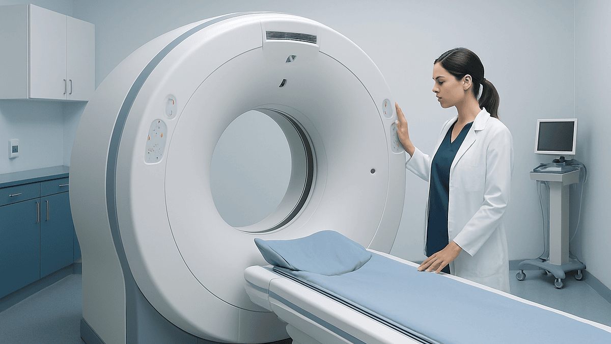
A CT scan (computed tomography) uses X-rays taken from many angles to build cross-sectional pictures. It is fast, shows most organs clearly and is the workhorse for checking chest, abdomen and pelvis for spread. Contrast dye, given by mouth or into a vein, helps blood vessels and tumors stand out.
MRI (magnetic resonance imaging) uses magnetic fields rather than radiation and gives finer detail of soft tissue. It is favored for brain, spine, pelvis, liver and prostate, and for judging how deeply a tumor has grown into nearby layers, which is the T question.
PET (positron emission tomography) works on chemistry rather than anatomy. A small amount of radioactive sugar is injected; cancer cells that burn glucose quickly light up. Mayo Clinic notes the tracer takes about 60 minutes to be absorbed before scanning, and the scan itself lasts roughly 30 minutes. PET is usually fused with CT so that a bright spot can be located precisely. It is especially useful for finding unexpected distant deposits, the M question, though inflammation and infection can also glow, so bright spots often need confirmation.
Ultrasound uses sound waves, involves no radiation and guides many biopsies. Endoscopic ultrasound, performed from inside the digestive tract, is used to stage esophageal, stomach and pancreatic cancers. Bone scans look for skeletal spread in cancers that favor bone, such as prostate and breast.
Which tests are ordered depends on the cancer type and on what earlier results showed. A radiologist reads each study and reports measurements that map directly onto TNM scores. Their report is a description, not a diagnosis; tissue still decides.
Why a biopsy is still essential: what pathology adds that scans cannot
Scans show shadows. Only tissue proves what those shadows are. A biopsy, the removal of a small sample of the suspected tumor, is the step that turns a suspicious mass into a named cancer, and MedlinePlus describes it as the way most cancers are definitively diagnosed.
Samples are taken in several ways. A needle biopsy, often guided by ultrasound or CT, draws out a core of tissue through the skin. An endoscopic biopsy collects tissue from inside the airway or digestive tract. An excisional biopsy removes the whole lump, common for skin lesions and some lymph nodes. Which method is chosen depends on where the tumor sits and how much tissue the pathologist needs.
In the laboratory the sample is preserved, sliced thinner than a sheet of paper, stained and examined. The pathologist confirms that cancer is present, identifies the cell type, assigns a grade and checks whether the tumor is invading blood or lymph vessels. Special stains and molecular tests can then look for features that steer treatment: hormone receptors in breast cancer, specific gene mutations in lung cancer or melanoma, and protein markers that predict whether particular drug classes are likely to be relevant.
Pathology also settles ambiguity in staging. A lymph node that looks enlarged on CT may simply be reacting to infection; a node that looks normal may harbor microscopic cancer. Sampling nodes, whether by needle or during surgery, converts an N guess into an N fact.
Sometimes a second biopsy is requested, often because the first sample was too small for molecular testing or because a distant spot on a scan needs confirmation before it changes the stage. This is not a sign that something went wrong. Proving spread before treating for spread is careful medicine.
Clinical staging vs pathological staging: why your stage may be revised
Staging happens in two passes, and understanding this saves considerable confusion later.
The first pass is clinical staging, written as cTNM. It combines everything known before any major treatment: physical examination, imaging, biopsy findings and blood tests. This is the stage the team uses to plan. Everyone has a clinical stage.
The second pass is pathological staging, written as pTNM. It becomes possible only if surgery removes the tumor and, usually, the surrounding lymph nodes. The pathologist measures the actual tumor, counts the nodes that contain cancer and checks whether the surgical margins are clear. Pathological staging is more accurate than clinical staging because it examines the tissue directly rather than inferring from pictures.
The two can disagree. Imaging may miss tiny deposits in lymph nodes, so a cancer staged clinically as N0 can become pathologically N1. Less often, an enlarged node that looked suspicious turns out to be benign, and the stage moves down. Neither situation means the first team made a mistake; it reflects the limits of what scans can resolve.
When a person receives chemotherapy or radiation before surgery, a practice called neoadjuvant treatment, a third notation appears: ypTNM, describing what the pathologist finds after that treatment has already shrunk the tumor. This is why some people hear two or even three stage descriptions for one cancer. All of them are correct for the moment they describe.
For record-keeping and research, the clinical stage at diagnosis is preserved as the original stage. If a later stage is different, the team will explain which one is guiding the current plan. If they do not, ask; it is a fair and common question.
Who is staged fully before treatment, and who is asked to wait, or to move faster?
Complete staging before any treatment is the standard path for most solid tumors where the plan hinges on extent. Breast, lung, colorectal, prostate, bladder, esophageal, stomach, pancreatic, head and neck, gynecologic cancers and melanoma typically follow this route. The team wants the full TNM picture before recommending surgery, radiation or systemic therapy, because the wrong first move is difficult to undo.
Some people are asked to wait longer than they expected. Common reasons include a first biopsy that yielded too little tissue for molecular testing, a PET finding that needs its own biopsy, or a scan that was inconclusive and must be repeated with a different technique. Waiting for a multidisciplinary meeting, where several specialists review the case at once, is another frequent source of a few extra days. Mayo Clinic describes this collaborative review as routine in modern cancer care.
Others are moved faster, with staging compressed or partly deferred. Cancers that are compressing the spinal cord, blocking the airway or bowel, or causing bleeding may need urgent treatment of the emergency before every scan is complete. Fast-growing blood cancers such as acute leukemia are often treated within days of diagnosis on the basis of blood and marrow tests, because their staging does not depend on imaging in the same way. Highly aggressive tumors such as some lymphomas may begin treatment with the first cycle while remaining scans are finished.
The middle group is people whose cancer is very small and low grade, where some teams offer active surveillance, meaning careful monitoring rather than immediate treatment. This applies to selected prostate, thyroid and kidney cancers and to certain in situ lesions. Surveillance is itself a decision that depends on complete staging; nobody is watched without first being mapped.
In every case the treating team weighs urgency against completeness and explains the reasoning. Ask them where you sit and why.
What do the days and weeks of staging usually look like?
There is no universal calendar, because tests are ordered in response to earlier results, but the sequence tends to follow a recognizable shape.
The first days usually belong to the biopsy and its report. The tissue must be processed, stained and read, and if molecular tests are added, those run on a separate timeline. Ask the team what turnaround they expect for your specific report rather than relying on general figures, since it varies by laboratory and by which tests were requested.
Imaging often runs in parallel. A CT of chest, abdomen and pelvis can usually be arranged quickly; MRI and PET may need scheduling around scanner availability and preparation requirements. Preparation is practical rather than dramatic: fasting for several hours before a PET scan, drinking contrast before some CTs, removing metal for MRI, and telling staff about kidney problems, allergies, pacemakers or pregnancy. Mayo Clinic advises that the radioactive tracer used for PET leaves the body within hours through urine, and that drinking water afterward helps.
Once results are in, the case goes to the multidisciplinary meeting. Surgeons, medical oncologists, radiation oncologists, radiologists and pathologists review the images and slides together and agree on a recommended approach. You then meet a clinician to hear the stage, the options and the reasoning, and to ask questions.
Emotionally these weeks are often harder than treatment itself. Uncertainty is heavier than a plan. Bring someone to appointments, write questions down beforehand, and ask for a single named contact who can tell you where each result stands. It is reasonable to ask how staging delays might affect the plan; for most solid tumors, the team will explain that a few weeks spent getting the map right does not change the strategy, while a wrong strategy would.
How much time does cancer take to go from stage 1 to 4?
This question is searched constantly, and the truthful answer is that no single number exists, because stage is not a timer that advances at a fixed rate.
Progression speed depends on the cancer type, its grade, its molecular features and the person carrying it. Some low-grade prostate and thyroid cancers can remain at stage 1 for many years, which is why active surveillance exists as a legitimate option. Some high-grade cancers, including certain lung, pancreatic and small cell tumors, can move from a localized state to distant spread over months. The same organ can host cancers at both ends of this range. Grade, discussed earlier, is the microscope clue to which end a particular tumor is closer to.
The stages are also not a staircase that every cancer climbs in order. A cancer can present at stage 4 without having been detected at any earlier stage, either because it grew in a location that produced no symptoms or because it spread early relative to its size. Others never progress beyond the stage at which they were found and treated.
This has two consequences for anyone worried about a wait. First, the interval between diagnosis and treatment is measured in weeks for most solid tumors, and stage transitions are generally not expected on that timescale; the team monitors for exceptions. Second, when symptoms have been present for a long time, that history is useful information for the team, not a reason for self-blame. Many cancers cause no symptoms until they are sizable, and mainstream sources including Mayo Clinic stress that this is a property of the disease rather than a failure of the person.
If you want a sense of how your specific cancer tends to behave, ask your oncologist. They can speak to the natural history of that type in a way no general figure can.
Is stage 4 cancer terminal? What terminal and advanced actually mean
Stage 4 means the cancer has spread to at least one distant organ. Terminal means a cancer that is not expected to respond to treatment and is expected to lead to death. These are different statements, and the words are often used as if they were the same.
Stage 4 is a description of extent. Terminal is a judgment about trajectory, made by a treating team about one person at one moment, taking into account the cancer type, its molecular features, how it has responded so far and the person’s overall health. Some stage 4 cancers are managed as long-term conditions for years with systemic therapy, particularly where targeted or hormonal treatments exist. A small number of stage 4 cancers with limited spread, sometimes called oligometastatic disease, are treated with the aim of removing every detectable deposit. Others are indeed life-limiting on a short horizon. Stage 4 tells you which conversation is coming; it does not tell you how it ends.
The NHS uses the word advanced for cancer that cannot be removed and has spread, and describes treatment in that setting as aimed at controlling the cancer, relieving symptoms and maintaining quality of life for as long as possible. Palliative care, meaning specialist support for symptoms and wellbeing, can run alongside active treatment from diagnosis onward and is not a signal that treatment has stopped.
If you or someone close to you has been told a cancer is stage 4, the useful questions are specific: what is the goal of the treatment being proposed, what would tell us it is working, and what would we do if it stopped working. Survival estimates for a given type and stage exist in published registries, but they describe large groups from past years and cannot be applied to one person without the treating team’s interpretation. Ask them to walk you through what the figures do and do not capture.
Does the stage change after treatment? Restaging, response and recurrence
The stage assigned at diagnosis does not officially change, but the cancer certainly can, and the team will repeatedly reassess it. This reassessment is usually called restaging or response assessment rather than a new stage.
During systemic therapy, scans are typically repeated after a set number of cycles to see whether the tumors have shrunk, stayed stable or grown. Radiologists often describe response using standardized measurement rules that compare the size of target lesions with the baseline scan. This is why the original staging images are so valuable: they are the yardstick.
If cancer returns after a period of no detectable disease, it is called a recurrence. A recurrence at the original site is local; in nearby nodes, regional; in a distant organ, distant or metastatic. Doctors will describe a recurrence in these terms rather than assigning a fresh stage number, though they may perform a full workup that looks very much like the original staging: imaging to define extent, and often a biopsy to confirm the cancer is the same one and to check whether its molecular features have shifted, which happens and can change which drug classes are relevant.
The phrase originally stage 2, now metastatic is therefore accurate and common. It preserves the reference point while describing the present.
Follow-up imaging schedules after treatment vary by cancer type and by guideline, and the team will set an interval that balances catching recurrence early against the radiation exposure and anxiety of frequent scans. If you are unsure why a scan is or is not being repeated, ask what the team is watching for and what would prompt them to look sooner.
What people often get wrong about cancer staging
Several misunderstandings surface again and again in clinic conversations. Correcting them makes the rest of the process easier to follow.
A bigger stage number means a faster cancer. Not necessarily. Stage measures extent; grade measures aggressiveness. A large, slow tumor can outrank a small, fast one on the stage scale.
Stage 4 means treatment is pointless. Stage 4 changes the goal of treatment for many cancers, from removal to long-term control, but it does not remove the options. Several common cancers are now managed at stage 4 for extended periods.
My stage went up, so the first doctors missed something. Pathological staging after surgery is more precise than imaging. Finding microscopic node involvement that CT could not see is expected, not negligent.
Waiting two weeks for scans lets the cancer advance a stage. For most solid tumors, staging intervals are short relative to how the disease progresses. The team watches for the exceptions and expedites when warranted.
Everyone with the same stage gets the same treatment. Molecular features, grade, other health conditions and personal priorities all shape the plan. Two people with identical TNM codes may reasonably receive different recommendations.
A clear PET scan proves there is no cancer anywhere. PET detects deposits above a certain size and metabolic activity. Small clusters of cells can be invisible, which is why lymph nodes are often sampled directly and why follow-up continues after treatment.
Lymphoma and leukemia use the same stages as solid tumors. They do not. Lymphoma has its own four-stage system based on lymph node regions, and leukemia is described by cell counts, genetics and risk category rather than anatomical spread.
If a phrase in your report or consultation does not match what you have read online, trust the report and ask the team to translate it.
Questions to ask your care team about staging
Good questions turn a confusing report into a shared plan. These are the ones clinicians most welcome, grouped by the moment they fit best.
Before or during the tests:
- Which scans and biopsies are you ordering, and what question is each one meant to answer?
- Is there anything I should stop eating, drinking or doing before each test, and should I mention any implants, allergies or kidney problems?
- Who will contact me with results, and roughly when should I expect each one?
When you receive the stage:
- What are my T, N and M scores, and what overall stage do they produce for this cancer type?
- Is this a clinical stage, or has pathology confirmed it? Could it change after surgery?
- What is the grade, and does it change how you would treat this stage?
- Were any molecular or receptor tests done, and how do the results affect the options?
When discussing the plan:
- What is the goal of the treatment you are recommending: removing all detectable cancer, controlling it long term, or relieving symptoms?
- What are the alternatives, including watchful waiting or a clinical trial, and why do you favor this one?
- Would you recommend a second opinion, and can you share the images and slides if I seek one?
- If the cancer does not respond as hoped, what would the next steps look like?
Bring a notebook or ask permission to record the conversation. Ask for a printed copy of the pathology report and the radiology reports; they are yours, and reading them at home with the definitions in this article will make the next appointment more productive. No question about your own stage is too basic to ask.
When to call your doctor during the staging period
Staging usually proceeds without incident, but the tests carry small risks and the cancer itself can cause problems that should not wait for the next scheduled appointment. Contact the team the same day, or seek emergency care, if any of the following occur.
After a biopsy: bleeding that does not stop with firm pressure after a few minutes, a rapidly enlarging or painful swelling at the site, fever or chills, spreading redness, or, after a lung biopsy, sudden shortness of breath or sharp chest pain, which can signal a collapsed lung.
After contrast dye: hives, swelling of the face or throat, wheezing or difficulty breathing, which can indicate an allergic reaction and needs emergency care; a marked drop in how much urine you pass over the following day should also be reported.
From the cancer itself, at any point: new weakness, numbness or difficulty walking, or loss of bladder or bowel control, which can indicate pressure on the spinal cord; severe or worsening headache with vomiting or confusion; coughing or vomiting blood, or black stools; a swollen, painful calf or sudden breathlessness, which can indicate a blood clot; inability to keep fluids down; or a fever with shaking, particularly if you have already begun any treatment that can lower white cell counts.
Less urgent but still worth a call: pain that is escalating despite the measures you were given, new jaundice (yellowing of the skin or eyes), or rapid unintended weight loss. These do not necessarily change the plan, but the team will want to know.
Ask at your first appointment for a direct number to use for these situations and for after-hours contact details. Writing that number on the fridge is a small act that makes an unsettling stretch of weeks a little safer. Every decision about how to respond rests with the treating team, and they would far rather hear from you early than late.
Frequently asked questions
Is stage 4 cancer terminal?
Not by definition. Stage 4 describes cancer that has spread to a distant organ, while terminal describes a cancer that is no longer expected to respond to treatment. Some stage 4 cancers are controlled for years with systemic therapy, and a few with limited spread are treated with the aim of removing all detectable disease. Only the treating team, knowing the specific cancer type and its response, can say where an individual sits.
How much time does cancer take to go from stage 1 to stage 4?
There is no fixed interval. Progression depends on cancer type, grade and molecular features; some low-grade prostate or thyroid cancers remain localized for years, while some high-grade lung or pancreatic cancers can spread within months. Cancers also do not necessarily pass through every stage in order. For most solid tumors, the weeks spent on staging are short relative to how the disease progresses, and the team expedites when a cancer appears aggressive.
What is the difference between stage 1 or 2 and stage 3 or 4 cancer?
Stages 1 and 2 describe cancer confined to its organ of origin or with limited local extension, often treated primarily with surgery or other local therapy. Stage 3 usually involves regional lymph nodes or invasion of nearby structures and often calls for combined treatments. Stage 4 means spread to a distant organ, where systemic therapy typically leads. The most consequential boundary is between 3 and 4.
What is terminal cancer?
Terminal cancer is cancer that is not expected to respond to treatment and is expected to lead to death, usually within a timeframe the treating team can only estimate. The NHS uses the term advanced for cancer that cannot be removed and has spread, and describes care in that setting as aimed at controlling the disease, relieving symptoms and preserving quality of life. Palliative care can run alongside active treatment throughout.
How is cancer staged if I have not had surgery yet?
Through clinical staging, which combines physical examination, imaging such as CT, MRI or PET, biopsy results and blood tests into a cTNM score. This is the stage used to plan treatment. If surgery later removes the tumor and lymph nodes, a pathological stage is assigned from the actual tissue and may differ slightly. Both are valid descriptions of the cancer at the moment each was assessed.
What is the TNM staging system in simple terms?
TNM is an international shorthand. T scores the size or depth of the primary tumor, N scores how many nearby lymph nodes contain cancer and where, and M records whether distant spread has been found. The three scores are combined, using rules specific to each cancer type, into an overall stage from 0 to 4. Letters such as c, p or y in front indicate whether the stage came from clinical tests, surgical pathology or assessment after preoperative treatment.
Why do I need a PET scan if I already had a CT scan?
The two tests answer different questions. CT shows anatomy in detail and measures tumors precisely. PET shows metabolic activity, highlighting cells that consume sugar rapidly, which helps find unexpected deposits that look normal in size on CT. Mayo Clinic notes that inflammation can also light up on PET, so bright spots often need confirmation. Together the scans give a more complete picture of extent than either alone.
Can my cancer stage go down after surgery?
Yes, though it happens less often than an upward revision. If a lymph node that appeared enlarged on imaging turns out under the microscope to contain no cancer, the pathological N score can be lower than the clinical estimate. The reverse, finding microscopic cancer in nodes that looked normal, is more common because imaging cannot detect very small deposits. Either way the original clinical stage stays on record as the reference point.
What is the difference between cancer stage and cancer grade?
Stage measures how far the cancer has spread in the body. Grade measures how abnormal the cancer cells look and how quickly they appear to be dividing, judged by a pathologist under a microscope, commonly on a scale of 1 to 3 or 1 to 4. A small stage 1 tumor can be high grade, and a stage 3 tumor can be low grade. Treatment planning uses both, and for some cancers grade carries the greater weight.
Does waiting for staging tests let the cancer spread?
For most solid tumors, the weeks needed to complete staging are short compared with the timescale over which these cancers progress, and treating without a complete map risks choosing the wrong approach. Teams monitor for exceptions: cancers causing emergencies, or fast-growing blood cancers and lymphomas, may begin treatment before every test is finished. If you are worried, ask your team directly how the wait relates to your specific cancer type.
References
This article is for general information only and is not a substitute for professional medical advice. Please consult a qualified doctor about your individual situation.

More from the Blog

When Does Leukemia Treatment Include a Stem Cell Transplant, and Who Is Assessed for It?
A stem cell transplant becomes part of leukemia treatment when the disease is judged likely to return after chemotherapy alone, or when it has…

Surgery or Radiation for Prostate Cancer? How Specialists Compare the Two Paths
For localized prostate cancer, surgery and radiation therapy are both standard treatments, and the largest long-term randomized trial found deaths from prostate cancer were…

How Is HIPEC Performed? Tumor Removal Followed by Heated Chemotherapy Inside the Abdomen
HIPEC is performed in two stages during a single operation. Surgeons first carry out cytoreductive surgery, removing every visible tumor deposit from the abdominal…
TURBT for Bladder Cancer: How the Endoscopic Tumor Removal Is Performed
A TURBT (transurethral resection of bladder tumor) removes a bladder growth through the urethra with a thin telescope and an electrified wire loop, so…

Life After Bladder Removal Surgery: Urinary Diversion Options and Daily Adjustments
After bladder removal (cystectomy), urine must leave the body by a new route called a urinary diversion: an ileal conduit draining into an external…

Getting a Second Opinion on a Melanoma Diagnosis: Pathology Review and Plan Questions
A second opinion on a melanoma diagnosis usually has two parts: a fresh read of the original biopsy slides by a pathologist who specializes…